

Clinical and genetic factors associated with contralateral progression in unilateral moyamoya disease: Longitudinal and Cross-Sectional Study
- PMID: 38404780
- PMCID: PMC10884840
- DOI: 10.1016/j.heliyon.2024.e26108
Clinical and genetic factors associated with contralateral progression in unilateral moyamoya disease: Longitudinal and Cross-Sectional Study
Abstract
Objective: This study aimed to explore the long-term outcome of unilateral moyamoya disease and predict the clinical and genetic factors associated with contralateral progression in unilateral moyamoya disease.
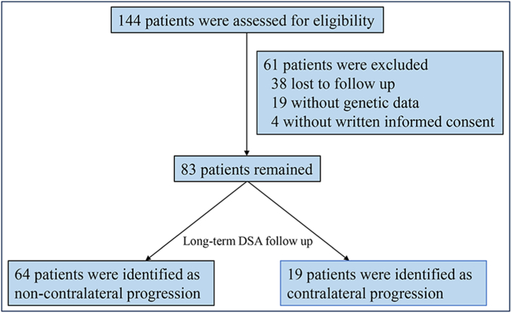
Methods: We retrospectively recruited unilateral moyamoya disease patients with available genetic data who underwent encephaloduroarteriosynangiosis (EDAS) surgery at our institution from January 2009 to November 2017. Long-term follow-up data, including clinical outcomes, angiographic features, and genetic information, were analyzed.
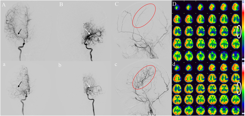
Results: A total of 83 unilateral moyamoya disease patients with available genetic data were enrolled in our study. The mean duration of clinical follow-up was 7.9 ± 2.0 years. Among all patients, 19 patients demonstrated contralateral progression to bilateral disease. Heterozygous Ring Finger Protein 213 p.R4810K mutations occurred significantly more frequently in unilateral moyamoya disease patients with contralateral progression. Furthermore, patients with contralateral progression typically demonstrated an earlier age of onset than those with non-progressing unilateral moyamoya disease. In the contralateral progression group, posterior circulation involvement was observed in 11 (11/19, 57.9%) patients compared to 12 (12/64, 18.8%) in the non-contralateral progression group (P = 0.001). The time to peak of cerebral perfusion and neurological status showed significant postoperative improvement.
Conclusion: Long-term follow-up revealed that the EDAS procedure might provide benefits for unilateral moyamoya disease patients. Ring Finger Protein 213 p.R4810K mutations, younger age, and posterior circulation involvement might predict the contralateral progression of unilateral moyamoya disease.
© 2024 The Authors.
Conflict of interest statement
The authors declare the following financial interests/personal relationships which may be considered as potential competing interests:Lian Duan reports article publishing charges was provided by National Natural Science Foundation of China. Qian-Nan Wang reports article publishing charges was provided by National Natural Science Foundation of China. If there are other authors, they declare that they have no known competing financial interests or personal relationships that could have appeared to influence the work reported in this paper.
Figures
Similar articles
-
RNF213 R4810K Variant in Suspected Unilateral Moyamoya Disease Predicts Contralateral Progression.J Am Heart Assoc. 2022 Aug 2;11(15):e025676. doi: 10.1161/JAHA.122.025676. Epub 2022 Jul 25. J Am Heart Assoc. 2022. PMID: 35876407 Free PMC article.
-
Genetic and nongenetic factors for contralateral progression of unilateral moyamoya disease: the first report from the SUPRA Japan Study Group.J Neurosurg. 2021 Sep 10;136(4):1005-1014. doi: 10.3171/2021.3.JNS203913. Print 2022 Apr 1. J Neurosurg. 2021. PMID: 34507293
-
A long-term study of posterior circulation changes after revascularization in patients with moyamoya disease.J Neurosurg. 2023 Apr 7;139(5):1281-1286. doi: 10.3171/2023.2.JNS222649. Print 2023 Nov 1. J Neurosurg. 2023. PMID: 37029668
-
Clinical Features and Surgical Outcomes of Patients With Moyamoya Disease and the Homozygous RNF213 p.R4810K Variant.J Child Neurol. 2019 Nov;34(13):793-800. doi: 10.1177/0883073819858264. Epub 2019 Jul 10. J Child Neurol. 2019. PMID: 31290353 Review.
-
[A case of adult moyamoya disease showing fulminant clinical course associated with progression from unilateral to bilateral involvement].No Shinkei Geka. 1997 Jan;25(1):79-84. No Shinkei Geka. 1997. PMID: 8990473 Review. Japanese.
Cited by
-
Research progress in unilateral moyamoya disease.Front Hum Neurosci. 2025 Jan 27;19:1503639. doi: 10.3389/fnhum.2025.1503639. eCollection 2025. Front Hum Neurosci. 2025. PMID: 39931047 Free PMC article. Review.
-
Vascular architecture characters and risk factors analysis of unstable moyamoya disease.Front Neurol. 2024 May 31;15:1398007. doi: 10.3389/fneur.2024.1398007. eCollection 2024. Front Neurol. 2024. PMID: 38882694 Free PMC article.
References
-
- Suzuki J., Takaku A. Cerebrovascular "moyamoya" disease. Disease showing abnormal net-like vessels in base of brain. Arch. Neurol. 1969;20(3):288–299. - PubMed
-
- Choi J.U., Kim D.S., Kim E.Y., Lee K.C. Natural history of moyamoya disease: comparison of activity of daily living in surgery and non surgery groups. Clin. Neurol. Neurosurg. 1997;99(Suppl 2):S11–S18. - PubMed
-
- Research committee on the pathology and treatment of spontaneous occlusion of the circle of willis; health labour sciences research grant for research on measures for infractable diseases. Guidelines for diagnosis and treatment of moyamoya disease (spontaneous occlusion of the circle of willis) Neurol. Med.-Chir. 2012;52(5):245–266. doi: 10.2176/nmc.52.245. - DOI - PubMed
-
- Smith E.R., Scott R.M. Progression of disease in unilateral moyamoya syndrome. Neurosurg. Focus. 2008;24(2):E17. - PubMed
-
- Houkin K., Abe H., Yoshimoto T., Takahashi A. Is "unilateral" moyamoya disease different from moyamoya disease? J. Neurosurg. 1996;85(5):772–776. - PubMed
LinkOut - more resources
Full Text Sources
