Case Reports
. 2023 Jul-Dec;13(2):232-235.
doi: 10.4103/ams.ams_49_23. Epub 2023 Sep 26.

Florid Cemento-osseous Dysplasia Associated with Secondary Infection - A Case Report

Affiliations
Case Reports

Florid Cemento-osseous Dysplasia Associated with Secondary Infection - A Case Report

Abrar Khalid Demyati. Ann Maxillofac Surg. 2023 Jul-Dec.

Abstract

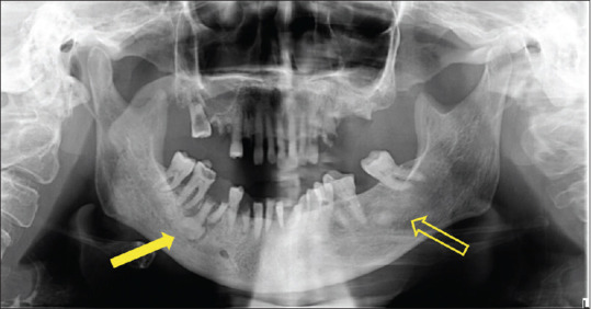
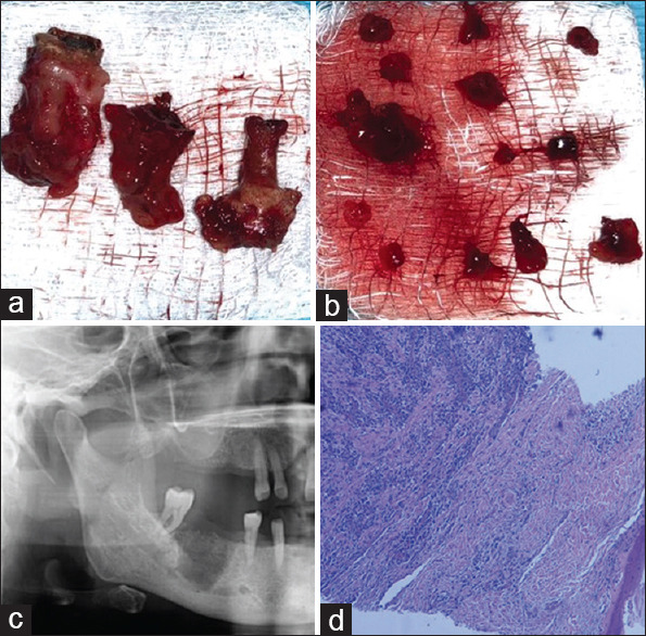
Rationale: The term cemento-osseous dysplasia (COD) refers to a bony fibro-osseous lesion, in which fibrous tissue and cementum-like tissue replace normal bone. There are three types of COD: periapical, focal and florid. The condition is usually asymptomatic and treatment is unnecessary; however, a secondary infection could occur, which requires treatment.

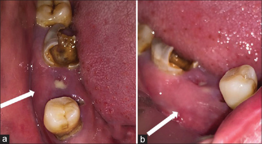
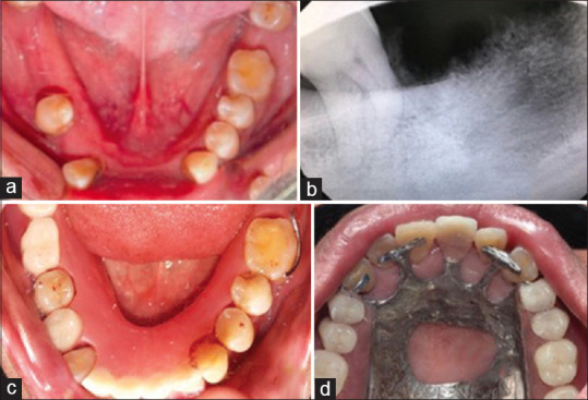
Patient concern: A 58-year-old female patient presented with symptoms in the mandibular posterior region of the right jaw for six months.

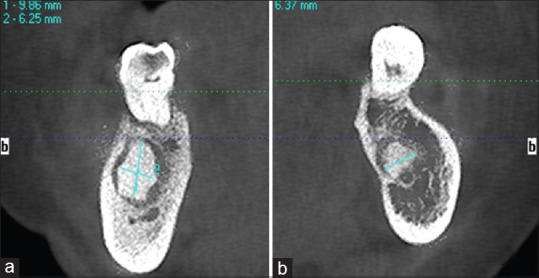
Diagnosis: Infected florid COD (FCOD).

Treatment: A pre-operative antibiotic, followed by extraction of non-restorable teeth, debridement of the infected tissue and necrotic bone removal.

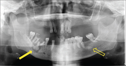
Outcome: The patient was followed for one year, during which all previously reported signs and symptoms were resolved.

Take-away lessons: Early lesion detection is essential. Treatment depends on the presence or absence of clinical and radiographic manifestations. The current case was treated surgically to minimise complications.

Keywords: Cemento-osseous dysplasia; florid; focal; infection; periapical.


Conflict of interest statement

There are no conflicts of interest.

Figures

Figure 1
Figure 1
The right lower quadrant with grossly decayed right lower molars. (a) Occlusal view with areas of suppuration (white solid arrow); (b) Lateral view showing the swelling (white solid arrow)
Figure 2
Figure 2
Pre-operative orthopantomogram. Radio-opaque lesions in the mandible localized on the periapical area of tooth #46 (solid arrow) and in the edentulous area corresponding to tooth #37 (hollow arrow). Multiple carious non-restorable teeth and periodontal disease can also be seen
Figure 3
Figure 3
Pre-operative panoramic radiograph reveals a radio-opaque lesion with radiolucent margin associated with grossly decayed tooth #46 (solid arrow). Radio-opaque lesion was related to the edentulous area of tooth #37 (hollow arrow)
Figure 4
Figure 4
(a) Cone-beam computed tomography of the coronal cross section shows a well-defined radio-opacity with the greatest dimension of 9.86 mm × 6.25 mm in the right lower area. (b) A well-defined radio-opacity with the greatest dimension of 6.37 mm × 5.32 mm in the left lower area
Figure 5
Figure 5
(a) Extracted teeth with the associated lesion. (b) Granulation tissue. (c) Immediate panoramic radiograph after surgical debridement. (d) Haematoxylin – eosin staining of a medium-power histologic cut revealing a transition of bone mass; the adjacent fibrous tissue presents an inflammatory infiltrate
Figure 6
Figure 6
(a) Clinical follow-up after six months. (b) Radiographic follow-up after six months. (c) Lower interim removable partial denture. (d) Upper metallic partial denture

Similar articles

Cited by

References

    1. Nelson BL, Phillips BJ. Benign fibro-osseous lesions of the head and neck. Head Neck Pathol. 2019;13:466–75. - PMC - PubMed
    1. El-Naggar AK, Chan JK, Grandis JR. Geneva, Switzerland: IARC Press, WHO Press, World Health Organization; 2017. WHO Classification of Head and Neck Tumours.
    1. Fokam ST, Kwedi GK, Messanga CB. Infected florid cemento-osseous dysplasia: About one clinical observation. Adv Oral Maxillofac Surg. 2022;7:100298.
    1. Min CK, Koh KJ, Kim KA. Recurrent symptomatic cemento-osseous dysplasia: A case report. Imaging Sci Dent. 2018;48:131–7. - PMC - PubMed
    1. Kato CN, de Arruda JA, Mendes PA, Neiva IM, Abreu LG, Moreno A, et al. Infected cemento-osseous dysplasia: Analysis of 66 cases and literature review. Head Neck Pathol. 2020;14:173–82. - PMC - PubMed

Publication types