[Preprint]. 2024 Feb 18:2023.11.26.568771.
doi: 10.1101/2023.11.26.568771.

Multiomic single-cell sequencing defines tissue-specific responses in Stevens-Johnson Syndrome and Toxic epidermal necrolysis

Affiliations

Multiomic single-cell sequencing defines tissue-specific responses in Stevens-Johnson Syndrome and Toxic epidermal necrolysis

Andrew Gibson et al. bioRxiv. .

Update in

Abstract

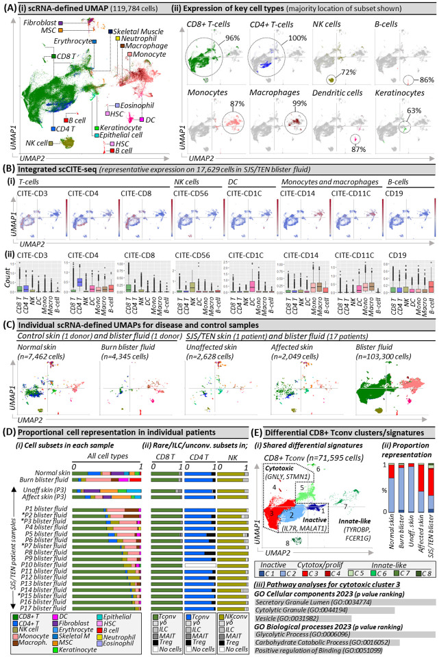
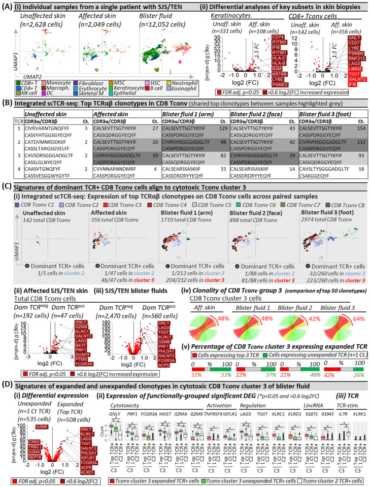
Stevens-Johnson syndrome and toxic epidermal necrolysis (SJS/TEN) is a rare but life-threatening cutaneous drug reaction mediated by human leukocyte antigen (HLA) class I-restricted CD8+ T-cells. To obtain an unbiased assessment of SJS/TEN cellular immunopathogenesis, we performed single-cell (sc) transcriptome, surface proteome, and TCR sequencing on unaffected skin, affected skin, and blister fluid from 17 SJS/TEN patients. From 119,784 total cells, we identified 16 scRNA-defined subsets, confirmed by subset-defining surface protein expression. Keratinocytes upregulated HLA and IFN-response genes in the affected skin. Cytotoxic CD8+ T-cell subpopulations of expanded and unexpanded TCRαβ clonotypes were shared in affected skin and blister fluid but absent or unexpanded in SJS/TEN unaffected skin. SJS/TEN blister fluid is a rich reservoir of oligoclonal CD8+ T-cells with an effector phenotype driving SJS/TEN pathogenesis. This multiomic database will act as the basis to define antigen-reactivity, HLA restriction, and signatures of drug-antigen-reactive T-cell clonotypes at a tissue level.


Figures

Figure 1.
Figure 1.. Unbiased multi-focal single-cell sequencing identifies expanded pathogenic CD8+ Tconv in affected skin and blister fluid during Stevens-Johnson syndrome and toxic epidermal necrolysis.
(A) (i) scRNA-defined uniform manifold approximation and projection (UMAP) of 119,784 live cells from normal skin, burn blister fluid, and unaffected skin, affected skin and/or blister fluid from 17 patients with acute SJS/TEN. (ii) Expression distribution for key cell types. The majority UMAP location of each subset is labeled with the percentage of each subset within that location indicated. Figure created using VGAS. (B) Integrated scCITE-seq expression of subset-defining cell surface proteins for T-cells (CD3, CD4, CD8), NK cells (CD56), DC (CD1c), monocytes and macrophages (CD14, CD11c), and B cells (CD19). (i) Heatmap expression (high to low, red to blue) for each surface protein is shown on a representative UMAP of 17,629 cells obtained within a single 10x run, and (ii) average box plot expression is shown between scRNA-defined subsets. Figure created using VGAS. (C) Individual scRNA-defined UMAPs for tissue-relevant controls from unrelated donors (burn blister fluid, normal skin) and skin and blister fluid from patients with SJS/TEN. The average expression is shown for patients with multiple samples from the same time point, indicated by an asterisk. Figure created using VGAS. (D) scRNA-defined cell representation of (i) all subsets in individual samples and (ii) rare, unconventional, and innate-like lymphoid cell (ILC) populations as a proportion of total CD8 T-cells, CD4 T-cells, or NK cells in each sample. (E) (i) Seurat-defined Tconv clusters 1–8 with shared naming according to differential gene expression signatures and (ii) proportional representation in control and disease sample phenotypes. (iii) Pathway analysis for the significant differentially expressed gene signature of CD8 Tconv cluster 3 using Enrichr and gene ontology (GO) cellular and biological terms ranked by p-value. MSC, Mesenchymal stromal cell; HSC, Hematopoietic stem cell; NK, Natural killer; DC, Dendritic cell; Tconv, T conventional cell; unconv., unconventional; ILC, innate-like lymphoid cell; Unaff, Unaffected; Aff, Affected.
Figure 2.
Figure 2.. Shared oligoclonal TCRαβ clonotypes on cytotoxic CD8 Tconv in affected skin and blister fluid are absent from or unexpanded in unaffected skin.
(A) (i) UMAPs for paired unaffected and affected skin and blister fluid samples from a single patient with SJS/TEN. (ii) Differential gene expression (Wilcoxon, Hochberg adj, p<0.05) between keratinocytes or CD8+ T-cells from unaffected and affected skin. Genes colored red are significantly (p<0.05) increased (light red <0.6log2FC, dark red >0.6log2FC). The top 10 genes are labeled. (B) Top functional TCR CDR3αβ pairings and counts (Ct.) in CD8 Tconv cells. The same top TCR clonotypes across affected samples are highlighted grey. (C) Signatures of dominant TCR+ CD8 Tconv align to cytotoxic Tconv cluster 3. (i) Expression of the top 3 dominantly-expanded TCR (black) on CD8 Tconv cells of each sample. The number of dominant TCR+ cells aligned to CD8 Tconv cluster 2 (control) and cluster 3 (cytotoxic) are indicated numerically. (ii-iii) Differential gene expression (Wilcoxon, Hochberg adj, p<0.05) between CD8 Tconv expressing dominant TCRs or other TCRs in (ii) affected skin or (iii) blister fluids. Genes colored red are significantly (p<0.05) increased (light red <0.6log2FC, dark red >0.6log2FC). The top 10 genes are labeled. (iv) Clonality of CD8 Tconv cluster 3 in affected skin and blister fluid samples. The top 50 TCRs are shown and the percentage indicates the proportion of counts aligned to the top 3 dominantly-expanded TCRs. Circos plot segment width is proportionate to dominance (increasing green to red). (v) Proportional expression of dominantly-expanded TCR+ cells (red) and unexpanded clonotypes (green, n=1 count) in CD8+ Tconv cluster 3 across affected skin and blister fluid samples. (D) Signatures of expanded and unexpanded clonotypes in Tconv cluster 3. (i) Differential gene expression (Wilcoxon, Hochberg adj, p<0.05) between expanded or unexpanded TCR+ cells of CD8 Tconv cluster 3 in SJS/TEN blister fluid. Genes colored red are significantly (p<0.05) increased (light red <0.6log2FC, dark red >0.6log2FC). The top 10 genes are labeled. (ii) DEG and (iii) interest TCR-activation-related gene expression between cells of Tconv cluster 3 expressing expanded (red) or unexpanded (green) TCR compared to TCR from control Tconv cluster 2 (white). *Indicates significant differential expression (Wilcoxon, Hochberg adj, p<0.05 and >0.6 log2FC). Figure created using VGAS. TCR, T-cell receptor; Dom TCR, Dominantly-expanded TCR; MSC, Mesenchymal stromal cell; HSC, Hematopoietic stem cell; NK, Natural killer; DC, Dendritic cell; Tconv, T conventional cell; CDR3, Complementary determining region 3; Ct., count; FDR, false discovery rate; FC, fold change; LincRNA, long intergenic non-coding RNA; Unaff, Unaffected; Aff, Affected.

Similar articles

References

    1. Marks M.E., et al. Updates in SJS/TEN: collaboration, innovation, and community. Front Med (Lausanne) 10, 1213889 (2023). - PMC - PubMed
    1. Chung W.H., et al. Medical genetics: a marker for Stevens-Johnson syndrome. Nature 428, 486 (2004). - PubMed
    1. Pan R.Y., et al. Identification of drug-specific public TCR driving severe cutaneous adverse reactions. Nature communications 10, 3569 (2019). - PMC - PubMed
    1. Chung W.-H., et al. Granulysin is a key mediator for disseminated keratinocyte death in Stevens-Johnson syndrome and toxic epidermal necrolysis. Nature Medicine 14, 1343–1350 (2008). - PubMed
    1. Bailin S.S., et al. Changes in subcutaneous white adipose tissue cellular composition and molecular programs underlie glucose intolerance in persons with HIV. Frontiers in immunology 14(2023). - PMC - PubMed

Publication types