[Preprint]. 2024 Apr 15:2024.02.10.579785.
doi: 10.1101/2024.02.10.579785.

Nuclear receptor interdomain communication is mediated by the hinge with ligand specificity

Affiliations

Nuclear receptor interdomain communication is mediated by the hinge with ligand specificity

Saurov Hazarika et al. bioRxiv. .

Update in

Abstract

Nuclear receptors are ligand-induced transcription factors that bind directly to target genes and regulate their expression. Ligand binding initiates conformational changes that propagate to other domains, allosterically regulating their activity. The nature of this interdomain communication in nuclear receptors is poorly understood, largely owing to the difficulty of experimentally characterizing full-length structures. We have applied computational modeling approaches to describe and study the structure of the full length farnesoid X receptor (FXR), approximated by the DNA binding domain (DBD) and ligand binding domain (LBD) connected by the flexible hinge region. Using extended molecular dynamics simulations (> 10 microseconds) and enhanced sampling simulations, we provide evidence that ligands selectively induce domain rearrangement, leading to interdomain contact. We use protein-protein interaction assays to provide experimental evidence of these interactions, identifying a critical role of the hinge in mediating interdomain contact. Our results illuminate previously unknown aspects of interdomain communication in FXR and provide a framework to enable characterization of other full length nuclear receptors.


Figures

Figure 1.
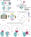
Figure 1.. Structural models of full-length FXR.
A) The fl-FXR model was generated by using full-length LXRβ (PDB 4NQA) as a template. Models of FXR DBD and LBD were aligned to LXRβ to predict initial arrangement of the domains. B) Modeller was used to insert the hinge region between the DBD and LBD. C) Two conformations of fl-FXR emerged following accelerated MD simulations to optimize the hinge conformation. D) Two fl-FXR conformations are designated as extended and compact. Interdomain angle (ω) and interdomain distance (r) parameters are illustrated on the models. E) Alignment of existing full length nuclear receptor crystal structures shows a range of interdomain (DBD-LBD) angles which encompass the angles of our extended and compact models.
Figure 2:
Figure 2:. Analysis of extended fl-FXR MD simulations.
Conformational changes, RMSF and changes in interdomain angle and distance are characterized for FXR-OCA (A-C), FXR-CDCA (D-F), FXR-LCA (G-I) and apo-FXR (J-L). DBD, hinge and LBD are colored magenta, grey and cyan respectively. FXR-OCA shifts from an extended to compact conformation over the 20 μs simulation (A, C). FXR-CDCA shifts into a partially compact conformation (D, F). FXR-LCA and apo-FXR do not undergo large conformational changes over the simulation (G, I, J, L). The highlighted region in RMSF plots is the hinge (B, E, H, K). For all complexes, largest fluctuations are observed in the DBD and hinge while the LBD remains stable.
Figure 3.
Figure 3.. Conformational classification of fl-FXR.
A) Two fl-FXR conformations with similar interdomain angle and interdomain distance have different 3D architectures. This illustrates that interdomain angle and distance are insufficient to describe the conformational ensemble of fl-FXR. B) We defined new parameters, namely rotational angle (θ) and vertical displacement (dv) to describe the 3D rotation of the DBD relative to the LBD. Combined with (r), these parameters can cluster the fl-FXR conformations from our simulations. C) To illustrate the rotational angle (θ), various interaction surfaces on the LBD are shown. For instance, a 180° rotation implies the DBD resides on the H10/H7 face, while a 90° rotation indicates that the DBD is located at the H5–H7 edge. D) Polar plot showing fl-FXR conformations described by rotational angle (θ), interdomain distance r (radially outward) and color-coded vertical displacement (dv). E-G) The conformations cluster into three groups (DLI-1, DLI-2, DLI-3), based on the interdomain interface (i.e. the LBD face/edge interacting with the DBD. E) In DLI-1, the interface is the H10/H7 LBD face. Two sub-groups are identified with positive or negative vertical displacement, respectively. F) The interdomain interface in DLI-2 is the H9 edge, all structures have positive dv. G) The interdomain interface in DLI-3 is the H5–H7 edge, all structures have negative dv.
Figure 4.
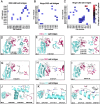
Figure 4.. Interdomain salt bridges in fl-FXR.
Salt bridges were identified from 38 accelerated MD trajectories of fl-FXR in various ligand bound states. Frequency of A) DBD-LBD, B) hinge-DBD, and C) hinge-LBD salt bridges are plotted as heat maps for comparison. Asterisks (*) indicate salt bridges illustrated in panels D-L. Of all three groups, hinge-LBD salt bridges are most prevalent across the 38 trajectories. D-E) DBD-LBD salt bridges are illustrated in DLI-1 and DL-3 conformations. G-I) Hinge-DBD salt bridges are illustrated in DLI-I conformations. J-L) Hinge-LBD salt bridges are illustrated in DLI-1, DLI-2 and DLI-3 conformations.
Figure 5.
Figure 5.. Experimental validation of interdomain interaction in FXR.
A) Four mammalian two hybrid fusion constructs were prepared for this study. The VP16 activation domain was fused to the FXR DBD (residues 120–196) and DBD plus hinge (residues 120–244). The Gal4DBD was fused to the FXR LBD (residues 247–476) and to the hinge plus LBD (residues 197–476). An unfused VP16 protein was used for experimental controls. B) Transcriptional activity of the luciferase gene under control of the UAS promoter is used to measure the interaction between Gal4DBD and VP16 fusion constructs. Data are reported as fold changes over control with no ligand (DMSO only). For all four ligands, transcription is measured under four conditions: LBD + no DBD control, LBD + DBD, LBD + DBD-hinge, and hinge-LBD + DBD. Background activation is observed in the control (no DBD) condition. No significant increase above background is observed for any ligand in the DBD + LBD condition. Similarly, no significant increase above background is observed in the DBD-hinge + LBD condition. A significant increase is only observed in the DBD + hinge-LBD condition, indicative of an interaction between the two fusion proteins.

Similar articles

References

    1. Mangelsdorf D.J.,’, Thummel C., Beato M., Herrlich P., Schiitq5 G., Umesono K., Blumberg B., Kastner P., Mark M., Chambon P., and Evan R.M.&’, The Nuclear Receptor Superfamily: The Second Decade (1995). - PMC - PubMed
    1. Weikum E.R., Liu X., and Ortlund E.A., “The nuclear receptor superfamily: A structural perspective,” Protein Science 27(11), 1876–1892 (2018). - PMC - PubMed
    1. Jiang L., Zhang H., Xiao D., Wei H., and Chen Y., “Farnesoid X receptor (FXR): Structures and ligands,” Comput Struct Biotechnol J 19, 2148–2159 (2021). - PMC - PubMed
    1. Lou X., Toresson G., Benod C., Suh J.H., Philips K.J., Webb P., and Gustafsson J.A., “Structure of the retinoid X receptor α-liver X receptor β (RXRα-LXRβ) heterodimer on DNA,” Nat Struct Mol Biol 21(3), 277–281 (2014). - PubMed
    1. Chandra V., Huang P., Hamuro Y., Raghuram S., Wang Y., Burris T.P., and Rastinejad F., “Structure of the intact PPAR-γ-RXR-α nuclear receptor complex on DNA,” Nature 456(7220), 350–356 (2008). - PMC - PubMed

Publication types