[Preprint]. 2024 Feb 18:2024.02.16.24302958.
doi: 10.1101/2024.02.16.24302958.

Associations between neuromelanin depletion and cortical rhythmic activity in Parkinson's disease

Affiliations

Associations between neuromelanin depletion and cortical rhythmic activity in Parkinson's disease

Alex I Wiesman et al. medRxiv. .

Update in

Abstract

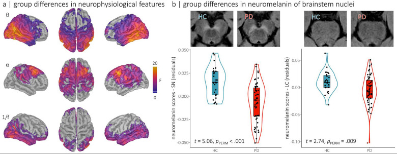
Background and objectives: Parkinson's disease (PD) is marked by the death of neuromelanin-rich dopaminergic and noradrenergic cells in the substantia nigra (SN) and the locus coeruleus (LC), respectively, resulting in motor and cognitive impairments. While SN dopamine dysfunction has clear neurophysiological effects, the impact of reduced LC norepinephrine signaling on brain activity in PD remains to be established.

Methods: We used neuromelanin-sensitive T1-weighted MRI (NPD = 58; NHC = 27) and task-free magnetoencephalography (NPD = 58; NHC = 65) to identify neuropathophysiological factors related to the degeneration of the LC and SN in patients with PD.

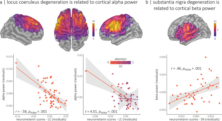
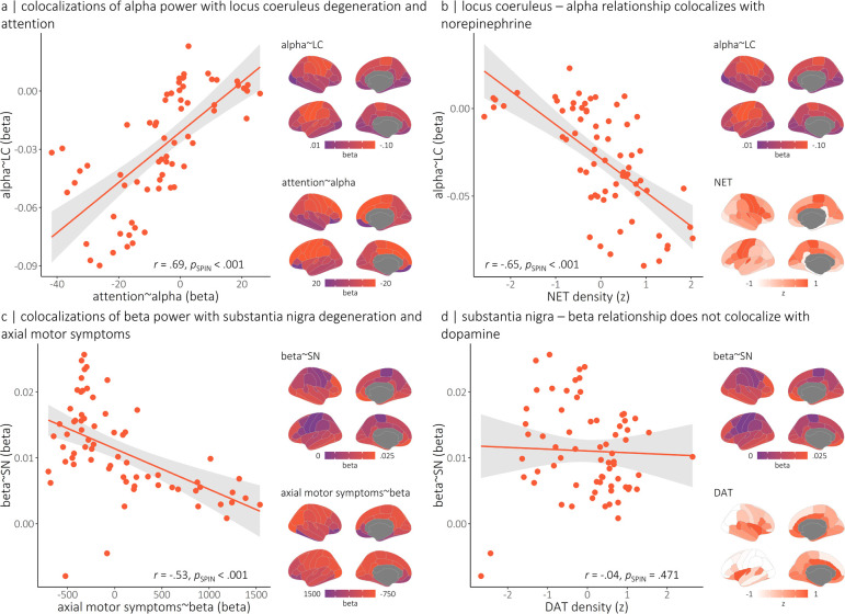
Results: We found pathological increases in rhythmic alpha (8 - 12 Hz) activity in patients with decreased LC neuromelanin, with a stronger association in patients with worse attentional impairments. This negative alpha-LC neuromelanin relationship is also stronger in fronto-motor cortices, which are regions with high densities of norepinephrine transporters in the healthy brain, and where alpha activity is negatively related to attention scores. These observations support a noradrenergic association between LC integrity and alpha band activity. Our data also show that rhythmic beta (15 - 29 Hz) activity in the left somato-motor cortex decreases with lower levels of SN neuromelanin; the same regions where beta activity reflects axial motor symptoms.

Discussion: Together, our findings clarify the association of well-documented alterations of rhythmic neurophysiology in PD with cortical and subcortical neurochemical systems. Specifically, attention-related alpha activity reflects dysfunction of the noradrenergic system, and beta activity with relevance to motor impairments reflects dopaminergic dysfunction.

Keywords: Parkinson’s disease; cortical rhythms; locus coeruleus; magnetoencephalography; neuromelanin; substantia nigra.


Figures

Figure 1.
Figure 1.. Alterations of regional cortical neurophysiology and neuromelanin depletion of brainstem nuclei in Parkinson’s disease.
(a) Cortical maps represent regional clusters of differences in rhythmic (i.e., theta and alpha oscillations) and arrhythmic (i.e., aperiodic 1/f slope) neurophysiological features between patients with Parkinson’s disease and healthy older adults. Colors indicate the strength of the statistical effect (in F values), thresholded based on the cluster limits identified using threshod-free cluster enhancement (pFWE < .05). No significant differences were observed in the delta and beta bands. (b) Top: representative neuromelanin-sensitive MRI data focused on the substantia nigra (left panel) and the locus coeruleus (right panel) for one participant with Parkinson’s disease (PD, in red) and one age-matched healthy control participant (HC, in blue). Bottom: group differences in neuromelanin scores for substantia nigra (left panel) and locus coeruleus (right panel), with individual points representing participants. Boxes-and-whiskers indicate the median, upper and lower quartiles, and minima/maxima for each group, and violin plots show the associated density distributions.
Figure 2.
Figure 2.. Regional cortical rhythmic neurophysiological features related to brainstem nuclei neuromelanin.
(a) The maps show the cortical regions where locus coeruleus neuromelanin scales with rhythmic alpha power, beyond the effects of substantia nigra neuromelanin and age. The nature of this relationship is displayed in scatter plots of peak-vertex alpha power values on the bottom left, with the partial correlation coefficient and permuted p-value overlaid. The line plots on the bottom right reflect the significant moderation of this relationship by attention abilities (indicated by colors of lines; orange = impaired attention, purple = preserved attention), such that individuals with worse attentional impairments exhibited a stronger relationship between locus coeruleus degeneration and rhythmic alpha power. (b) The map shows the cortical regions where substantia nigra neuromelanin scores are associated with rhythmic beta power, beyond the effects of locus coeruleus neuromelanin, disease duration, and age. The nature of this relationship is displayed in the scatter plot below using peak-vertex beta power values, with the partial correlation coefficient and permuted p-value overlaid. Colors on the cortical maps indicate the strength of the statistical effect (in F values), thresholded based on the cluster limits identified using threshold-free cluster enhancement (pFWE < .05).We used nonparametric permutation testing to account for the influence of outliers in these associations. Also note that the subjective outlier in the top left of the scatterplots in panel (a) did not exert undue influence on the model (defined as a Cook’s distance > 3 SD from the group mean).
Figure 3.
Figure 3.. Colocalization of cortical associations between rhythmic neurophysiological features, clinical symptoms and brainstem nuclei neuromelanin.
(a) Cortical maps indicate the region-wise linear relationships, in unstandardized regression (i.e., beta) weights, between alpha power and locus coeruleus neuromelanin (top) and attention (bottom). The scatter plot to the left indicates the alignment between these maps (each dot is from a parcel of the Desikan-Killiany cortical atlas), with r-values and p-values overlaid. (b) Colocalization of the region-wise alpha-LC relationships shown in (a) with the density of cortical norepinephrine transporter. (c) Similar to the effects shown in (a), but concerning relationships between rhythmic beta power, substantia nigra neuromelanin, and axial motor symptoms. (d) Similar to the effects shown in (b), but concerning colocalization of the region-wise beta-SN relationships with the density of cortical dopamine transporter. Note that null distributions for estimating p-values were generated using 5,000 autocorrelation-preserving spatial permutations of the data.

Similar articles

References

    1. Jenkinson N, Brown P. New insights into the relationship between dopamine, beta oscillations and motor function. Trends in neurosciences 2011;34:611–618. - PubMed
    1. Braak H, Del Tredici K, Rüb U, De Vos RA, Steur ENJ, Braak E. Staging of brain pathology related to sporadic Parkinson’s disease. Neurobiology of aging 2003;24:197–211. - PubMed
    1. Del Tredici K, Braak H. Dysfunction of the locus coeruleus–norepinephrine system and related circuitry in Parkinson’s disease-related dementia. Journal of Neurology, Neurosurgery & Psychiatry 2013;84:774–783. - PubMed
    1. Aston-Jones G, Rajkowski J, Cohen J. Locus coeruleus and regulation of behavioral flexibility and attention. Progress in brain research 2000;126:165–182. - PubMed
    1. Smith A, Nutt D. Noradrenaline and attention lapses. Nature 1996. - PubMed

Publication types