[Preprint]. 2024 Nov 17:2024.02.16.580675.
doi: 10.1101/2024.02.16.580675.

Molecular glues of the regulatory ChREBP/14-3-3 complex protect beta cells from glucolipotoxicity

Affiliations

Molecular glues of the regulatory ChREBP/14-3-3 complex protect beta cells from glucolipotoxicity

Liora S Katz et al. bioRxiv. .

Update in

Abstract

The Carbohydrate Response Element Binding Protein (ChREBP) is a glucose-responsive transcription factor (TF) with two major splice isoforms (α and β). In chronic hyperglycemia and glucolipotoxicity, ChREBPα-mediated ChREBPβ expression surges, leading to insulin-secreting β-cell dedifferentiation and death. 14-3-3 binding to ChREBPα results in cytoplasmic retention and suppression of transcriptional activity. Thus, small molecule-mediated stabilization of this protein-protein interaction (PPI) may be of therapeutic value. Here, we show that structure-based optimizations of a 'molecular glue' compound led to potent ChREBPα/14-3-3 PPI stabilizers with cellular activity. In primary human β-cells, the most active compound retained ChREBPα in the cytoplasm, and efficiently protected β-cells from glucolipotoxicity while maintaining β-cell identity. This study may thus not only provide the basis for the development of a unique class of compounds for the treatment of Type 2 Diabetes but also showcases an alternative 'molecular glue' approach for achieving small molecule control of notoriously difficult to target TFs.


Conflict of interest statement

The authors declare the following compeng financial interest(s): L.B. and C.O. are founders of Ambagon Therapeucs. L.B. is a member of Ambagon’s scienfic advisory board, C.O. is employee of Ambagon.

Figures
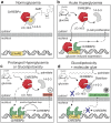
Fig. 1:
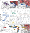
Fig. 1:. Protein-Protein Interaction between 14-3-3 and ChREBPα regulates β-cell fate.
a. Under normoglycemic conditions, ChREBPα remains mostly cytoplasmic by binding to 14-3-3. ChREBPα is one of very few phosphorylation-independent 14-3-3 partner proteins and binds via a pocket containing a phosphate or sulfate ion, ketone, or AMP. b. In acute hyperglycemia, ChREBPα dissociates from 14-3-3 and transiently translocates into the nucleus where it binds multiple carbohydrate response elements (ChoREs) and promotes adaptive β-cell expansion. c. In prolonged hyperglycemia or hyperglycemia combined with hyperlipidemia (glucolipotoxicity), ChREBPα initiates and maintains a feed-forward surge in ChREBPβ expression, leading to β-cell demise. d. A novel class of molecular glue drugs specifically stabilize ChREBPα/14-3-3 interaction, prevent surge of ChREBPβ expression in glucolipotoxicity, and protect β-cell identity and survival.
Fig. 2:
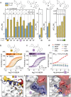
Fig. 2:. SAR around analog 1 results in improved stabilizer 30.
a. Crystal structure of compound 1 (blue sticks) in complex with 14-3-3σ (white surface) and ChREBPα (red sticks and surface). Final 2Fo-Fc electron density contoured at 1.0σ. b. Interactions of 1 (blue sticks) with 14-3-3σ (white) and ChREBPα (red) residues (relevant side chains are displayed in stick representation, polar contacts are shown as black dashed lines). c. FA 2D protein titration of 14-3-3β in FITC-labeled ChREBPα peptide (10 nM) and varied but fixed concentrations of 1 (0–500 μM), including the cooperativity factor (α, determined, by fitting, using a thermodynamic equilibrium model) and intrinsic affinity of 1 to 14-3-3 (KDII). d. Structure and activity analogs of 1. The two best compounds are marked in cyan and yellow. EC50 in parenthesis with mean ± SD, n = 2. For FA titration graphs see Fig. S4, S5. e, f. Crystallographic overlay of 1 (blue sticks) with 30 (yellow sticks) in complex of 14-3-3σ (white cartoon) and ChREBPα (red cartoon). g. Interactions of 30 (yellow) with 14-3-3 σ (white) and ChREBPα (red) (relevant side chains are displayed in stick representation, polar contacts are shown as black dashed lines).
Fig. 3:
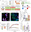
Fig. 3:. Fluorination of compounds enhances stabilizing potency.
a. Structures and bar graphs of pEC50 values derived from FA compound titrations, for Y=H (blue bars) and Y=F (yellow bars). (For graphs see Fig. S4, S5, for EC50 values see Table S3) (mean ± SD, n=2). b, c. Titration of 14-3-3β to FITC-labeled ChREBPα peptide (10 nM) against varying fixed concentrations of 30 or 43 (0–500 μM) (mean ± SD, n = 2), including the cooperativity factor (α, determined, by fitting, using a thermodynamic equilibrium model) and intrinsic affinity of the stabilizers to 14-3-3 (KDII). d. Selectivity studies by titrating 43 to 14-3-3β and eight different 14-3-3 interaction FITC-labeled peptides (all 10 nM) (mean ± SD, n = 2). e. Crystallographic overlay 30 (yellow) and 43 (purple) in complex with 14-3-3σ (white cartoon) and ChREBPα (red cartoon). Helix 9 of 14-3-3σ is colored in the same color as the corresponding compound, showing a helical ‘clamping’ effect when 43 (purple) is present. f. Surface representation of 43 (purple) in complex with 14-3-3σ (white) and ChREBPα (red), showing the distances (black dashes) of the 43 m-F substitution to the residues (sticks) of 14-3-3σ and ChREBPα. g. Interactions of 43 (purple) with 14-3-3σ (white) and ChREBPα (red) (relevant side chains are displayed in stick representation, polar contacts are shown as black dashed lines).
Fig 4:
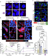
Fig 4:. Active compounds protect human β-cells from glucolipotoxicity.
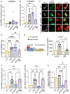
a. Overview of compounds included in cellular assays. Table shows results of cytotoxicity and β-cell rescue from glucolipotoxicity in the presence of the compounds (green indicates positive outcome and red cytotoxicity). b. Schematic of adaptation to the SPARKL assay in human islets to specifically monitor β-cells. c. Representative figures from d at 48 h with 43. The results are representative from 4 different human cadaveric donors. d. Representative kinetics of β-cell death in glucolipotoxicity (20 mM glucose+500 μM palmitate), in the presence of 10 μM of the indicated compounds. e. Quantification of β-cell death (assessed by Yoyo3+% of GFP+ cells) at 24 h from d. f. Human islets were treated for 24 h as indicated, followed by quantification of glucose-stimulated insulin secretion (GSIS) in KREBS buffer (2.8 mM glucose, 1% BSA) over 30 min. The corresponding GSIS-stimulation index (SI) was obtained by determining the ratio of insulin release at high vs low glucose. Data are means +/−SEM; n=4; p<0.01**, p<0.01; ***, p<0.005; ****, pP<0.001.
Fig. 5:
Fig. 5:. 43 stabilizes ChREBPα/14-3-3 interaction and thus retains cytoplasmic ChREBPα localization in response to glucose and glucolipotoxicity.
a. Proximity ligation assay demonstrating increased interaction between 14-3-3 and ChREBPα. INS-1 cells were cultured overnight (ON) at low (5.5 mM) glucose and exposed to high (20 mM) glucose for the indicated times b, d. Representative figures showing the nuclear localization of ChREBPα after exposure to high glucose (b) or glucolipotoxic (d) conditions. c, e. Time course of nuclear localization of ChREBPα based on figures b, d, respectively in addition to 1mM AICAR. f, g. CRISPR/Cas9 engineered INS-1 cells treated with the indicated compounds for 24 h; Low-5.5 mM glucose, High-20 mM glucose, glucolipotoxicity-20 mM glucose+500 μM palmitate. f. Representative images at 24 h. g. Quantification of % nuclear ChREBP at 24 h. Data are the means +/− SEM, n=3–5, *p<0.05, **p<0.01.
Fig. 6:
Fig. 6:. 43 preserves β-cell identity in glucolipotoxicity and prevents upregulation of ChREBPβ in high glucose and glucolipotoxicity.
a,b,f,g. mRNA fold-enrichment over control low glucose in human islets, treated with 10 μM 43 for 24 h; Low-5.5 mM glucose, High-20 mM glucose, glucotox-20 mM glucose+500 μM palmitate. c, d, h. Immunostaining for Pdx1, c-term ChREBP, and insulin in dispersed islets treated for 48 h with the indicated treatments. e. INS-1 cells expressing luciferase driven by the human TXNIP promoter were incubated for 24 h at the indicated glucose concentrations, in the presence or absence of 10 μM 43. Data are the means +/− SEM, n=3–7, *p<0.05, **p<0.01***, p<0.005, ****, p<0.001.

Similar articles

References

    1. Ogurtsova K. et al. IDF diabetes Atlas: Global esmates of undiagnosed diabetes in adults for 2021. Diabetes Res Clin Pract 183, 109118 (2022). - PubMed
    1. Collaborators G.B.D.D. Global, regional, and naonal burden of diabetes from 1990 to 2021, with projecons of prevalence to 2050: a systemac analysis for the Global Burden of Disease Study 2021. Lancet 402, 203–234 (2023). - PMC - PubMed
    1. Tancredi M. et al. Excess mortality among persons with type 2 diabetes. New England Journal of Medicine 373, 1720–1732 (2015). - PubMed
    1. Chaterjee S., Khun K. & Davies M.J. Type 2 diabetes. The lancet 389, 2239–2251 (2017). - PubMed
    1. Schellenberg E.S., Dryden D.M., Vandermeer B., Ha C. & Korownyk C. Lifestyle intervenons for paents with and at risk for type 2 diabetes: a systemac review and meta-analysis. Annals of internal medicine 159, 543–551 (2013). - PubMed

Publication types