[Preprint]. 2024 Feb 17:2024.02.15.580553.
doi: 10.1101/2024.02.15.580553.

Structural and biochemical characterization of the mitomycin C repair exonuclease MrfB

Affiliations

Structural and biochemical characterization of the mitomycin C repair exonuclease MrfB

Kelly A Manthei et al. bioRxiv. .

Update in

Abstract

Mitomycin C (MMC) repair factor A (mrfA) and factor B (mrfB), encode a conserved helicase and exonuclease that repair DNA damage in the soil-dwelling bacterium Bacillus subtilis. Here we have focused on the characterization of MrfB, a DEDDh exonuclease in the DnaQ superfamily. We solved the structure of the exonuclease core of MrfB to a resolution of 2.1 Å, in what appears to be an inactive state. In this conformation, a predicted α-helix containing the catalytic DEDDh residue Asp172 adopts a random coil, which moves Asp172 away from the active site and results in the occupancy of only one of the two catalytic Mg2+ ions. We propose that MrfB resides in this inactive state until it interacts with DNA to become activated. By comparing our structure to an AlphaFold prediction as well as other DnaQ-family structures, we located residues hypothesized to be important for exonuclease function. Using exonuclease assays we show that MrfB is a Mg2+-dependent 3'-5' DNA exonuclease. We show that Leu113 aids in coordinating the 3' end of the DNA substrate, and that a basic loop is important for substrate binding. This work provides insight into the function of a recently discovered bacterial exonuclease important for the repair of MMC-induced DNA adducts.


Figures

Figure 1.
Figure 1.. Exonuclease assays with 5' labeled DNA confirm directionality and metal-dependence.
(a-b) Initial biochemical assays performed with full-length WT MrfB. All assays used 20-mer oligos with various labels as depicted above. NaOH refers to the 3’-labeled RNA substrate treated with alkaline hydrolysis for a ladder. (a) Assays stopped at 20 min with 1 mM of the indicated metal show that MrfB can use Mg2+ or Mn2+. (b) Comparison of 20-mer oligos labeled on different ends and a third with RNA at the 3’ end showing that, MrfB is a 3’-5’ exonuclease with a preference for DNA. (c) Left: domain architecture of full length MrfB. TPR stands for tetratricopeptide repeat. Right: AlphaFold model of full-length MrfB (entry P50837), showing the three truncation constructs that were cloned and purified with brackets. (d) Purified exonuclease core (residues 33-279, 28.6 kDa) electrophoresed on an SDS-PAGE gel (left), and on a Superdex 200 Increase 10/300 GL column on right (blue trace). The gray traces show standards with their molecular weight indicated above each peak, confirming that the exonuclease core is a monomer. (e) The same assay as in (a) with increasing concentrations of full-length MrfB and the exonuclease core.
Figure 2.
Figure 2.. The structure of the MrfB exonuclease core.
(a) The 2.1 Å structure appears to be an inactive state due to an unwound helix that pulls D172 away from the active site, and likely leading to only one observed Mg2+ ion (green sphere). DEDDh residue side chains are shown as sticks. Density was not observed for loops spanning residues 112-118 and 204-212 (dashed lines). (b) An AlphaFold model of residues 33-279, modeled using the Colab notebook that uses no homologous templates (https://colab.research.google.com/github/deepmind/alphafold/blob/main/notebooks/AlphaFold.ipynb) (22). The AlphaFold model is more consistent with an active state, with a helix containing Asp172 spanning residues 171-183 (see arrow), which positions all DEDDh residues at the active site. Mg2+ ions are modeled from PDB 5YWU. (c) A consistent model for the exonuclease core is observed from different software, aside from the loop containing Leu113. Models are from AlphaFold (Leu113 in dashed circle), I-TASSER and SWISS-MODEL (22,37,38). (d) The AlphaFold model is consistent with other exonuclease structures that position Leu113 at the active site. The DNA is modeled from alignment with an ExoX structure (PDB 4FZZ)(40). Potential DNA-interacting residues are shown with side chains as sticks.
Figure 3.
Figure 3.. MrfB variant exonuclease activity on ssDNA.
(a) DEDDh variants reveal that D107, E109, and H258A are required for catalytic activity. (b) DNA-interacting variants show the importance of the basic loop and Leu113. (a-b) Both panels show a gel-based assay with 5' labeled DNA on top, and quantification of the remaining DNA from the top of the gel at the bottom as compared to the buffer control (see Methods). In all assays exonuclease activity is measured using a 20-mer ssDNA substrate. Data are mean ± s.e.m. of 3-4 independent experiments. * 0.01

Figure 4.
Figure 4.. PicoGreen assay examining MrfB variants on various dsDNA substrates.
Fluorescence was monitored for 2 h to examine nuclease activity on (a) an 80 bp blunt or (b) a 3’ overhang, nicked, and gap of 1 or 3 nucleotide substrate (see Figure S2 for substrates). The data in the top panels were fit to a one phase decay model to determine the rate, which was then compared in the bottom panels. Data are mean ± s.e.m. of 3 independent experiments. * 0.01

Figure 5.
Figure 5.
The mrfB L113A and RRKR are unable to complement ΔmrfB in vivo. The addition of MMC is more deleterious in a ΔmrfB background, with growth restored by complementation with WT, and partially restored by complementation with R203A, and R212A mrfB when overexpressed with xylose. The L113A and RRKR mutants are unable to complement ΔmrfB even when induced with xylose.
Figure 6.
Figure 6.
MrfB structural modeling and comparisons. (a-b) A comparison of the deposited AlphaFold model with 5 models determined without a structural template, showing some uncertainty for the α-helix preceding Phe171 and Asp172. (22). AlphaFold models are colored by pLDDT, a model confidence score. Very high confidence (>90) scores are dark blue, high confidence is light blue (70-90), low scores are yellow (50-70), and very low scores are orange (<50). (c) Different crystal structures aligned at the active site to show a variety of DEDD exonucleases use a leucine wedge at the active site. Structures shown: MrfB AlphaFold model, blue; ExoX (PDB 4FZZ) light blue; TREX2 (PDB 6A47) tan, TREX1 (PDB 2O4I) dark green; Klenow fragment (PDB 1KLN), teal; NrnC (PDB 7MPO), light green (22,34,39-41,50).
Figure 7.
Figure 7.. DEDD exonuclease structural comparisons.
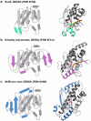
(a-c) In all 3, the fold topology is shown on the left, and structural features colored the same on the right. The canonical fold is in gray. DEDDh/y residues are shown with the single letter in their approximate location, and a yellow wedge for any leucine or other potential wedge location. In the structural models, the same highlighted active site residues are shown with sticks. (a) ExoX, PDB 4FZZ, DEDDh (40). 3 bases of the 3’ end are shown for clarity, but the structure contains dsDNA. Green spheres are Na+. (b) The proofreading exonuclease domain from Pyrococcus abyssi B-family polymerase bound to dsDNA, which is melted so only ssDNA is at the exonuclease active site (54). There is no need for a leucine wedge as the pink β-hairpin stabilizes the other strand of the duplex and only ssDNA is fed into the active site. (c) MrfB exo core as described in this paper, with part of the N-terminal insertion starting at residue 33 shown in blue.

Similar articles

References

    1. Friedberg E.C., Walker G.C., Siede W., Wood R.D., Schultz R.A. and Ellenberger T. (2006) DNA Repair and Mutagenesis: Second Edition. American Society for Microbiology, Washington, DC.
    1. Procopio R.E., Silva I.R., Martins M.K., Azevedo J.L. and Araujo J.M. (2012) Antibiotics produced by Streptomyces. Braz J Infect Dis, 16, 466–471. - PubMed
    1. Hata T., Sano Y., Sugawara R., Matsumae A., Kanamori K., Shima T. and Hoshi T. (1956) Mitomycin, a new antibiotic from Streptomyces. I. The Journal of Antibiotics, Series A, 9, 141–146. - PubMed
    1. Tomasz M. (1995) Mitomycin C: small, fast and deadly (but very selective). Chemistry & biology, 2, 575–579. - PubMed
    1. Iyer V. and Szybalski W. (1963) A molecular mechanism of mitomycin action: linking of complementary DNA strands. Proceedings of the National Academy of Sciences, 50, 355–362. - PMC - PubMed

Publication types