[Preprint]. 2024 Feb 14:2024.02.14.580389.
doi: 10.1101/2024.02.14.580389.

Translational response to mitochondrial stresses is orchestrated by tRNA modifications

Affiliations

Translational response to mitochondrial stresses is orchestrated by tRNA modifications

Sherif Rashad et al. bioRxiv. .

Abstract

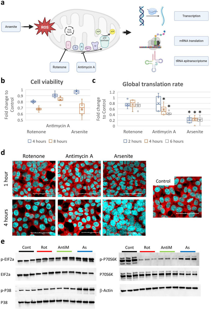
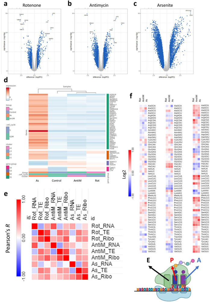
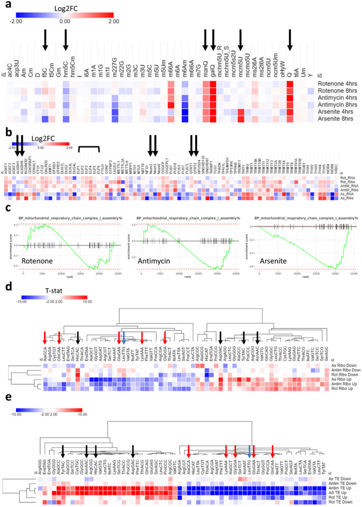
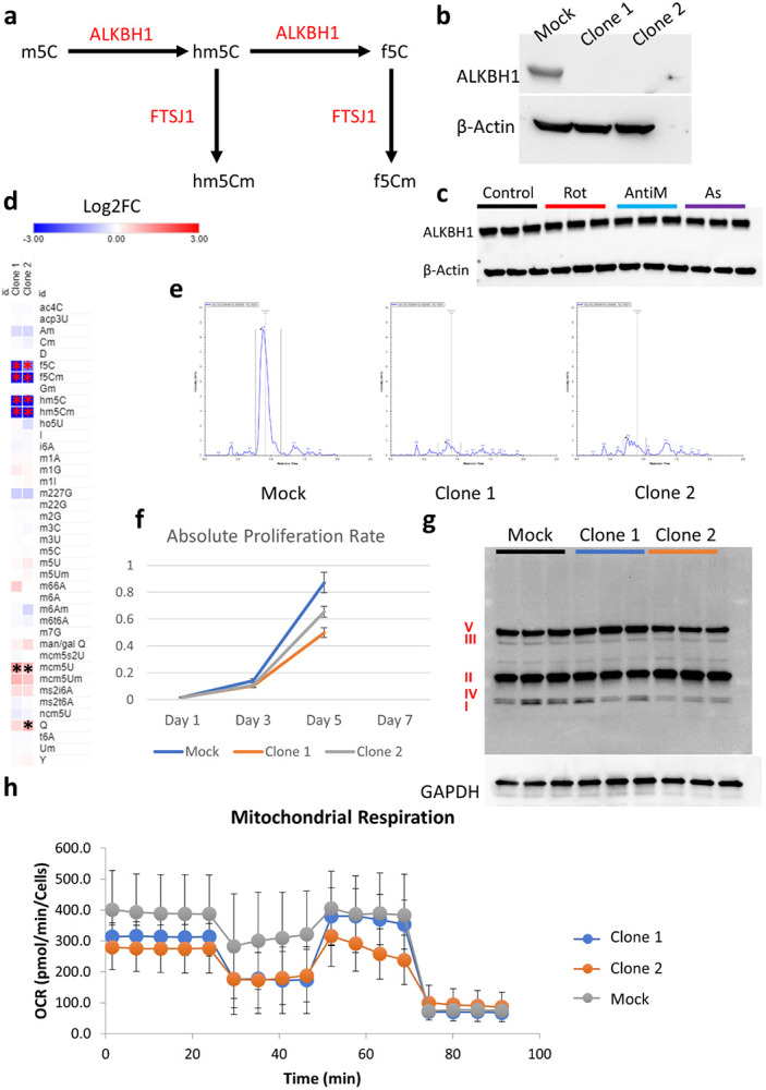
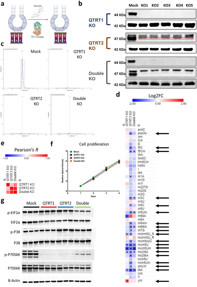
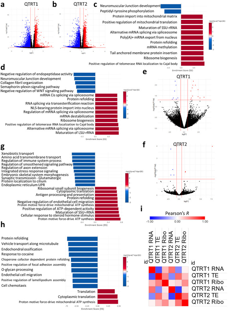
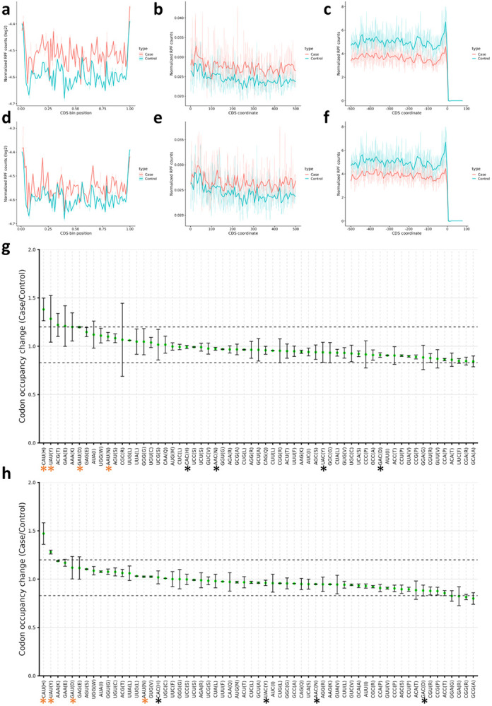
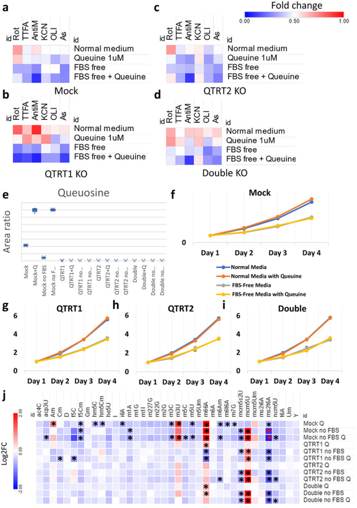
Mitochondrial stress and dysfunction play important roles in many pathologies. However, how cells respond to mitochondrial stress is not fully understood. Here, we examined the translational response to electron transport chain (ETC) inhibition and arsenite induced mitochondrial stresses. Our analysis revealed that during mitochondrial stress, tRNA modifications (namely f5C, hm5C, queuosine and its derivatives, and mcm5U) dynamically change to fine tune codon decoding, usage, and optimality. These changes in codon optimality drive the translation of many pathways and gene sets, such as the ATF4 pathway and selenoproteins, involved in the cellular response to mitochondrial stress. We further examined several of these modifications using targeted approaches. ALKBH1 knockout (KO) abrogated f5C and hm5C levels and led to mitochondrial dysfunction, reduced proliferation, and impacted mRNA translation rates. Our analysis revealed that tRNA queuosine (tRNA-Q) is a master regulator of the mitochondrial stress response. KO of QTRT1 or QTRT2, the enzymes responsible for tRNA-Q synthesis, led to mitochondrial dysfunction, translational dysregulation, and metabolic alterations in mitochondria-related pathways, without altering cellular proliferation. In addition, our analysis revealed that tRNA-Q loss led to a domino effect on various tRNA modifications. Some of these changes could be explained by metabolic profiling. Our analysis also revealed that utilizing serum deprivation or alteration with Queuine supplementation to study tRNA-Q or stress response can introduce various confounding factors by altering many other tRNA modifications. In summary, our data show that tRNA modifications are master regulators of the mitochondrial stress response by driving changes in codon decoding.

Keywords: Codon usage; Mitochondrial stress; Oxidative stress; Queuine; Queuosine; RNA modifications; mRNA translation; tRNA; tRNA modifications.


Figures

Figure 1:
Figure 1:. Induction of mitochondrial stress.
a: Schematic of the study analysis. b: Cell viability analysis via MTT assay after exposure to 10μM Rotenone (Rot), 10μg/ml Antimycin A (AntiM), or 100μM sodium metaArsenite (As). c: Puromycin incorporation assay after stress exposure. d: Staining with anti-G3BP1 (in red) for analysis of stress granules (SG) assembly after stress. e: Western blot analysis of ISR and RSR markers after 8 hours of stress.
Figure 2:
Figure 2:. Mitochondrial stress induces transcriptional and translational dysregulation.
a-c: Volcano plots for differentially expressed genes (DEGs) in the RNA-seq analysis after 8 hours of stress exposure. d: Cluster heatmap analysis of the RNA-seq datasets. e: Ribosome dwelling times after stress exposure across different ribosome sites. f: Pearson’s correlation analysis between stresses across different datasets.
Figure 3:
Figure 3:. tRNA modifications drive translational changes after mitochondrial stress.
a:Analysis of tRNA modifications after exposure to 10μM Rotenone, 10μg/ml Antimycin A, or 100μM Arsenite. Black arrow indicates fold change > 1.5 and p < 0.05. Data is normalized to controls. b: Analysis of all tRNA modifying enzymes from the Ribo-seq and RNA-seq datasets. Black arrows indicate enzymes related to significant modifications in a. c: GOBP pathway analysis of mitochondrial respiratory chain complex I assembly in all stresses. d: Isoacceptors codon frequency analysis of the Ribo-seq datasets in all stresses. e: Isoacceptors codon frequency analysis of the translational efficiency (TE) datasets in all stresses.
Figure 4:
Figure 4:. ALKBH1 regulates mitochondrial function through its dioxygenase activity.
a: Schematic for the synthesis pathway of hm5C and f5C modifications and the responsible enzymes. b: Validation of ALKBH1 knockout (KO) in the two generated clones. c: Analysis of ALKBH1 expression after 8 hours of stress exposure. d: LCMS/MS analysis of tRNA modifications after ALKBH1 KO. Data is normalized to Mock. Asterisk indicates fold change > 1.5 and p < 0.05. e: Analysis of hm5C peaks showing the complete depletion of hm5C after ALKBH1 KO. f: Cell proliferation after ALKBH1 KO. g: Analysis of OXPHOS related proteins in all 5 complexes using western blot. h: Mitochondrial respiration analysis using Seahorse.
Figure 5:
Figure 5:. tRNA-Q loss induces translational stress.
a: Schematic of tRNA-Q mediated codon recognition. b: Validation of QTRT1, QTRT2, and double KO using western blots. c: Validation of tRNA-Q peak loss using LC-MS/MS. d: Analysis of the impact of tRNA-Q loss on other RNA modifications in the three KO cell lines. Arrows: significant on ANOVA analysis. Asterisk: fold change > 1.5, p < 0.05. Data normalized to Mock. e: Pearson’s correlation analysis using data from d. f: Proliferation rates of the three KO cell lines. g: Analysis of markers of ISR and RSR using western blotting.
Figure 6:
Figure 6:. Impact of QTRT1 and QTRT2 KO on mRNA expression and translation.
a-b:Volcano plots for RNA-seq datasets. c: RNA-seq GOBP analysis after QTRT1 KO. d: RNA-seq GOBP analysis after QTRT2 KO. e-f: Volcano plots for Ribo-seq dataset. g: Ribo-seq GOBP analysis after QTRT1 KO. h: Ribo-seq GOBP analysis after QTRT2 KO. i: Pearson’s correlation analysis between different datasets and cell lines.
Figure 7:
Figure 7:. tRNA-Q loss impacts NAU codon decoding.
a-c: Occupancy metagene plots in QTRT1 KO cells (Across CDS, downstream from start codon, and upstream from end codon respectively). d-f: Occupancy metagene plots in QTRT1 KO cells (Across CDS, downstream from start codon, and upstream from end codon respectively). g: Ribosome A-site pausing in QTRT1 KO. h: Ribosome A-site pausing in QTRT2 KO. Black asterisk: NAC codons, orange asterisk: NAU codons.
Figure 8:
Figure 8:. tRNA-Q loss as well as Queuine availability influence stress response and the epitranscriptome.
a-d: Heatmaps of cell viability analysis after exposure to mitochondrial ETC inhibitors for 4 hours (Rotenone 80μM, TTFA 1.5mM, Antimycin A 50μg/ml, Potassium Cyanide (KCN) 15mM, and Oligomycin 20μM) as well as Arsenite 600μM, in normal media, in serum (FBS) free media, and with Queuine supplementation (1μM in either media). Data were normalized to controls for each cell line and expressed as fold change. e: Validation of tRNA-Q levels after serum deprivation and Queuine supplementation in all cell lines. f-i: Proliferation rates of all cell lines in different media used. j: Analysis of the epitranscriptional changes of serum deprivation and/or Queuine supplementation in all cell lines using LC-MS/MS. Asterisk: fold change > 1.5, p < 0.05. Data normalized to normal media values for each cell line and presented as Log2FC.

References

    1. Afgan E., Baker D., Batut B., van den Beek M., Bouvier D., Cech M., Chilton J., Clements D., Coraor N., Grüning B.A., et al. (2018). The Galaxy platform for accessible, reproducible and collaborative biomedical analyses: 2018 update. Nucleic Acids Res 46, W537–w544. - PMC - PubMed
    1. Alim I., Caulfield J.T., Chen Y., Swarup V., Geschwind D.H., Ivanova E., Seravalli J., Ai Y., Sansing L.H., Ste Marie E.J., et al. (2019). Selenium Drives a Transcriptional Adaptive Program to Block Ferroptosis and Treat Stroke. Cell 177, 1262–1279 e1225. - PubMed
    1. Ando D., Rashad S., Begley T., Endo H., Aoki M., Dedon P., and Niizuma K. (2023). Mammalian tissue specific translation regulation; role of tRNA epitranscriptome in regulating codon optimality patterns across tissues. bioRxiv, 2023.2010.2024.563884.
    1. Arguello A.E., Li A., Sun X., Eggert T.W., Mairhofer E., and Kleiner R.E. (2022). Reactivity-dependent profiling of RNA 5-methylcytidine dioxygenases. Nature Communications 13, 4176. - PMC - PubMed
    1. Asano K., Suzuki T., Saito A., Wei F.Y., Ikeuchi Y., Numata T., Tanaka R., Yamane Y., Yamamoto T., Goto T., et al. (2018). Metabolic and chemical regulation of tRNA modification associated with taurine deficiency and human disease. Nucleic Acids Res 46, 1565–1583. - PMC - PubMed

Publication types