. 2024 Feb 8:18:1361590.
doi: 10.3389/fnins.2024.1361590. eCollection 2024.

Enhancing regeneration and repair of long-distance peripheral nerve defect injuries with continuous microcurrent electrical nerve stimulation

Affiliations

Enhancing regeneration and repair of long-distance peripheral nerve defect injuries with continuous microcurrent electrical nerve stimulation

Junjie Kong et al. Front Neurosci. .

Abstract

Introduction: Peripheral nerve injuries, especially those involving long-distance deficits, pose significant challenges in clinical repair. This study explores the potential of continuous microcurrent electrical nerve stimulation (cMENS) as an adjunctive strategy to promote regeneration and repair in such cases.

Methods: The study initially optimized cMENS parameters and assessed its impact on Schwann cell activity, neurotrophic factor secretion, and the nerve regeneration microenvironment. Subsequently, a rat sciatic nerve defect-bridge repair model was employed to evaluate the reparative effects of cMENS as an adjuvant treatment. Functional recovery was assessed through gait analysis, motor function tests, and nerve conduction assessments. Additionally, nerve regeneration and denervated muscle atrophy were observed through histological examination.

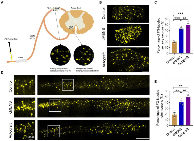
Results: The study identified a 10-day regimen of 100uA microcurrent stimulation as optimal. Evaluation focused on Schwann cell activity and the microenvironment, revealing the positive impact of cMENS on maintaining denervated Schwann cell proliferation and enhancing neurotrophic factor secretion. In the rat model of sciatic nerve defect-bridge repair, cMENS demonstrated superior effects compared to control groups, promoting motor function recovery, nerve conduction, and sensory and motor neuron regeneration. Histological examinations revealed enhanced maturation of regenerated nerve fibers and reduced denervated muscle atrophy.

Discussion: While cMENS shows promise as an adjuvant treatment for long-distance nerve defects, future research should explore extended stimulation durations and potential synergies with tissue engineering grafts to improve outcomes. This study contributes comprehensive evidence supporting the efficacy of cMENS in enhancing peripheral nerve regeneration.

Keywords: Schwann cell activity; continuous microcurrent electrical nerve stimulation; functional recovery; long-distance nerve defects; peripheral nerve injury; tissue engineering.


Conflict of interest statement

The authors declare that the research was conducted in the absence of any commercial or financial relationships that could be construed as a potential conflict of interest.

Figures

Figure 1
Figure 1
Effect of different electrical stimulation parameters on axon regeneration. (A) Schematic illustrating the experimental setup. A 5-mm sciatic nerve defect in rats was repaired using a silicone tube bridge. Flexible electrodes were attached to both tube ends, linked to the proximal and distal nerve stumps. These electrodes were threaded under the skin, securing their opposite ends to the dorsal neck skin. While receiving electrical stimulation, anesthetized rats were connected to a multi-channel stimulator via this electrode pair. (B) Representative immunofluorescence images depicting axon regeneration at the bridging site 10 days post-nerve repair. Arrows indicate the anterior part of the regenerating axon. The right images are magnification of the boxed areas in the left images. NF-H labels axons, P75 labels Schwann cells, and DAPI labels nuclei. Scale bar: left, 500 μm; right, 20 μm. (C) Quantitative analysis of the relative length of regenerated axons (n = 3). Data are presented as mean ± SEM and were statistically analyzed using one-way ANOVA, followed by Tukey post hoc test. *p < 0.05, **p < 0.01, ***p < 0.001, ns, not significant.
Figure 2
Figure 2
Effects of cMENS Treatment on the Proliferation and Migration of Schwann Cells (SCs) and Secretion of Neurotrophic Factors in the Local Microenvironment. (A) Gross observation of sciatic nerve at 10 days after 5 mm defection and bridging repair (the silicone tube has been removed). (B) Representative immunofluorescence image illustrating the migration of Schwann cells (SCs) in the bridging segment. P75 labels Schwann cells. Scale bar: 500 μm. (C) Quantification of SCs migrated distance (n = 5). (D) Quantification of proliferated SCs in the distal nerve stump (n = 5). (E) Representative immunofluorescence images depicting Schwann cell proliferation in the distal nerve stumps. Ki67 labels the proliferating cells, P75 labels Schwann cells, and DAPI labels nuclei. Scale bar: 200 μm. (F) Quantitative polymerase chain reaction (qPCR) analysis of neurotrophic factor secretion in the bridging segment. Data are presented as mean ± SEM and were statistically analyzed using Student’s t test (two-tailed, unpaired) (C,D), and Multiple t tests (F). *p < 0.05, **p < 0.01, ***p < 0.001, ns, not significant.
Figure 3
Figure 3
Effect of cMENS Treatment on the Improvement of Sciatic Nerve Function. (A) Representative footprint images 12 weeks after sciatic nerve 10 mm defect-bridging repair. LH represents the left hind paw (injured side), and RH represents the right hind paw (uninjured side). (B) Measurement of 3D footprint intensities 12w after nerve repair (n = 5). (C) Quantification of sciatic functional index (SFI) at 4, 8, and 12 weeks post-injury (n = 5). (D) Representative compound muscle action potential (CMAP) recordings at 12 weeks post-injury. (E) Quantification of peak amplitude of action potentials (n = 5). (F) Quantification of motor conduction velocity (MCV) (n = 5). Data are presented as mean ± SEM and were statistically analyzed using one-way ANOVA, followed by Tukey post hoc test (B,E,F), and two-way ANOVA, followed by Tukey post hoc test (C). *p < 0.05; **p < 0.01; ***p < 0.001; ns, not significant.
Figure 4
Figure 4
Effect of cMENS Treatment on Motor Function Recovery of the Injured Hindlimb. (A) Representative stick view decomposition of rat hindlimb movements during stance and swing at 12 weeks post sciatic nerve 10 mm defect-bridging repair. Left indicates the left hind paw (injured side), and Right indicates the right hind paw (uninjured side). (B) Quantification of iliac crest height (n = 5). (C) Quantification of hip joint swing angle during movement (n = 5). (D) Quantification of knee joint swing angle during movement (n = 5). (E) Quantification of ankle joint swing angle during movement (n = 5). Data are presented as mean ± SEM and were statistically analyzed using one-way ANOVA, followed by Tukey post hoc test (B,C,D,E). *p < 0.05; **p < 0.01; ***p < 0.001; ns, not significant.
Figure 5
Figure 5
Effect of cMENS Treatment on the Regeneration of Sensory and Motor Neurons. (A) Schematic diagram of retrograde tracing of neurons. (B) Representative images displaying retrogradely labeled sensory neurons within the dorsal root ganglia (DRG) 12 weeks after nerve repair. Scale bar: 200 μm. (C) Quantification of FG-labeled sensory neurons (n = 3). (D) Representative images showing retrogradely labeled motor neurons within the ventral horn of the spinal cord 12 weeks after nerve repair. Magnified view of boxed areas displayed on the left side. Scale bar: left, 100 μm; right, 500 μm. (E) Quantification of FG-labeled motor neurons (n = 3). Data are presented as mean ± SEM and were statistically analyzed using one-way ANOVA, followed by Tukey post hoc test (C,E). **p < 0.01; ***p < 0.001; ns, not significant.
Figure 6
Figure 6
Effect of cMENS Treatment on Axon Regeneration and Myelination of Regenerating Axons. (A) Representative immunofluorescence images of transverse section of the distal part of the bridge segment at 12 weeks after sciatic nerve 10 mm defect-bridging repair. NF-H labels axons, P75 labels Schwann cells, and DAPI labels nuclei. Scale bar: left, 200 μm; right, 10 μm. (B) Quantification of the number of regenerated axons (n = 5). (C) Representative transmission electron microscopy images of myelinated nerve fibers at 12 weeks post-nerve repair. Scale bar: upper, 5 μm; lower, 500 nm. (D) Quantification of thickness of the myelin sheath (n = 3). (E) Quantification of axonal diameter of myelinated nerve fibers (n = 3). (F) G-ratio of myelinated nerve fibers (n = 3). Data are presented as mean ± SEM and were statistically analyzed using one-way ANOVA, followed by Tukey post hoc test (B,D,E,F). *p < 0.05; **p < 0.01; ***p < 0.001; ns, not significant.
Figure 7
Figure 7
Effect of cMENS Treatment on Atrophy of Denervated Muscle. (A) Gross observation of tibialis anterior muscles on the injured and intact sides at 12 weeks after sciatic nerve 10 mm defect-bridging repair. (B) Representative images of Masson trichrome staining of the anterior tibial muscle on the injured side. Scale bar: upper, 50 μm; lower, 10 μm. (C) Wet weight ratio of the gastrocnemius (GAS) and tibialis anterior (TA) muscles (n = 5). (D) Quantification of the cross-sectional area (CSA) of muscle fibers in the tibialis anterior muscles (n = 5). (E) Average percentage of collagen fiber area in tibialis anterior muscle (n = 5). Data are presented as mean ± SEM and were statistically analyzed using two-way ANOVA, followed by Tukey post hoc test (C), and one-way ANOVA, followed by Tukey post hoc test (D,E). *p < 0.05; **p < 0.01; ***p < 0.001; ns, not significant.

Similar articles

Cited by

References

    1. Avendaño-Coy J., López-Muñoz P., Serrano-Muñoz D., Comino-Suárez N., Avendaño-López C., Martin-Espinosa N. (2022). Electrical microcurrent stimulation therapy for wound healing: a meta-analysis of randomized clinical trials. J. Tissue Viability 31, 268–277. doi: 10.1016/j.jtv.2021.12.002, PMID: - DOI - PubMed
    1. Bain J. R., Mackinnon S. E., Hunter D. A. (1989). Functional evaluation of complete sciatic, peroneal, and posterior tibial nerve lesions in the rat. Plast. Reconstr. Surg. 83, 129–136. doi: 10.1097/00006534-198901000-00024, PMID: - DOI - PubMed
    1. Calvey C., Zhou W., Stakleff K. S., Sendelbach-Sloan P., Harkins A. B., Lanzinger W., et al. . (2015). Short-term electrical stimulation to promote nerve repair and functional recovery in a rat model. J. Hand Surg. Am. 40, 314–322. doi: 10.1016/j.jhsa.2014.10.002, PMID: - DOI - PubMed
    1. Chen C., Bai X., Ding Y., Lee I. S. (2019). Electrical stimulation as a novel tool for regulating cell behavior in tissue engineering. Biomater. Res. 23:25. doi: 10.1186/s40824-019-0176-8, PMID: - DOI - PMC - PubMed
    1. Chen Q., Liu Q., Zhang Y., Li S., Yi S. (2021). Leukemia inhibitory factor regulates Schwann cell proliferation and migration and affects peripheral nerve regeneration. Cell Death Dis. 12:417. doi: 10.1038/s41419-021-03706-8, PMID: - DOI - PMC - PubMed

LinkOut - more resources