. 2023 Nov;16(11):1678-1684.
doi: 10.25122/jml-2023-0089.

Rebastinib attenuates liver injury following cecal ligation and puncture in male mice

Affiliations

Rebastinib attenuates liver injury following cecal ligation and puncture in male mice

Aula Zaini et al. J Med Life. 2023 Nov.

Abstract

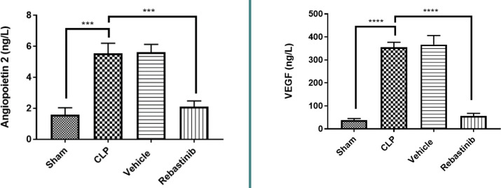
Sepsis remains a public health issue with high morbidity and mortality. Liver injury due to sepsis is associated with poor outcomes and an increased risk of death among septic patients. Rebastinib is a small molecule inhibiting the Tie2 receptor and vascular endothelial growth factor receptor-2 (VEGFR2). The current study aimed to reveal the potential protective impact of Rebastinib against sepsis-induced liver injury. Twenty-four adult male mice were allocated into four groups (six per group) as follows: Sham group was exposed to anesthesia and laparotomy with no cecal ligation and puncture procedure (CLP); CLP group was subjected CLP procedure; vehicle-treated group was pretreated with vehicle (oral route) one hour prior to CLP procedure; Rebastinib group was pretreated with oral Rebastinib one hour before induction of CLP. Collected blood was used to measure the serum levels of AST, ALT, and angiopoietin 2. Homogenized liver tissues were used to investigate IL-6, TNF-α, ICAM-1, MIF, VEGF, F2-isoprostanes, and caspase-11 levels. Histological examination was used to determine the severity of liver damage. Compared to the sham group, mice subjected to CLP had high levels of these biomarkers with a high degree of liver injury. In contrast, Rebastinib markedly reduced these levels and mitigated the liver damage. Rebastinib may be a hepatoprotective agent against sepsis-associated liver injury.

Keywords: Rebastinib-treated group; Sepsis; Tie2 receptor; inflammation; liver injury.


Conflict of interest statement

The authors declare no conflict of interest.

Figures

Figure 1
Figure 1
Impact of Rebastinib on serum ALT and AST levels post-CLP Serum ALT and AST levels across groups are shown as mean±SEM (n=6). One-way ANOVA and Bonferroni test; significance levels: **p≤0.01, ***p≤0.001, ****p≤0.0001.
Figure 2
Figure 2
Effect of Rebastinib on IL-6 and TNF-α levels in the liver Tissue levels of IL-6 and TNF-α across groups, shown as mean±SEM (n=6). One-way ANOVA and Bonferroni test; *p≤0.05, **p≤0.01.
Figure 3
Figure 3
Effect of Rebastinib on F2-isoprostane and caspase 11 levels in the liver Tissue levels of F2-isoprostane and caspase 11 across groups, shown as mean±SEM (n=6). One-way ANOVA followed by Bonferroni test; *p≤0.05, **p≤0.01, ***p≤0.001.
Figure 4
Figure 4
Effect of Rebastinib on tissue levels of MIF and ICAM-1 Tissue levels of MIF and ICAM-across groups are shown as mean±SEM (n=6). One-way ANOVA followed by Bonferroni test; **p≤0.01, ***p≤0.001, ****p≤0.0001.
Figure 5
Figure 5
Effect of Rebastinib on angiopoietin 2 and VEGF levels Serum levels of Ang2 and tissue levels of VEGF across groups are shown as mean±SEM (n=6). One-way ANOVA followed by Bonferroni test; ***p≤0.001, ****p≤0.0001.
Figure 6
Figure 6
Effect of Rebastinib on liver injury Liver injury scores across groups are shown as mean±SEM (n=6). Kruskal Wallis test and Bonferroni test; *p≤0.05, ***p≤0.001.
Figure 7
Figure 7
Sham group: normal liver histology Liver tissue sections displaying a central vein (red arrow) and normal hepatocytes (black arrows), stained with H&E. Magnifications: A - 100x, B - 400x.
Figure 8
Figure 8
CLP group: severe liver damage (score 3) Liver sections show cytoplasmic eosinophilia, apoptotic cells with chromatin condensation, pyknotic nuclei (black arrows), necrotic cells with nuclear fading (red arrows), steatosis (blue arrows), focal mild inflammation (yellow arrow), and vascular congestion (green arrow). Stained with H&E. Magnifications: A - 100x, B/C/D - 400x.
Figure 9
Figure 9
Vehicle group: severe liver damage (Score 3) Liver sections characterized by cytoplasmic eosinophilia, apoptotic cells with chromatin condensation, and pyknotic nuclei (black arrows). Necrotic cells with nuclear fading (red arrows). H&E. Magnifications: A - 100x, B - 400x.
Figure 10
Figure 10
Effect of Rebastinib on liver tissue post-CLP A: Mild liver damage (Score 1) with cytoplasmic vacuoles (400x magnification). B: Vascular congestion with erythrocyte stasis (black arrows) and mild inflammation (red arrows) (400x magnification). C: Moderate liver damage (Score 2) featuring cytoplasmic eosinophilia, apoptotic cells with chromatin condensation, pyknotic nuclei (black arrows), and cytoplasmic vacuoles (red arrows) (400x magnification)

Similar articles

References

    1. DeMerle KM, Angus DC, Baillie JK, Brant E, et al. Sepsis Subclasses: A Framework for Development and Interpretation. Crit Care Med. 2021;49:748–59. doi: 10.1097/CCM.0000000000004842. - DOI - PMC - PubMed
    1. Liang H, Song H, Zhang X, Song G, et al. Metformin attenuated sepsis-related liver injury by modulating gut microbiota. Emerg Microbes Infect. 2022;11:815–28. doi: 10.1080/22221751.2022.2045876. - DOI - PMC - PubMed
    1. Cecconi M, Evans L, Levy M, Rhodes A. Sepsis and septic shock. Lancet. 2018;392:75–87. doi: 10.1016/S0140-6736(18)30696-2. - DOI - PubMed
    1. Zhang P, Peng J, Ren Y-Q, Zheng H, Yan H. Dexmedetomidine protects against endothelial injury in septic rats induced by cecal ligation and puncture by decreasing angiopeiotin 2 and increasing vascular endothelial cadherin levels. Exp Ther Med. 2021;21:111. doi: 10.3892/etm.2020.9543. - DOI - PMC - PubMed
    1. Gotts JE, Matthay MA. Sepsis: pathophysiology and clinical management. BMJ. 2016;353:i1585. doi: 10.1136/bmj.i1585. - DOI - PubMed