Case Reports
. 2024 Mar;30(3):555-559.
doi: 10.3201/eid3003.231402.

Taenia martis Neurocysticercosis-Like Lesion in Child, Associated with Local Source, the Netherlands

Case Reports

Taenia martis Neurocysticercosis-Like Lesion in Child, Associated with Local Source, the Netherlands

Hendriekje Eggink et al. Emerg Infect Dis. 2024 Mar.

Abstract

A neurocysticercosis-like lesion in an 11-year-old boy in the Netherlands was determined to be caused by the zoonotic Taenia martis tapeworm. Subsequent testing revealed that 15% of wild martens tested in that region were infected with T. martis tapeworms with 100% genetic similarity; thus, the infection source was most likely local.

Keywords: Cestoda; Mustelidae; Neurocysticercosis; Taenia; parasites; the Netherlands; zoonoses.


Figures
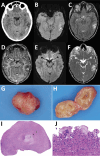
Figure 1
Figure 1
Diagnostic imaging of the brain and cystic lesion resected from boy with neurocysticercosis-like lesion, the Netherlands. A) Axial computed tomography showing edema in the left temporal lobe with a barely visible hypointense round lesion with a noncalcified, isointense rim (arrow). B–F) Axial magnetic resonance images at slightly different levels through the cystic lesion with surrounding edema in the left temporal lobe, showing a hypointense ring on susceptibility-weighted image (B) using minimum intensity projection (arrow) and on T2-weighted image (C), suggestive of a fibrotic capsule. D) Three-dimensional T1-weighted image showing a slightly irregular enhancement of the rim. E, F) On diffusion-weighted image (E) and apparent diffusion coefficient map (F), the rim is isointense and central diffusion restriction is absent, excluding a bacterial abscess. G, H) Macroscopic picture of the lesion showing a round nodule (G) and a cyst-like lesion (H) on cut section with a white-greyish central area surrounded by a thin capsule. I, J) Microscopic images showing a necrotic core (1) surrounded by a rim of fibrosis (2) and a mixed inflammatory response (3) (I) and multinuclear foreign-body-type giant cells (J).
Figure 2
Figure 2
Phylogenetic analysis of the partial CO1 gene of Taenia martis tapeworm samples from a patient, martens, and a squirrel in the Netherlands and reference sequences. GenBank accession numbers are shown when available. The tree is based on multiple alignment with Jukes and Cantor correction and neighbor-joining cluster analysis. Branch quality was determined by bootstrap analysis with 10,000 simulations. Reference sequences were from patients from Italy (GenBank accession no. KJ459910.1), Croatia (accession no. AB731758), France (accession no. KP198618.1), and Germany (accession no. JX415821). Moreover, T. saginata (accession nos. AB645845 and JQ756972), T. solium (accession nos. EF0767752 and AB033408), T. polyacantha (accession nos. EU544587 and EU544588), and T. taeniaeformis (accession no. EF090612) were included in the phylogenetic analysis. The cestode Spirometra erinaceieuropaei (accession no. AB278576) was included as outgroup. Scale bar indicates nucleotide substitutions/site.

References

    1. Deplazes P, Eichenberger RM, Grimm F. Wildlife-transmitted Taenia and Versteria cysticercosis and coenurosis in humans and other primates. Int J Parasitol Parasites Wildl. 2019;9:342–58. 10.1016/j.ijppaw.2019.03.013 - DOI - PMC - PubMed
    1. Brunet J, Benoilid A, Kremer S, Dalvit C, Lefebvre N, Hansmann Y, et al. First case of human cerebral Taenia martis cysticercosis. J Clin Microbiol. 2015;53:2756–9. 10.1128/JCM.01033-15 - DOI - PMC - PubMed
    1. Eberwein P, Haeupler A, Kuepper F, Wagner D, Kern WV, Muntau B, et al. Human infection with marten tapeworm. Emerg Infect Dis. 2013;19:1152–4. 10.3201/eid1907.121114 - DOI - PMC - PubMed
    1. Koch T, Schoen C, Muntau B, Addo M, Ostertag H, Wiechens B, et al. Molecular diagnosis of human Taenia martis eye infection. Am J Trop Med Hyg. 2016;94:1055–7. 10.4269/ajtmh.15-0881 - DOI - PMC - PubMed
    1. Mueller A, Förch G, Zustin J, Muntau B, Schuldt G, Tappe D. Case report: molecular identification of larval Taenia martis infection in the pouch of Douglas. Am J Trop Med Hyg. 2020;103:2315–7. 10.4269/ajtmh.20-0782 - DOI - PMC - PubMed

Publication types

LinkOut - more resources