. 2024 May 8;52(8):4313-4327.
doi: 10.1093/nar/gkae105.

DNA-PK controls Apollo's access to leading-end telomeres

Affiliations

DNA-PK controls Apollo's access to leading-end telomeres

Ceylan Sonmez et al. Nucleic Acids Res. .

Abstract

The complex formed by Ku70/80 and DNA-PKcs (DNA-PK) promotes the synapsis and the joining of double strand breaks (DSBs) during canonical non-homologous end joining (c-NHEJ). In c-NHEJ during V(D)J recombination, DNA-PK promotes the processing of the ends and the opening of the DNA hairpins by recruiting and/or activating the nuclease Artemis/DCLRE1C/SNM1C. Paradoxically, DNA-PK is also required to prevent the fusions of newly replicated leading-end telomeres. Here, we describe the role for DNA-PK in controlling Apollo/DCLRE1B/SNM1B, the nuclease that resects leading-end telomeres. We show that the telomeric function of Apollo requires DNA-PKcs's kinase activity and the binding of Apollo to DNA-PK. Furthermore, AlphaFold-Multimer predicts that Apollo's nuclease domain has extensive additional interactions with DNA-PKcs, and comparison to the cryo-EM structure of Artemis bound to DNA-PK phosphorylated on the ABCDE/Thr2609 cluster suggests that DNA-PK can similarly grant Apollo access to the DNA end. In agreement, the telomeric function of DNA-PK requires the ABCDE/Thr2609 cluster. These data reveal that resection of leading-end telomeres is regulated by DNA-PK through its binding to Apollo and its (auto)phosphorylation-dependent positioning of Apollo at the DNA end, analogous but not identical to DNA-PK dependent regulation of Artemis at hairpins.


Figures

Graphical Abstract
Graphical Abstract
Figure 1.
Figure 1.
DNA-PK inhibition prevents Apollo-dependent leading-end telomere protection. (A) Representative chromosome orientation fluorescence in situ hybridization (CO-FISH) of metaphase spreads of ApolloF/F MEFs immortalized with SV40LT 96 h after Hit & Run Cre treatment and/or after 24 h treatment with DNA-PKcs inhibitor NU7441 (DNA-PKi). Leading and lagging-end telomeres were detected with Cy3-(TTAGGG)3 (red) and Alexa488-(CCCTAA)3 (green) probes, respectively. DNA was stained with DAPI (blue). Arrows indicate leading-end telomere fusions. (B) Quantification of telomere fusions as shown in (A). Each dot represents the percentage of telomeres fused in one metaphase, aggregated for chromatid-type (involving two leading-end telomeres or two lagging-end telomeres) or chromosome-type fusions. Bars represent the median of telomere fusions in 40 metaphases across n = 4 independent experiments (10 metaphases per experiment). (C) Telomeric overhang assay on ApolloF/F MEFs treated as in (A). Genomic DNA was digested with MboI, and the single-stranded telomeric DNA was detected in-gel with end-labeled 32P-(AACCCT)4 (native, left panel). DNA was then denatured, and the gel was rehybridized with the same probe to determine the total telomeric signal (denatured, right panel). The ssTTAGGG signal was normalized to the total telomeric DNA in the same lane. The normalized no Cre value was set to 1 and all the other values were calculated relative to it. (D) Quantification of the relative overhang signal as detected in (C) for n = 4 independent experiments (indicated by different shades), with means and SDs. Statistical analysis from non-parametric Kruskal–Wallis ANOVA test for multiple comparisons (B) or ordinary one-way ANOVA for multiple comparisons (D), ****P< 0.0001, ***P< 0.001, **P< 0.01, *P< 0.05; n.s., not significant.
Figure 2.
Figure 2.
DNA-PK kinase activity promotes Apollo-dependent leading-end telomere protection. (A) Immunoblots for Apollo and actin loading control in HT1080 human cancer cells 120 h after lentiviral transduction with sgRNA targeting human Apollo and/or after 24 h treatment with DNA-PKi. Graph represents Apollo levels normalized over actin in n = 4 independent experiments, with mean and SD. (B) Representative CO-FISH of metaphase spreads of HT1080 cells treated as described in (A). Leading and lagging-end telomeres were detected with Alexa Fluor 647-OO-(TTAGGG)3 (red) and Cy3-OO-(CCCTAA)3 (green), respectively. DNA was stained with DAPI (blue). Arrows indicate leading-end telomere fusions. (C) Quantification of leading-end telomere fusions as described in (B). Each dot represents the percentage of leading-end telomere fusions in one metaphase. Bars represent the median of telomere fusions in 40 metaphases across n = 4 independent experiments (10 metaphases each). (D and E) CO-FISH metaphase analysis and quantification of leading-end telomere fusions as described in (B) and (C) for DNA-PKcs+/+ or DNA-PKcsKD/KD MEFs 108 h after lentiviral transduction with an sgRNA targeting mouse Apollo or the empty vector control. Bars represent the median of telomere fusions in 45 metaphases across n = 3 independent experiments (15 metaphases per experiment). Statistical analysis by ordinary one-way ANOVA for multiple comparisons (A) or by non-parametric Kruskal–Wallis ANOVA test for multiple comparisons (C,E). Statistical significance was indicated by ****P < 0.0001, ***P < 0.001, **P < 0.01, *P < 0.05; n.s., not significant.
Figure 3.
Figure 3.
DNA-PK kinase activity is not required for Apollo recruitment to telomeres. (A) Immunoblots for HA-Apollo (anti-HA), TRF2 and phosphorylated Chk2 in SV40LT-immortalized TRF2F/F Lig4 -/− MEFs after retroviral transduction transduced with an empty vector (EV) or HA-Apollo, 108 h after Hit & Run Cre-mediated deletion of TRF2 and/or 24 h DNA-PKi treatment. (B and C) Representative immunofluorescence-fluorescence in-situ hybridization (IF-FISH) images and quantification of HA-Apollo localization at telomeres in the same MEFs as described in (A) 120 h after Hit & Run Cre transduction or without any treatment. Apollo and telomeres were detected using anti-HA (red) or Alexa488-OO-(TTAGGG)3 probe (green), respectively. Approximately 300 nuclei were analyzed for each condition over n = 3 independent experiments (100 nuclei per experiment). Median bar is indicated. (D) Immunoblots for HA-Apollo (anti-DCLRE1B, Atlas) and actin in SV40-LT-immortalized DNA-PKcs+/+, DNA-PKcs−/− or DNA-PKcsKD/KD MEFs transduced with an empty vector (EV) or HA-Apollo. * aspecific band. (E and F) IF-FISH analysis and quantification of HA-Apollo localization at telomeres in the SV40-LT-immortalized DNA-PKcs+/+, DNA-PKcs−/− or DNA-PKcsKKD/KD MEFs transduced with an empty vector (EV) or HA-Apollo, analyzed as described in (B, C). For HA-Apollo, 300 nuclei over n = 3 independent experiments are shown (100 nuclei per experiment), with median bar. As negative control, 296 nuclei (at least 90 nuclei per experiment) were analyzed in parallel for EV. (G) Co-immunoprecipitation (Co-IP) of Myc-mTRF2 (WT or F120A) with HA-mApollo in 293T cells without or after 24 h treatment with DNA-PKi. Pull down was performed with anti-HA antibody. HA IP (first and second panels) and Input (third and fourth panels) were analyzed with anti Myc (first and third panels) or HA (second and fourth panels) antibodies. MYC-mTRF2-F120A (F120A) was used as negative control. Statistical analysis by non-parametric Kruskal–Wallis ANOVA test for multiple comparisons (C, F). Statistical significance was indicated by ****P< 0.0001, ***P< 0.001, **P< 0.01, *P< 0.05; n.s., not significant.
Figure 4.
Figure 4.
DNA-PK, not Apollo S/T-Q sites, is the target of DNA-PK kinase activity. (A and B) CO-FISH metaphase analysis and quantification of leading-end telomere fusions in SV40LT-immortalized ApolloF/F MEFs transduced with an empty vector (EV), HA-Apollo (WT) or HA-Apollo-AA (AA), 96 h after Hit & Run Cre-mediated deletion of Apollo. The graph represents 45 metaphases over n = 3 independent experiments, with median bars. (C and D) CO-FISH and quantification of leading-end telomere fusions in DNA-PKcs+/+ or DNA-PKcs3A/3A MEFs with or without 24 h treatment with DNA-PKi for 45 metaphases over n = 3 independent experiments, with median bar. Statistical analysis by non-parametric Kruskal–Wallis ANOVA test for multiple comparisons (B, D). Statistical significance was indicated by ****P< 0.0001, ***P< 0.001, **P< 0.01, *P< 0.05; n.s., not significant.
Figure 5.
Figure 5.
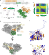
Phosphorylation on the DNA-PK ABCDE/Thr2609 cluster is predicted to allow Apollo access to telomere ends. (A) Schematics of mApollo and mDNA-PKcs. The metallo β-lactamase (MBL) and the β-CASP domains of Apollo and the N-terminal unit, the Circular cradle unit, the FAT and the Kinase domains of DNA-PKcs are indicated. Highlighted, the Apollo region (residues 1–344) predicted with high confidence by AlphaFold-Multimer modeling using AFsample and the DNA-PKcs region (residues 1981–2760) used in the modeling, with PQR and the ABCDE clusters. (B) The predicted Local Distance Difference Test (pLDDT) per residue position. (C) A model for the interaction of full-length mApollo and mDNA-PKcs (amino acids 1981–2760) as predicted by AFsample. Residues 345–541 in Apollo were removed from the model due to low confidence. (D) Predicted aligned error (PAE), as expected position error (x,y) at residue x if the predicted and true structures were aligned on residue y. (E) Superposition of (C) to the cryo-EM structure of autophosphorylated DNA-PK in complex with DNA (PBD: 7SGL) (7). (F,G) Comparison between the catalytic domains of hArtemis as in (PBD:7SGL) and mApollo model as superposed to PBD:7SGL (7) in (E), and enlargement on the catalytic residues. The key catalytic residues in Apollo and Artemis are indicated in orange and blue, respectively.
Figure 6.
Figure 6.
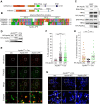
Apollo interaction with DNA-PK mediates telomere protection. (A) Schematics of mArtemis and mApollo. The MBL, the β-CASP, the known Lig4, PTIP (Pax transactivation-domain interacting protein) and TRF2-interacting domains, and the DNA-PKcs interacting regions are indicated. (B) MUSCLE alignment of human and mouse Artemis and Apollo C-terminal tails. Amino acids are colored according to physico-chemical properties (Zappo). Highlighted, the contact points (CP) of Artemis with DNA-PKcs (17) and the predicted DNA-PK-interacting region of Apollo, with a patch of positively charged amino acids followed by a patch of negatively charged ones. (C) Co-IP of endogenous DNA-PKcs and TRF2 with HA-mApollo-WT (WT) or HA-mApollo-ΔPK (ΔPK) in 293T cells. Pull-down was performed with anti-HA antibody. HA IP and input were analyzed by immunoblotting with anti-DNA-PKcs (top panels), TRF2 (middle panels) or HA (lower panel) antibodies. (D) Immunoblot of SV40LT-immortalized ApolloF/F MEFs transduced with either EV, HA-Apollo-WT or HA-Apollo-ΔPK 108 h after Hit & Run Cre-mediated deletion of endogenous Apollo. (E and F) IF-FISH analysis and quantification of Apollo localization at telomeres in the same MEFs as in (D). Graph represents 100 nuclei for each condition from n = 3 independent experiments (300 nuclei in total), with medians. (G and H) CO-FISH metaphase analysis and quantification of leading-end telomere fusions on MEFs treated as in (D). Graph represents 30 metaphases over n = 3 independent experiments with medians. Statistic by non-parametric Kruskal–Wallis ANOVA test for multiple comparisons (F, H). Statistical significance was indicated by ****P< 0.0001, ***P< 0.001, **P< 0.01, *P< 0.05; n.s., not significant.
Figure 7.
Figure 7.
Proposed model for Apollo recruitment and access to leading-end telomeres after replication. (A) Apollo is recruited at telomeres by TRF2 binding. The physical presence of inactive DNA-PK at the blunt DNA end prevents any nucleolytic attack of the ends, including Apollo’s. After autophosphorylation of DNA-PKcs on the ABCDE/Thr2609 cluster and Apollo interaction with DNA-PKcs, Apollo gains access to the telomere ends and perform the initial resection required for the generation of the 3′overhang and for telomere protection. (B) In the absence of Apollo, when Apollo cannot be recruited by TRF2 or cannot interact with DNA-PKcs, the leading-end telomeres are not resected and are fused via alt-EJ. (C) In the absence of DNA-PK autophosphorylation, due to inhibition of the kinase activity of DNA-PK and/or to mutations affecting the kinase activity and/or the ABCDE/Thr2609 cluster of DNA-PK, Apollo cannot get access to the telomere ends, the leading-end telomeres are not resected and are fused via the alt-EJ.

References

    1. Wu Q., Liang S., Ochi T., Chirgadze D.Y., Huiskonen J.T., Blundell T.L.. Understanding the structure and role of DNA-PK in NHEJ: how X-ray diffraction and cryo-EM contribute in complementary ways. Prog. Biophys. Mol. Biol. 2019; 147:26–32. - PubMed
    1. Zhao B., Rothenberg E., Ramsden D.A., Lieber M.R.. The molecular basis and disease relevance of non-homologous DNA end joining. Nat. Rev. Mol. Cell Biol. 2020; 21:765–781. - PMC - PubMed
    1. Buehl C.J., Goff N.J., Hardwick S.W., Gellert M., Blundell T.L., Yang W., Chaplin A.K., Meek K.. Two distinct long-range synaptic complexes promote different aspects of end processing prior to repair of DNA breaks by non-homologous end joining. Mol. Cell. 2023; 83:698–714. - PMC - PubMed
    1. Chaplin A.K., Hardwick S.W., Stavridi A.K., Buehl C.J., Goff N.J., Ropars V., Liang S., De Oliveira T.M., Chirgadze D.Y., Meek K.et al. .. Cryo-EM of NHEJ supercomplexes provides insights into DNA repair. Mol. Cell. 2021; 81:3400–3409. - PMC - PubMed
    1. Chen X., Xu X., Chen Y., Cheung J.C., Wang H., Jiang J., de Val N., Fox T., Gellert M., Yang W.. Structure of an activated DNA-PK and its implications for NHEJ. Mol. Cell. 2021; 81:801–810. - PMC - PubMed

Publication types

MeSH terms