RESUSCITATIVE ENDOVASCULAR BALLOON OCCLUSION OF THE AORTA: ZONE 1 REPERFUSION-INDUCED COAGULOPATHY
- PMID: 38407818
- PMCID: PMC10955717
- DOI: 10.1097/SHK.0000000000002299
RESUSCITATIVE ENDOVASCULAR BALLOON OCCLUSION OF THE AORTA: ZONE 1 REPERFUSION-INDUCED COAGULOPATHY
Abstract
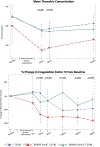
Objective: We sought to identify potential drivers behind resuscitative endovascular balloon occlusion of the aorta (REBOA) induced reperfusion coagulopathy using novel proteomic methods. Background: Coagulopathy associated with REBOA is poorly defined. The REBOA Zone 1 provokes hepatic and intestinal ischemia that may alter coagulation factor production and lead to molecular pathway alterations that compromises hemostasis. We hypothesized that REBOA Zone 1 would lead to reperfusion coagulopathy driven by mediators of fibrinolysis, loss of coagulation factors, and potential endothelial dysfunction. Methods: Yorkshire swine were subjected to a polytrauma injury (blast traumatic brain injury, tissue injury, and hemorrhagic shock). Pigs were randomized to observation only (controls, n = 6) or to 30 min of REBOA Zone 1 (n = 6) or REBOA Zone 3 (n = 4) as part of their resuscitation. Thromboelastography was used to detect coagulopathy. ELISA assays and mass spectrometry proteomics were used to measure plasma protein levels related to coagulation and systemic inflammation. Results: After the polytrauma phase, balloon deflation of REBOA Zone 1 was associated with significant hyperfibrinolysis (TEG results: REBOA Zone 1 35.50% versus control 9.5% vs. Zone 3 2.4%, P < 0.05). In the proteomics and ELISA results, REBOA Zone 1 was associated with significant decreases in coagulation factor XI and coagulation factor II, and significant elevations of active tissue plasminogen activator, plasmin-antiplasmin complex complexes, and syndecan-1 (P < 0.05). Conclusion: REBOA Zone 1 alters circulating mediators of clot formation, clot lysis, and increases plasma levels of known markers of endotheliopathy, leading to a reperfusion-induced coagulopathy compared with REBOA Zone 3 and no REBOA.
Copyright © 2023 by the Shock Society.
Conflict of interest statement
EEM has patents pending related to coagulation and fibrinolysis diagnostics and therapeutic fibrinolytics and was a cofounder of ThromboTherepeutics. EEM has received grant support from Haemonetics, Hemosonics, Werfen, Stago, and Prytime outside the submitted work. CJF is a clinical consultant for Prytime Medical. The remaining authors report no conflict of interests.
Figures


References
-
- Brenner M, Bulger EM, Perina DG, et al. Joint statement from the American College of Surgeons Committee on Trauma (ACS COT) and the American College of Emergency Physicians (ACEP) regarding the clinical use of Resuscitative Endovascular Balloon Occlusion of the Aorta (REBOA). Trauma Surg Acute Care Open. 2018;3(1):e000154. - PMC - PubMed
-
- Butler FK Jr, Holcomb JB, Shackelford SA, et al. Advanced Resuscitative Care in Tactical Combat Casualty Care: TCCC Guidelines Change 18–01:14 October 2018. J Spec Oper Med. 2018;18:37–55 - PubMed
-
- Morrison JJ, Ross JD, Rasmussen TE, Midwinter MJ, Jansen JO. Resuscitative endovascular balloon occlusion of the aorta: a gap analysis of severely injured UK combat casualties. Shock. 2014; 41:388–93. - PubMed
Publication types
MeSH terms
Substances
Grants and funding
LinkOut - more resources
Full Text Sources
Medical
Research Materials

