. 2024 Feb 26;19(2):e0297080.
doi: 10.1371/journal.pone.0297080. eCollection 2024.

Nutraceuticals known to promote hair growth do not interfere with the inhibitory action of tamoxifen in MCF7, T47D and BT483 breast cancer cell lines

Affiliations

Nutraceuticals known to promote hair growth do not interfere with the inhibitory action of tamoxifen in MCF7, T47D and BT483 breast cancer cell lines

Richard Baker et al. PLoS One. .

Abstract

Background: Hair loss/thinning is a common side effect of tamoxifen in estrogen receptor (ER) positive breast cancer therapy. Some nutraceuticals known to promote hair growth are avoided during breast cancer therapy for fear of phytoestrogenic activity. However, not all botanical ingredients have similarities to estrogens, and in fact, no information exists as to the true interaction of these ingredients with tamoxifen. Therefore, this study sought to ascertain the effect of nutraceuticals (+/- estrogen/tamoxifen), on proliferation of breast cancer cells and the relative expression of ERα/β.

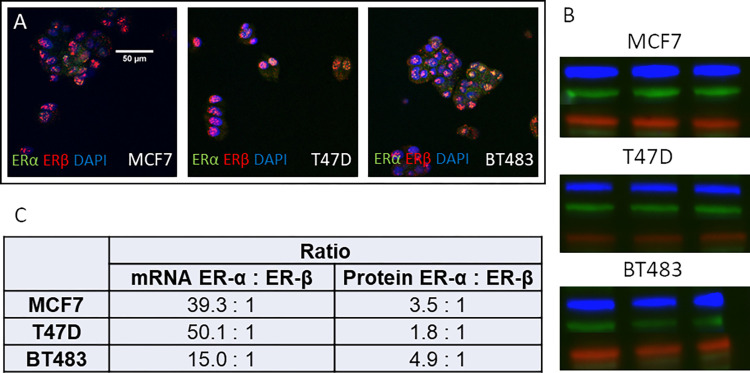
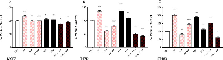
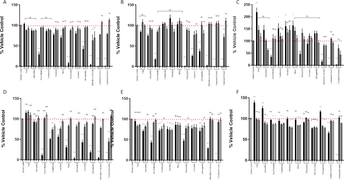
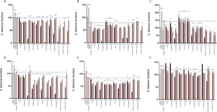
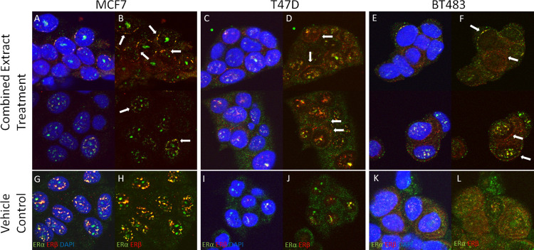
Methods: Kelp, Astaxanthin, Saw Palmetto, Tocotrienols, Maca, Horsetail, Resveratrol, Curcumin and Ashwagandha were assessed on proliferation of MCF7, T47D and BT483 breast cancer cell lines +/- 17β-estradiol and tamoxifen. Each extract was analysed by high performance liquid chromatography (HPLC) prior to use. Cellular ERα and ERβ expression was assessed by qRT-PCR and western blot. Changes in the cellular localisation of ERα:ERβ and their ratio following incubation with the nutraceuticals was confirmed by immunocytochemistry.

Results: Estradiol stimulated DNA synthesis in three different breast cancer cell lines: MCF7, T47D and BT483, which was inhibited by tamoxifen; this was mirrored by a specific ERa agonist in T47D and BT483 cells. Overall, nutraceuticals did not interfere with tamoxifen inhibition of estrogen; some even induced further inhibition when combined with tamoxifen. The ERα:ERβ ratio was higher at mRNA and protein level in all cell lines. However, incubation with nutraceuticals induced a shift to higher ERβ expression and a localization of ERs around the nuclear periphery.

Conclusions: As ERα is the key driver of estrogen-dependent breast cancer, if nutraceuticals have a higher affinity for ERβ they may offer a protective effect, particularly if they synergize and augment the actions of tamoxifen. Since ERβ is the predominant ER in the hair follicle, further studies confirming whether nutraceuticals can shift the ratio towards ERβ in hair follicle cells would support a role for them in hair growth. Although more research is needed to assess safety and efficacy, this promising data suggests the potential of nutraceuticals as adjuvant therapy for hair loss in breast cancer patients receiving endocrine therapy.


Conflict of interest statement

This study was supported by Nutraceutical Wellness, Inc. New York, USA This does not alter our adherence to PLOS ONE policies on sharing data and materials.

Figures

Fig 1
Fig 1. Cellular localization of ERα:ERβ and quantification of ERα:ERβ mRNA and protein expression in MCF7, T47D & BT483 cells.
(A) Immunocytochemistry: ERα = green, ERβ = red, DAPI = blue, co-localisation = yellow (B) Western blots: ERα = blue, ERβ = green, GAPDH = red. Full western blot image available in S1 Fig. (C) Quantification of mRNA transcripts by qRT-PCR (n = 3 per cell line) and protein expression by quantification of Western blot band density (n = 3 per cell line).
Fig 2
Fig 2. Cell proliferation is stimulated by 17b-estradiol and an ERa agonist which is negated by tamoxifen: Incorporation of BrdU by breast cancer cells MCF7, T47D and BT483 as a percentage of the vehicle control (+/- SEM of 3 separate assays with 8 experimental replicates).
CON = vehicle control (0.1% DMSO); E2 = 17b-estradiol (10nM); TAM = tamoxifen (2μM); PPT (100nM), ERB = ERB-041 (100nM) Cells were incubated for 48 hours. ANOVA (Dunnett’s multiple comparison) statistical significance is displayed on the graph above the bars (* denotes significance vs CON. # denotes significance vs E2.). *p ≤ 0.05, **p ≤ 0.01, ***p ≤ 0.001, ****p ≤ 0.0001, ns non-significant.
Fig 3
Fig 3
Nutraceuticals do not stimulate breast cancer cell proliferation at lower concentrations: Cellular proliferation assessed by incorporation of BrdU by breast cancer cells MCF7 (A), T47D (B), BT483 (C) as a percentage of the vehicle control. Cellular proliferation assessed by Alamar Blue of MCF7 (D), T47D €, BT483 (F) as a percentage of the vehicle control (+/- SEM of 3 separate assays). Vehicle control (0.1% DMSO), Kelp (black bar = 50 μg/ml, dark grey = 5 μg/ml, light grey = 0.5μg/ml), Astaxanthin (black bar = 10 μg/ml, dark grey = 1 μg/ml, light grey = 0.1 μg/ml), Saw Palmetto (black bar = 50 μg/ml, dark grey = 5 μg/ml, light grey = 0.5 μg/ml), Tocotrienols (black bar = 10 μg/ml, dark grey = 1 μg/ml, light grey = 0.1 μg/ml), Maca (black bar = 10 μg/ml, dark grey = 1 μg/ml, light grey = 0.1 μg/ml), Horsetail (black bar = 10 μg/ml, dark grey = 1 μg/ml, light grey = 0.1 μg/ml), Resveratrol (black bar = 10 μg/ml, dark grey = 1 μg/ml, light grey = 0.1 μg/ml), Curcumin (black bar = 1 μg/ml, dark grey = 0.1 μg/ml, light grey = 0.01 μg/ml), Ashwagandha (black bar = 100 μg/ml, dark grey = 10 μg/ml, light grey = 1 μg/ml), Alternative Curcumin (black bar = 10 μg/ml, dark grey = 1 μg/ml, light grey = 0.1 μg/ml) Combined Extract 1 = All extracts combined except Alternative Curcumin (black bar = 25%, dark grey = 10%, light grey = 1%), Combined Extract 2 = All extracts combined except Curcumin (black bar = 25%, dark grey = 10%, light grey = 1%). Cells were incubated with the nutraceuticals for 48 hours. ANOVA (Dunnett’s multiple comparison) statistical significance is displayed on the graph above the bars (* denotes significance vs vehicle control.) *p ≤ 0.05, **p ≤ 0.01, ***p ≤ 0.001, ns non-significant.
Fig 4
Fig 4. Nutraceuticals do not interfere with the inhibitory effect of tamoxifen on cell proliferation stimulated by 17b-estradiol at lower concentrations.
Cellular proliferation assessed by incorporation of BrdU by breast cancer cells MCF7 (A), T47D (B), BT483 (C) as a percentage of the vehicle control. Cellular proliferation assessed by Alamar Blue of MCF7 (D), T47D (E), BT483 (F) as a percentage of the vehicle control (+/- SEM of 3 separate assays): Vehicle control (0.1% DMSO); E2 = 17b-estradiol (10nM); Tam = tamoxifen (2μM); Kelp (black bar = 50 μg/ml, dark grey = 5 μg/ml, light grey = 0.5μg/ml), Astaxanthin (black bar = 10 μg/ml, dark grey = 1 μg/ml, light grey = 0.1 μg/ml), Saw Palmetto (black bar = 50 μg/ml, dark grey = 5 μg/ml, light grey = 0.5 μg/ml), Tocotrienols (black bar = 10 μg/ml, dark grey = 1 μg/ml, light grey = 0.1 μg/ml), Maca (black bar = 10 μg/ml, dark grey = 1 μg/ml, light grey = 0.1 μg/ml), Horsetail (black bar = 10 μg/ml, dark grey = 1 μg/ml, light grey = 0.1 μg/ml), Resveratrol (black bar = 10 μg/ml, dark grey = 1 μg/ml, light grey = 0.1 μg/ml), Curcumin (black bar = 1 μg/ml, dark grey = 0.1 μg/ml, light grey = 0.01 μg/ml), Ashwagandha (black bar = 100 μg/ml, dark grey = 10 μg/ml, light grey = 1 μg/ml), Alternative Curcumin (black bar = 10 μg/ml, dark grey = 1 μg/ml, light grey = 0.1 μg/ml) Combined Extract 1 = All extracts combined except Alternative Curcumin (black bar = 25%, dark grey = 10%, light grey = 1%), Combined Extract 2 = All extracts combined except Curcumin (black bar = 25%, dark grey = 10%, light grey = 1%). Cells were incubated with the nutraceuticals for 48 hours in the presence of E2 and tamoxifen. ANOVA (Dunnett’s multiple comparison) statistical significance is displayed on the graph above the bars (* denotes significance vs E2 + TAM. # Denotes significance vs vehicle control. ^ Denotes significance vs E2 treated control). *p ≤ 0.05, **p ≤ 0.01, ***p ≤ 0.001.
Fig 5
Fig 5. Cellular localization of ERα:ERβ in presence of combined nutraceuticals.
Cellular localization of ERα:ERβ in presence of combined nutraceuticals. Note the distribution of ERs around the nuclear periphery. MCF7 cells treated with 10% Combined Extract (without Curcumin) with DAPI overlay (A) and without DAPI (B) and vehicle control (G, H). T47D cells treated with 10% Combined Extract (without Alternative Curcumin) with DAPI overlay (C) and without DAPI (D) and vehicle control (I, J). BT483 cells treated with Combined Extract (without Alternative Curcumin) with DAPI overlay (E) and without DAPI (F) and vehicle control (K, L) ERα = green, ERβ = red, Magnification = x630.

Similar articles

Cited by

References

    1. Li J, Chen Z, Su K, Zeng J. Clinicopathological classification and traditional prognostic indicators of breast cancer. Int J Clin Exp Pathol. 2015. Jul 1;8(7):8500–5. - PMC - PubMed
    1. Dey P, Wang A, Ziegler Y, Kumar S, Yan S, Kim SH, et al.. Estrogen Receptor Beta 1: A Potential Therapeutic Target for Female Triple Negative Breast Cancer. Endocrinology. 2022. Oct 23;163(12). doi: 10.1210/endocr/bqac172 - DOI - PMC - PubMed
    1. Hartman J, Lindberg K, Morani A, Inzunza J, Strom A, Gustafsson JA. Estrogen Receptor β Inhibits Angiogenesis and Growth of T47D Breast Cancer Xenografts. Cancer Res.2006. Dec; 66 (23): 11207–11213 - PubMed
    1. Song W., Tang L., Xu Y.,Sun Q et al.. ERβ1 inhibits metastasis of androgen receptor-positive triple-negative breast cancer by suppressing ZEB1. J Exp Clin Cancer Res. 2017; 36, 75. - PMC - PubMed
    1. Song P, Li Y, Dong Y, Liang Y, Qu H, Qi D et al.. Estrogen receptor β inhibits breast cancer cells migration and invasion through CLDN6-mediated autophagy. J Exp Clin Cancer Res. 2019. Aug 14;38(1):354. - PMC - PubMed