. 2024 Feb 26;20(2):e1011718.
doi: 10.1371/journal.ppat.1011718. eCollection 2024 Feb.

TRIM72 restricts lyssavirus infection by inducing K48-linked ubiquitination and proteasome degradation of the matrix protein

Affiliations

TRIM72 restricts lyssavirus infection by inducing K48-linked ubiquitination and proteasome degradation of the matrix protein

Baokun Sui et al. PLoS Pathog. .

Abstract

The tripartite motif (TRIM) protein family is the largest subfamily of E3 ubiquitin ligases, playing a crucial role in the antiviral process. In this study, we found that TRIM72, a member of the TRIM protein family, was increased in neuronal cells and mouse brains following rabies lyssavirus (RABV) infection. Over-expression of TRIM72 significantly reduced the viral titer of RABV in neuronal cells and mitigated the pathogenicity of RABV in mice. Furthermore, we found that TRIM72 over-expression effectively prevents the assembly and/or release of RABV. In terms of the mechanism, TRIM72 promotes the K48-linked ubiquitination of RABV Matrix protein (M), leading to the degradation of M through the proteasome pathway. TRIM72 directly interacts with M and the interaction sites were identified and confirmed through TRIM72-M interaction model construction and mutation analysis. Further investigation revealed that the degradation of M induced by TRIM72 was attributed to TRIM72's promotion of ubiquitination at site K195 in M. Importantly, the K195 site was found to be partially conserved among lyssavirus's M proteins, and TRIM72 over-expression induced the degradation of these lyssavirus M proteins. In summary, our study has uncovered a TRIM family protein, TRIM72, that can restrict lyssavirus replication by degrading M, and we have identified a novel ubiquitination site (K195) in lyssavirus M.


Conflict of interest statement

The authors have declared that no competing interests exist.

Figures
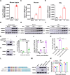
Fig 1
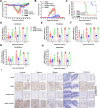
Fig 1. RABV infection induces up-regulation of TRIM72 in mouse brains and neuronal cells, and TRIM72 reduced RABV replication in neuronal cells.
(A-B) 8-week-old female C57 BL/6 mice were intranasally infected with 100 fluorescence focus units (FFU) RABV (CVS-B2c) or mock-infected. The mouse brains were collected at 12th d post-infection, the mRNA level of TRIM72 was analyzed by qPCR (A), and the protein levels of TRIM72 and RABV-N were analyzed by western blotting with anti-TRIM72 antibody and anti-RABV-N antibody (B). (C-D) Primary mouse neuron cells were isolated and cultured, then infected with RABV at different MOI for 36 h. The mRNA level of TRIM72 was analyzed by qPCR (C), and the protein levels of TRIM72 and RABV-N were analyzed by western blotting with anti-TRIM72 antibody and anti-RABV-N antibody (D). (E-F) N2a cells were infected with RABV at different MOI for 36 h. The mRNA level of TRIM72 was analyzed by qPCR (E), and the protein levels of TRIM72 and RABV-N were analyzed by western blotting with anti-TRIM72 antibody and anti-RABV-N antibody (F). (G) Empty vector (pCAGGS) or TRIM72-flag over-expression vector (pCA-Trim72-flag) were transfected into N2a cells respectively for 48 h, and then the TRIM72-flag level was analyzed by western blotting with anti-flag antibody. (H) Trim72-/- N2a cells were constructed, then the TRIM72 level was analyzed in WT and Trim72-/- N2a cells by western blotting with an anti-TRIM72 antibody. (I) Empty vector or TRIM72-flag over-expression vectors were transfected into N2a cells respectively for 12 h, then infected with RABV (MOI = 0.01) for the indicated time, and the viral titers in the supernatant were analyzed. (J) WT and Trim72-/- N2a cells were infected with RABV (MOI = 0.01) for the indicated time respectively, and the viral titers in the supernatant were analyzed. (K) WT and Trim72-/- N2a cells were transfected with empty vector or TRIM72-flag over-expression vectors for 12 h, then infected with RABV (MOI = 0.01) for 48 h and the viral titers in the supernatant were analyzed. (L) Trim72 and Trim72(-) were inserted into the genome of a recombinant RABV (rRABV), named rRABV-TRIM72 and rRABV-TRIM72(-) respectively. (M) N2a cells were infected with different types of rRABVs (MOI = 3) for 36 h, then the TRIM72 level was analyzed by western blotting with an anti-TRIM72 antibody. (N) N2a cells were infected with different types of rRABVs (MOI = 0.01) and their growth kinetics were compared. Statistical analysis of grouped comparisons was carried out by student’s t-test (*P < 0.05; **P<0.01; ***P<0.001; ****P<0.0001). The bar graph represents means ± SD, n = 3. Western blot data are representative of at least two independent experiments.
Fig 2
Fig 2. TRIM72 attenuates RABV pathogenicity in vivo.
(A-C) Female C57 BL/6 mice (8-week-old, n = 10) were intranasally infected with 100 FFU rRABV, rRABV-Trim72, rRABV-Trim72(-), or were mock-infected. Body weight change (A), clinical score (B), and survival ratio (C) were monitored daily for continuous 3 weeks. (means ± SEM; ***P<0.001; ***P<0.0001; body weight change and the clinical score were analyzed by Two-way ANOVA test; survival ratio was analyzed by log-rank test). (D-H) At indicated time points, different brain parts of the infected mice were collected to analyze the levels of RABV N mRNA by qPCR. (n = 5; means ± SEM; *P < 0.05; **P<0.01; ***P<0.001; ***P<0.0001 by student’s Two-way ANOVA test). (I) At the 12th day post-infection, mouse brains were collected, sectioned, and analyzed by immunohistochemistry using antibodies against RABV P. Scale bar, 100 μm. IHC data are representative of three sections.
Fig 3
Fig 3. TRIM72 restricts RABV infection at the viral release stage.
(A) TRIM72-flag over-expression vector or empty vector were transfected into N2a cells respectively for 36 h, then infected with RABV (MOI = 0.1) for 1 h at 4°C, the cells were collected and the viral genomic RNA (vRNA) levels were analyzed by qPCR. (B) TRIM72-flag over-expression vector or empty vector was transfected into N2a cells individually for 36 h then infected with RABV (MOI = 0.1) for 2 h at 37°C, the cells were collected and the vRNA levels were analyzed by qPCR. (C-D) TRIM72-flag over-expression vector or empty vector were transfected into N2a cells individually for 12 h and then infected with RABV (MOI = 0.01) for the indicated time points, the cells were collected and the levels of RABV-N-mRNA (D) and vRNA (E) were analyzed by qPCR. (E-F) N2a cells were infected with rRABV, rRABV-Trim72, or rRABV-Trim72(-) at MOI 0.001. Then the cells were covered with culture medium containing 1% agar and incubated for 48 h. The viral spread was compared and the infected cell numbers were calculated within the fluorescence focus. Scale bar, 100 μm. (G) pCAGGS or pCA-Trim72-myc together with pCA-M-HA and pCA-G-flag were co-transfected into HEK-293T cells respectively for 48 h, and the protein levels of M-HA and G-flag in the supernatants and cells were analyzed by western blotting. Western blot data are representative of at least two independent experiments.
Fig 4
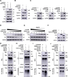
Fig 4. TRIM72 induces proteasomal degradation of RABV-M by promoting K48-linked ubiquitination.
(A) pCAGGS or pCA-Trim72-myc together with pCA-N, pCA-P, pCA-M-HA, and pCA-G-flag were co-transfected into N2a cells respectively for 48 h, and the protein levels of N, P, M-HA and G-flag were analyzed by western blotting. (B) pCAGGS or pCA-Trim72-flag were transfected into N2a cells for 12 h, then infected with RABV (MOI = 1) for 36h and RABV-N and RABV-M protein levels were analyzed by western blotting. (C) pCAGGS or pCA-Trim72-flag together with pCA-M-HA were co-transfected into N2a cells respectively. The specific inhibitors for proteasome and lysosome, Mg132 (10 μM) and NH4Cl (5 mM) were applied. Then the protein levels of TRIM72 and M were analyzed by western blotting. (D) pCA-M-HA together with pCAGGS or different concentrations of pCA-Trim72-flag were co-transfected into N2a cells for 48 h. Then the protein levels of TRIM72-flag and M-HA were analyzed by western blotting. (E) pCA-M-HA together with pCAGGS or different concentrations of pCA-Trim72-flag were co-transfected into N2a cells. Then Mg132 (10 μM) was applied and the protein levels of TRIM72-flag and M-HA were analyzed by western blotting. (F) pCA-M-HA together with pCAGGS, pCA-Trim72-flag, or Trim72-ΔRING-flag were co-transfected into N2a cells respectively for 48 h, then M-HA level was analyzed by western blotting. (G) pCAGGS or pCA-Trim72-myc together with pCA-M-flag and UB-HA over-expression vectors were co-transfected into N2a cells respectively. Then treated with Mg132 and Co-IP assays were performed with anti-flag antibody. The protein levels of TRIM72-myc, M-flag, and UB-HA were analyzed by western blotting. (H) pCAGGS or pCA-Trim72-flag were transfected into N2a cells respectively. Then infected with RABV (MOI = 1) for 36 h and treated with Mg132 and Co-IP assays were performed with anti-M antibody. The protein levels of TRIM72-flag, M, and UB were analyzed by western blotting. (I) pCAGGS or pCA-Trim72-myc together with pCA-M-flag and UB-K48-HA over-expression vectors were co-transfected into N2a cells respectively. Then treated with Mg132 and Co-IP assays were performed with anti-flag antibody post transfection for 48 h. Then the protein levels of TRIM72-myc, M-flag, and UB-K48-HA were analyzed by western blotting. (J) pCAGGS or pCA-Trim72-myc together with pCA-M-flag and UB-K63-HA over-expression vectors were co-transfected into N2a cells respectively. Then treated with Mg132 and Co-IP assays were performed with anti-flag antibody. The protein levels of TRIM72-myc, M-flag, and UB-K63-HA were analyzed by western blotting. Western blot data are representative of at least two independent experiments.
Fig 5
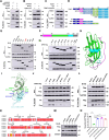
Fig 5. TRIM72 directly interacts with RABV-M via the SPRY domain.
(A) pCAGGS or pCA-Trim72-flag together with pCA-M-HA were co-transfected into N2a cells respectively. Then Mg132 (10 μM) was treated, and Co-IP assays were performed with anti-flag antibody post-transfection for 48 h. The protein levels of TRIM72-flag and M-HA were analyzed by western blotting. (B) pCAGGS or pCA-M-flag together with pCA-Trim72-HA were co-transfected into N2a cells respectively for 48 h. Then Co-IP assays were performed with anti-flag antibody. The protein levels of TRIM72-flag and M-HA were analyzed by western blotting. (C) N2a cells were infected with RABV (MOI = 1) for 36 h. Then Co-IP assays were performed with anti-TRIM72 antibody. The protein levels of TRIM72 and M were analyzed by western blotting. (D) TRIM72 truncations were designed and constructed based on its functional domain. (E) The over-expression vectors of TRIM72-flag truncations together with pCA-M-HA were co-transfected into N2a cells respectively for 48 h. Then Co-IP assays were performed with anti-flag antibody. The protein levels of TRIM72 truncations and M-HA were analyzed by western blotting. (F) RABV-M truncations were designed and constructed based on its secondary structure. (G) The over-expression vectors of M-HA truncations together with pCA-Trim72-SPRY-flag were co-transfected into N2a cells for 48 h. Then Co-IP assays were performed with anti-HA antibody. The protein levels of M truncations and SPRY-flag were analyzed by western blotting. (H) A structural model of RABV-M was built using SWISS-MODEL online software (https://swissmodel.expasy.org/interactive) based on the crystal structure of Lagos bat virus M protein (PDB code: 2W2S). (I) An interaction model of the human TRIM72-SPRY domain and RABV-M was built with GalaxyWEB online software (https://galaxy.seoklab.org/) based on hTRIM72 structure (PDB code: 6NPY), the potential interaction sites were labeled. (J) Over-expression vectors of RABV-M-HA mutations together with TRIM72-SPRY-flag over-expression vector were co-transfected into N2a cells respectively for 48 h. Then Co-IP assays were performed with anti-HA antibody. The protein levels were analyzed by western blotting. (K) The protein sequences of SPRY domain between hTRIM72 and mouse TRIM72 were compared and analyzed with ESPript 3.0 online software (https://espript.ibcp.fr/ESPript/cgi-bin/ESPript.cgi). (L) Over-expression vectors of TRIM72-SPRY-flag mutations together with M-HA over-expression vectors were co-transfected into N2a cells respectively for 48 h. Then Co-IP assays were performed with anti-flag antibody and protein levels were analyzed by western blotting. (M) Over-expression vectors of TRIM72-flag mutations together with M-HA over-expression vector were co-transfected into N2a cells respectively for 48 h. The protein levels were analyzed by western blotting. (N) Over-expression vectors of TRIM72-flag mutations or empty vectors were transfected into N2a cells respectively for 12 h, then infected with rRABV (MOI = 0.01) for 48 h and the viral titers in the supernatants were analyzed. Statistical analysis of grouped comparisons was carried out by student’s t-test (*P < 0.05; **P<0.01; ***P<0.001; ****P<0.0001). The bar graph represents means ± SD, n = 3. Western blot data are representative of at least two independent experiments.
Fig 6
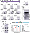
Fig 6. TRIM72 induces ubiquitination of RABV-M at K195.
(A) Illustration of the lysine (K) sites in RABV-M. (B) The lysine residues in RABV-M were mutated to alanine and over-expression vectors were constructed and together with pCAGGS or pCA-Trim72-flag were co-transfected into N2a cells respectively for 48 h. The protein levels of M-HA mutations and TRIM72-flag were analyzed by western blotting. (C) WT M-HA or M-K195A-HA was over-expressed in N2a cells respectively for 48 h, and the protein level was analyzed by western blotting. (D) pCA-M-flag or pCA-M-K195A-flag together with UB-HA and pCAGGS or pCA-Trim72-myc were co-transfected into N2a cells and treated with Mg132 (10 μM), then Co-IP assays were performed with anti-flag antibody. The protein levels were analyzed by western blotting. (E) pCAGGS or pCA-Trim72-myc together with pCA-M-K195A-HA and pCA-G-flag were co-transfected into HEK-293T cells respectively for 48 h, the protein levels of M-K195A-HA and G-flag in the supernatants and cells were analyzed by western blotting. (F) Schematic diagram of the recombinant rRABV-M-K195A. (G) N2a cells were infected with rRABV or rRABV-M-K195A (MOI = 0.01) for the indicated time and their growth kinetics were compared. (H) pCAGGS or pCA-Trim72-flag was transfected into N2a cells respectively for 12 h, then infected with WT rRABV (MOI = 0.01) or rRABV-M-K195A (MOI = 0.01) for 48 h. The supernatants were collected and viral titers were analyzed. Statistical analysis of grouped comparisons was carried out by student’s t-test (*P < 0.05; **P<0.01; ***P<0.001; ****P<0.0001). The bar graph represents means ± SD, n = 3. Western blot data are representative of at least two independent experiments.
Fig 7
Fig 7. K195 is the conserved ubiquitination site among lyssavirus targeted by TRIM72.
(A-D) HA-tagged lyssavirus M proteins (DRV-M-HA, ABLV-M-HA, DUVV-M-HA and EBLV-M-HA) together with empty vector or TRIM72-flag were co-overexpressed in N2a cells for 48 h, then protein levels of DRV-M-HA (A), ABLV-M-HA (B), DUVV-M-HA (C) and EBLV-M-HA (D) were analyzed by western blotting. (E-H) HA-tagged lyssavirus M proteins (DRV-M-HA, ABLV-M-HA, DUVV-M-HA, and EBLV-M-HA) together with empty vector or TRIM72-flag were co-overexpressed in N2a cells respectively, followed by treatment with Mg132 (10 μM). Co-IP assays were performed with anti-flag antibody post-transfection for 48 h, and protein levels of DRV-M-HA (E), ABLV-M-HA (F), DUVV-M-HA (G), and EBLV-M-HA (H) were analyzed by western blotting. (I-L) The K195 sites (K205 site in ABLV-M) in lyssavirus M proteins were mutated to Alanine, then together with empty vector or TRIM72-flag were co-overexpressed in N2a cells for 48 h. The protein levels of DRV-M-K195A-HA (I), ABLV-M-K205A-HA (J), DUVV-M-K195A-HA (K), and EBLV-M-K195A-HA (L) were analyzed by western blotting. Western blot data are representative of at least two independent experiments.
Fig 8
Fig 8. Model for TRIM72 restricts lyssavirus assembly and/or release by inducing K48-linked ubiquitination and proteasome degradation of the matrix protein.

Similar articles

Cited by

References

    1. Walker PJ, Blasdell KR, Calisher CH, Dietzgen RG, Kondo H, Kurath G, et al.. ICTV Virus Taxonomy Profile: Rhabdoviridae. The Journal of general virology. 2018;99(4):447–8. Epub 2018/02/22. doi: 10.1099/jgv.0.001020 . - DOI - PubMed
    1. Rupprecht CE, Mshelbwala PP, Reeves RG, Kuzmin IV. Rabies in a postpandemic world: resilient reservoirs, redoubtable riposte, recurrent roadblocks, and resolute recidivism. Animal diseases. 2023;3(1):15. Epub 2023/05/30. doi: 10.1186/s44149-023-00078-8 ; PubMed Central PMCID: PMC10195671. - DOI - PMC - PubMed
    1. Rupprecht CE, Turmelle A, Kuzmin IV. A perspective on lyssavirus emergence and perpetuation. Current opinion in virology. 2011;1(6):662–70. Epub 2012/03/24. doi: 10.1016/j.coviro.2011.10.014 . - DOI - PubMed
    1. Hampson K, Coudeville L, Lembo T, Sambo M, Kieffer A, Attlan M, et al.. Estimating the global burden of endemic canine rabies. PLoS neglected tropical diseases. 2015;9(4):e0003709. Epub 2015/04/17. doi: 10.1371/journal.pntd.0003709 ; PubMed Central PMCID: PMC4400070 does not alter our adherence to all PLOS NTDs policies on sharing data and materials. The findings and conclusions in this report are those of the authors and do not necessarily represent the views of the Centers for Disease Control and Prevention. - DOI - PMC - PubMed
    1. Fisher CR, Streicker DG, Schnell MJ. The spread and evolution of rabies virus: conquering new frontiers. Nature reviews Microbiology. 2018;16(4):241–55. Epub 2018/02/27. doi: 10.1038/nrmicro.2018.11 ; PubMed Central PMCID: PMC6899062. - DOI - PMC - PubMed

MeSH terms