Dinickel enzyme evolved to metabolize the pharmaceutical metformin and its implications for wastewater and human microbiomes
- PMID: 38408229
- PMCID: PMC10927577
- DOI: 10.1073/pnas.2312652121
Dinickel enzyme evolved to metabolize the pharmaceutical metformin and its implications for wastewater and human microbiomes
Abstract
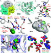
Metformin is the first-line treatment for type II diabetes patients and a pervasive pollutant with more than 180 million kg ingested globally and entering wastewater. The drug's direct mode of action is currently unknown but is linked to effects on gut microbiomes and may involve specific gut microbial reactions to the drug. In wastewater treatment plants, metformin is known to be transformed by microbes to guanylurea, although genes encoding this metabolism had not been elucidated. In the present study, we revealed the function of two genes responsible for metformin decomposition (mfmA and mfmB) found in isolated bacteria from activated sludge. MfmA and MfmB form an active heterocomplex (MfmAB) and are members of the ureohydrolase protein superfamily with binuclear metal-dependent activity. MfmAB is nickel-dependent and catalyzes the hydrolysis of metformin to dimethylamine and guanylurea with a catalytic efficiency (kcat/KM) of 9.6 × 10³ M-1s-1 and KM for metformin of 0.82 mM. MfmAB shows preferential activity for metformin, being able to discriminate other close substrates by several orders of magnitude. Crystal structures of MfmAB show coordination of binuclear nickel bound in the active site of the MfmA subunit but not MfmB subunits, indicating that MfmA is the active site for the MfmAB complex. Mutagenesis of residues conserved in the MfmA active site revealed those critical to metformin hydrolase activity and its small substrate binding pocket allowed for modeling of bound metformin. This study characterizes the products of the mfmAB genes identified in wastewater treatment plants on three continents, suggesting that metformin hydrolase is widespread globally in wastewater.
Keywords: biodegradation; human gut microbiome; metallohydrolase; metformin; nickel.
Conflict of interest statement
Competing interests statement:The authors declare no competing interest.
Figures


Similar articles
-
Discovery of a Ni2+-dependent heterohexameric metformin hydrolase.Nat Commun. 2024 Jul 20;15(1):6121. doi: 10.1038/s41467-024-50409-7. Nat Commun. 2024. PMID: 39033196 Free PMC article.
-
Metformin hydrolase is a recently evolved nickel-dependent heteromeric ureohydrolase.Nat Commun. 2024 Sep 14;15(1):8045. doi: 10.1038/s41467-024-51752-5. Nat Commun. 2024. PMID: 39271653 Free PMC article.
-
Filling in the Gaps in Metformin Biodegradation: a New Enzyme and a Metabolic Pathway for Guanylurea.Appl Environ Microbiol. 2021 May 11;87(11):e03003-20. doi: 10.1128/AEM.03003-20. Print 2021 May 11. Appl Environ Microbiol. 2021. PMID: 33741630 Free PMC article.
-
Co-occurrence, toxicity, and biotransformation pathways of metformin and its intermediate product guanylurea: Current state and future prospects for enhanced biodegradation strategy.Sci Total Environ. 2024 Apr 15;921:171108. doi: 10.1016/j.scitotenv.2024.171108. Epub 2024 Feb 22. Sci Total Environ. 2024. PMID: 38395159 Review.
-
Occurrence, Impact, Analysis and Treatment of Metformin and Guanylurea in Coastal Aquatic Environments of Canada, USA and Europe.Adv Mar Biol. 2018;81:23-58. doi: 10.1016/bs.amb.2018.09.005. Epub 2018 Nov 7. Adv Mar Biol. 2018. PMID: 30471658 Review.
Cited by
-
Discovery of a Ni2+-dependent heterohexameric metformin hydrolase.Nat Commun. 2024 Jul 20;15(1):6121. doi: 10.1038/s41467-024-50409-7. Nat Commun. 2024. PMID: 39033196 Free PMC article.
-
Biochemical and genomic evidence for converging metabolic routes of metformin and biguanide breakdown in environmental Pseudomonads.J Biol Chem. 2024 Dec;300(12):107935. doi: 10.1016/j.jbc.2024.107935. Epub 2024 Oct 28. J Biol Chem. 2024. PMID: 39476966 Free PMC article.
-
Metformin hydrolase is a recently evolved nickel-dependent heteromeric ureohydrolase.Nat Commun. 2024 Sep 14;15(1):8045. doi: 10.1038/s41467-024-51752-5. Nat Commun. 2024. PMID: 39271653 Free PMC article.
-
Insights into the action of the pharmaceutical metformin: Targeted inhibition of the gut microbial enzyme agmatinase.iScience. 2024 Jan 12;27(2):108900. doi: 10.1016/j.isci.2024.108900. eCollection 2024 Feb 16. iScience. 2024. PMID: 38318350 Free PMC article.
-
A prescription for engineering PFAS biodegradation.Biochem J. 2024 Dec 4;481(23):1757-1770. doi: 10.1042/BCJ20240283. Biochem J. 2024. PMID: 39585294 Free PMC article. Review.
References
-
- Bradley P. M., et al. , Metformin and other pharmaceuticals widespread in wadeable streams of the southeastern United States. Environ. Sci. Technol. Lett. 3, 243–249 (2016).
-
- Forslund K., et al. , Disentangling type 2 diabetes and metformin treatment signatures in the human gut microbiota. Nature 545, 116 (2015). - PubMed
MeSH terms
Substances
Grants and funding
LinkOut - more resources
Full Text Sources
Medical
Research Materials
Miscellaneous

