. 2024 Jun 1;102(3):238-251.
doi: 10.1139/bcb-2023-0363. Epub 2024 Feb 26.

Protamines and the sperm nuclear basic proteins Pandora's Box of insects

Affiliations

Protamines and the sperm nuclear basic proteins Pandora's Box of insects

Melissa R Leyden et al. Biochem Cell Biol. .

Abstract

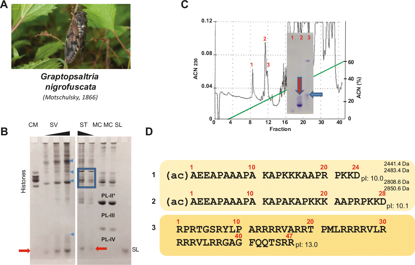
Insects are the largest group of animals when it comes to the number and diversity of species. Yet, with the exception of Drosophila, no information is currently available on the primary structure of their sperm nuclear basic proteins (SNBPs). This paper represents the first attempt in this regard and provides information about six species of Neoptera: Poecillimon thessalicus, Graptosaltria nigrofuscata, Apis mellifera, Nasonia vitripennis, Parachauliodes continentalis, and Tribolium castaneum. The SNBPs of these species were characterized by acetic acid urea gel electrophoresis (AU-PAGE) and high-performance liquid chromatography fractionated. Protein sequencing was obtained using a combination of mass spectrometry sequencing, Edman N-terminal degradation sequencing and genome mining. While the SNBPs of several of these species exhibit a canonical arginine-rich protamine nature, a few of them exhibit a protamine-like composition. They appear to be the products of extensive cleavage processing from a precursor protein which are sometimes further processed by other post-translational modifications that are likely involved in the chromatin transitions observed during spermiogenesis in these organisms.

Keywords: insects; mass spectrometry/Edman N-terminal sequencing; protamines; sperm nuclear basic proteins (SNBPs).


Conflict of interest statement

All authors have read and agreed to the published version of the manuscript and there is no conflict of interest, including specific financial interest and relationships and affiliations relevant to the subject of the manuscript.

Figures

Fig. 1.
Fig. 1.
Insect phylogeny (Tihelka et al. 2021) with the names of the species used in this work and for which the sperm nuclear basic proteins were determined experimentally (red) or obtained from data mining of existing genomes (pink). The number of species belonging to several of the different lineages and orders (Engel 2015) is also indicated. Figures modified from (Tihelka et al. 2021) and reproduced with permission from Elsevier.
Fig. 2.
Fig. 2.
Spermiogenesis and protamine of the honeybee. (A) The honeybee Apis mellifera. (B) AU-PAGE of the sperm nuclear basic proteins (SNBPs) of A. mellifera (AM) sperm in comparison to the SNBPs from salmon Oncorhynchus keta (salmine protamine, SL), California mussel (Mytilus californianus, MC), and somatic histones from chicken erythrocytes (CE). The PL-II*, PL-III, and PL-IV from M. californianus (Carlos et al. 1993a) are indicated. (C) Reversed phase HPLC analysis of the HCl-extracted SNBPs from sperm of A. mellifera. An AU-PAGE of the fractions (I–V) collected is also shown. (D) Protein sequence of the protamine P2 precursor identified with a BLAST search of the BeeBase (https://hymenoptera.elsiklab.missouri.edu/beebase) against the experimentally determined (N-terminal Edman degradation sequencing) protein sequence of A. mellifera P1. (E) Protamine sequence of P1a, P1b, and P2 using Edman N-terminal degradation sequencing and mass spectrometry analysis. The black arrow represents an LCMS identified cleavage site. (F) Electron micrographs at different stages of spermiogenesis. I = longitudinally and cross-sectioned nuclei from early spermatids in which chromatin fibers start to coalesce. Chromatin fiber thickening (II–IV) progresses in late spermatids until the sperm are mature (V) with electron dense nuclei. At this stage, microtubules longitudinally aligned above and below the sectioned nuclei can be observed. The acrosomes (a) and tails (t) are indicated within the image. Scale bars = 500 nm.
Fig. 3.
Fig. 3.
Bee protamine 2 evolution. (A) Evolutionary rates (aminoacidchangeper100 sites) of bee protamine P2 and bee somatic histone H1 compared to Drosophila PL (protamines) (Alvi et al. 2013) and primate pP1/pP2 protamines (Retief et al. 1993) and to histones (Isenberg 1978). (B) Protamine sequences and accession numbers for Apis florea (red dwarf honey bee) (Aflorea_gi|380025480|ref|XM_003696454.1|:1267–1599 predicted: uncharacterized LOC100871426, mRNA); Bombus impatiens (bumble bee) Bimpatiens PROTAMINEnt_(gnl|Bimp_2.0|scf_0083 NT_176502.1); and Megachile rotundata (Alfalfa Leafcutter bee) (Mrotundata_gi|383852691|ref|XM_003701811.1|:1684–2010 PREDICTED: uncharacterized LOC100880031, mRNA). (C) Representative protamine sequences from invertebrate and vertebrate organisms. Loligo opalescence (opalescent squid) (Lewis et al. 2004); Drosophila melanogaster protamines A and B (Jayaramaiah Raja and Renkawitz-Pohl 2005); Homo sapiens protamines P1 and P2. The boxed parts of the sequences correspond to protein regions (in blue) of the precursors that become processed by cleavage during spermiogenesis. The underlined regions in yellow indicate the arginine/cysteine clusters characteristic of protamines.
Fig. 4.
Fig. 4.
Protamines of the jewel wasp. (A) The jewel wasp Nasonia vitripennis (picture provided by Hans Smid). (B) AU-PAGE analysis of the SNBPs of N. vitripennis (NV) in comparison to the sperm nuclear basic proteins (SNBPs) of the mussel M. californianus (MC), salmon protamine (SL), and chicken erythrocyte histones (CM). (C) Mass sectrometry identified SNBPS from a mature gonad protein extract as shown in (B) NV. (D) Electron micrographs at different stages of spermiogenesis. I and II = longitudinal nuclear sections and III transversal cross section of nuclei of late spermatids. IV is a longitudinal section of mature sperm. Scale bars = 500 nm.
Fig. 5.
Fig. 5.
Sperm nuclear basic proteins (SNBPs) of the bush cricket Poecillimon thessalicus (A). (B) AU_PAGE comparative analysis of P. thessalicus SNBPs. (CM, MC, and SL as in the legend of previous figures). The blue arrows point to potential SNBP precursor proteins. (C) Amino acid sequence determined by N-terminal Edman sequencing of the Reversed phase HPLC purified protein shown by the red arrow in (B).
Fig. 6.
Fig. 6.
Partial analysis of the sperm nuclear basic proteins (SNBPs) of the large brown cicada Graptosaltria nigrofuscata (A). (C) Reversed phase HPLC chromatogram of the sample (SV) shown in (B). The inset shows an electrophoretic analysis of elution peaks 1–3. (B) AU-PAGE of HCl extracts from seminal vesicles (SV) and from spermatheca (ST) in comparison to chicken erythrocyte histones (CM), mussel (MC), and salmine (SL). The blue square indicates the histones from the spermathecal tissue and the blue arrows point to potential SNBP precursor proteins. (D) The fraction indicated by the red arrow in (C) was analyzed by mass spectrometry and the SNBPs identified are shown in (D). The proteins in 1 and 2 were both present in their acetylated (ac) and not acetylated form. The arginine-rich protein in 3 might correspond to the band highlighted by a blue arrow in (C).
Fig. 7.
Fig. 7.
Characterization of the sperm nuclear basic proteins (SNBPs) of the fishfly Paracauliodes continentalis (A). (B) Electrophoretic (AU-PAGE) analysis of the HCl-extracted proteins from seminal vesicles (PC) Compared to M. californianus SNBPs (MC). (C and D) A representative depiction of the LCMS approach used for the characterization of P. continentalis SNBPs. (C) Precursor ions at 485.56 m/z (z = 12) were fragmented by electron transfer dissociation (ETD) to produce the MS2 and selected abundant fragment ions are labeled. (D) Sequence coverage of 485.56 m/z (z = 12) P. continentalis protamine with molecular mas of 5812.6 Da by ETD. The cleavages depict the c and z ions observed, giving unambiguous sequence coverage. (E) Table summarizing all the SNBPs detected using the mass spectrometry approach. The blue circles highlight some of the amino acid variations.
Fig. 8.
Fig. 8.
Genome mining identification of the red flour beetle Trilobium castaneum (A). (B) Protein identified using Apis mellifera sperm nuclear basic proteins (SNBP) (Fig. 2E) in recurrent BLAST searches of the (Tribolium Genome Sequencing et al. 2008) genome on GenBank (project accession AAJJ00000000.2). (C) T. castaneum (short) SNBP. (D) Electron microscopy of Tribolium corretto showing: I, early spermatids; II, details of the nucleus (N) and acrosome organization (Ac); II, Mature sperm nuclei (N) and IV, an enlarged magnification of the sperm nuclei (N).

Similar articles

Cited by

References

    1. Adamson SW, Browning RE, Chao CC, Bateman RC Jr., Ching WM, and Karim S 2013. Molecular characterization of tick salivary gland glutaminyl cyclase. Insect. Biochem. Mol. Biol. 43(9): 781–793. doi: 10.1016/j.ibmb.2013.05.011. - DOI - PMC - PubMed
    1. Alvi ZA, Chu TC, Schawaroch V, and Klaus AV 2013. Protamine-like proteins in 12 sequenced species of Drosophila. Protein Pept. Lett. 20(1): 17–35. Available from https://www.ncbi.nlm.nih.gov/pubmed/22789106. doi: 10.2174/092986613804096847. - DOI - PubMed
    1. Ando T, Yamasaki M, and Suzuki K 1973. Protamines: isolation, characterization and function. Springer-Verlag, NY. - PubMed
    1. Ausió J 1992. Presence of a highly specific histone H1-like protein in the chromatin of the sperm of the bivalve mollusks. Mol. Cell. Biochem. 115(2): 163–172. Available from http://research.bmn.com/medline/search/record?uid=MDLN.93078720. doi: 10.1007/BF00230327. - DOI - PubMed
    1. Ausió J 1995. Histone H1 and the evolution of the nuclear sperm specific proteins. Memoires de Museum National d’Histoire Naturelle, Paris.

Publication types

LinkOut - more resources