. 2024 Feb 26;11(3):ENEURO.0002-24.2024.
doi: 10.1523/ENEURO.0002-24.2024. Online ahead of print.

Expression of endogenous epitope-tagged GPR4 in the mouse brain

Affiliations

Expression of endogenous epitope-tagged GPR4 in the mouse brain

Elizabeth C Gonye et al. eNeuro. .

Abstract

GPR4 is a proton-sensing G protein-coupled receptor implicated in many peripheral and central physiological processes. GPR4 expression has previously been assessed only via detection of the cognate transcript or indirectly, by use of fluorescent reporters. In this work, CRISPR/Cas9 knock-in technology was used to encode a hemagglutinin (HA) epitope tag within the endogenous locus of Gpr4 and visualize GPR4-HA in the mouse central nervous system using a specific, well characterized HA antibody; GPR4 expression was further verified by complementary Gpr4 mRNA detection. HA immunoreactivity was found in a limited set of brain regions, including in the retrotrapezoid nucleus (RTN), serotonergic raphe nuclei, medial habenula, lateral septum, and several thalamic nuclei. GPR4 expression was not restricted to cells of a specific neurochemical identity as it was observed in excitatory, inhibitory, and aminergic neuronal cell groups. HA immunoreactivity was not detected in brain vascular endothelium, despite clear expression of Gpr4 mRNA in endothelial cells. In the RTN, GPR4 expression was detected at the soma and in proximal dendrites along blood vessels and the ventral surface of the brainstem; HA immunoreactivity was not detected in RTN projections to two known target regions. This localization of GPR4 protein in mouse brain neurons corroborates putative sites of expression where its function has been previously implicated (e.g., CO2-regulated breathing by RTN), and provides a guide for where GPR4 could contribute to other CO2/H+ modulated brain functions. Finally, GPR4-HA animals provide a useful reagent for further study of GPR4 in other physiological processes outside of the brain.Significance Statement GPR4 is a proton-sensing G-protein coupled receptor whose expression is necessary for a number of diverse physiological processes including acid-base sensing in the kidney, immune function, and cancer progression. In the brain, GPR4 has been implicated in the hypercapnic ventilatory response mediated by brainstem neurons. While knockout studies in animals have clearly demonstrated its necessity for normal physiology, descriptions of GPR4 expression have been limited due to a lack of specific antibodies for use in mouse models. In this paper, we implemented a CRISPR/Cas9 knock-in approach to incorporate the coding sequence for a small epitope tag into the locus of GPR4. Using these mice, we were able to describe GPR4 protein expression directly for the first time.


Conflict of interest statement

The authors declare no competing financial interest.

Figures
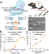
Figure 1.
Figure 1.
Incorporating an HA epitope tag into GPR4 does not alter receptor function in vitro or the hypercapnic ventilatory reflex in vivo. A, Illustration of the CRISPR/Cas9 knock-in strategy, generated in BioRender, and a representative agarose gel of the diagnostic PCR used to detect the HA tag at the C-terminal end of GPR4. B, Activation by acidification of wild-type and HA-tagged GPR4 in HEK293T cells detected using the luminescent GloSensor assay for cAMP production (normalized to pH-independent, forskolin-activated cAMP production). Note that acidification does not increase cAMP in cells expressing a nonsignaling GPR4(R117A). C, Minute ventilation of Gpr4HA/HA and wild-type Gpr4+/+ mice assessed by whole body plethysmography.
Figure 2.
Figure 2.
GPR4 transcript and protein expression in the retrotrapezoid nucleus (RTN). A, RNAscope multiplex in situ hybridization (ISH) labeling for Gpr4 and the RTN marker Nmb at bregma level −6.48 mm. Arrowheads indicate RTN neurons that coexpress Nmb and Gpr4; arrows indicate more dorsally located neurons with high levels of Nmb that do not express Gpr4 (Shi et al., 2017). B, C, HA immunostaining in the RTN of Gpr4HA/HA (B) and wild-type Gpr4+/+ (C) mice; RTN neurons are identified by expression of PHOX2B. D, Representative maps of PHOX2B+/HA+ cells and PHOX2B-only cells through the rostrocaudal extent of the RTN (top, bregma −5.8 to −7.08), and average distribution of HA+ cells through the RTN (bottom). Data were averaged (±SEM) from four mice; scale bar, 50 µm.
Figure 3.
Figure 3.
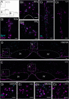
GPR4 transcript and protein expression in the caudal raphe. A, Representative diagram of three caudal raphe nuclei and notable landmarks, based on the Paxinos and Franklin atlas, bregma −6.64 mm. ROb, raphe obscurus; RPa, raphe pallidus; PPy, parapyramidal nucleus; py, pyramidal tract. Bi–iii, RNAscope ISH labeling of Gpr4 expression in serotonergic raphe neurons identified by Tph2 expression. Ci,ii, HA staining in the raphe obscurus serotonergic (TPH+) nucleus of Gpr4HA/HA (i) and wild-type Gpr4+/+ (ii) mice. D, E, HA staining in the parapyramidal (i) and raphe pallidus (ii) nuclei of Gpr4HA/HA (D) and wild-type Gpr4+/+ (E) mice. Scale bar, 50 µm.
Figure 4.
Figure 4.
Location and proportion of serotonergic caudal raphe neurons that express GPR4. A, Representative maps of HA+/TPH+ cells and TPH+ cell locations through the caudal raphe, bregma levels −5.80 to −7.12. B, Average percentage of TPH+ cells that are also HA+ (% HA+/TPH+) within ROb (33 ± 4 TPH+ cells/section), RPa/RMg (23 ± 3 TPH+ cells/section), and PPy (26 ± 3 TPH+ cells/section). Data were averaged (±SEM) from four mice.
Figure 5.
Figure 5.
GPR4 transcript and protein expression in the dorsal and median raphe. A, Representative diagram of key nuclei and landmarks at bregma −4.84 mm, including the nuclei of the dorsal and median raphe, according to the Paxinos and Franklin atlas. Aq, aqueduct (Silvius); DRD, dorsal raphe nucleus, dorsal part; DRVL, dorsal raphe, ventrolateral; DRV, dorsal raphe nucleus, ventral; mlf, medial longitudinal fasciculus; scp, superior cerebellar peduncle (brachium conjunctivum); VTg, ventral tegmental nucleus; MnR, median raphe nucleus; ts, tectospinal tract. B, RNAscope ISH labeling demonstrating Gpr4 expression in Tph2+ cells of the dorsal and median raphe. C, D, GPR4-HA immunolabeling in the dorsal and median raphe of Gpr4HA/HA (C) and wild-type Gpr4+/+ (D) mice; (i) represents section of the image viewed in higher magnification in Ci–Di. Scale bar, 50 µm.
Figure 6.
Figure 6.
Gpr4 transcript expression in multiple nuclei of the thalamus. A, Representative diagram of key landmarks and thalamic nuclei at bregma −1.34 mm; inset rectangles represent regions presented in panels B–F. CA3, hippocampus CA3; DG, dentate gyrus; D3V, dorsal third ventricle; LD, laterodorsal thalamus; Hb, habenula; PV, paraventricular thalamus; Po, posterior thalamic group; MD, mediodorsal thalamus; VPT, ventroposterior thalamus; VMT, ventromedial thalamus; Re, reuniens thalamic nucleus; mt, mammillary tract; ic, internal capsule; ZI, zona incerta; DM, dorsomedial hypothalamus. B–F, RNAscope in situ labeling of Gpr4 expression in glutamatergic (Slc17a6) cells of the laterodorsal (B), mediodorsal (C, F), medial habenula (E), and ventromedial (D) thalamic nuclei. A few GABAergic (Slc32a1) cells visible at the ventral border of VMT appear negative for Gpr4. Scale bar, 50 µm.
Figure 7.
Figure 7.
GPR4 protein expression in the thalamus. A, Composite image of the thalamus of a Gpr4HA/HA mouse stained for HA and ChAT, with general landmarks delineated by dashed lines. DG, dentate gyrus; D3V, dorsal third ventricle; LD, laterodorsal thalamus; LHb, lateral habenula; MHb, medial habenula; PV, paraventricular thalamus; VM, ventromedial thalamus. B–D, HA staining (with DAPI labeling) in the laterodorsal (B), mediodorsal (C), and ventromedial (D) thalamus of Gpr4HA/HA (i) and wild-type Gpr4+/+ (ii) mice. Ei, HA and ChAT staining in the medial habenula of Gpr4HA/HA and wild-type Gpr4+/+ mice (Eii, inset). Scale bar, 50 µm; dotted lines identify blood vessels (in Bii,Di).
Figure 8.
Figure 8.
GPR4 transcript and protein expression in the geniculate nucleus. A, Representative diagram of key landmarks around the medial geniculate nucleus at bregma level −3.28 mm. MGD, medial geniculate nucleus, dorsal part; MGV, medial geniculate nucleus, ventral part; MGM, medial geniculate nucleus, medial part; SG, suprageniculate nucleus; PIL, posterior intralaminar thalamic nucleus. B, RNAscope ISH labeling of Gpr4 expression in the geniculate nucleus, together with markers for glutamatergic (Slc17a6) and GABAergic neurons (Slc32a1). C, HA staining in the Calbindin B (CALB) expressing cells of the dorsal aspect of the geniculate nucleus of Gpr4HA/HA mice; dotted line designates region viewed at higher power in (Ci). D, Lack of HA staining in a wild-type mouse at the region analogous to that displayed in (Ci).
Figure 9.
Figure 9.
GPR4 transcript and protein expression in the lateral septum. A, Representative diagram of key landmarks around the lateral septum at bregma level 0.38 mm. LV, lateral ventricle; cc, corpus callosum; LSD, dorsal lateral septal nucleus; LSI, intermediate lateral septal nucleus; LSV, ventral septal nucleus; MS, medial septal nucleus; BST, bed nucleus of the stria terminalis; aca, anterior commissure anterior part. B, RNAscope ISH labeling of Gpr4 expression in GABAergic (Slc32a1) neurons of the lateral septum. C, HA staining throughout the lateral septum of a Gpr4HA/HA mouse; the indicated region is displayed at higher magnification in (Ci). D, Lack of HA staining in a wild-type mouse at a region analogous to that displayed in (Ci). Scale bar, 50 µm.
Figure 10.
Figure 10.
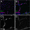
GPR4 transcript, but not protein, is detectable in brain endothelial cells. A, RNAscope in situ labeling of Gpr4 expression in endothelial (Pecam1+) cells in the wild-type mouse brainstem. B, C, HA staining in RTN neurons from a Gpr4HA/HA mouse, with adjacent small vessels labeled by PECAM expression; vessel cross sections (walls and lumen) are delineated by dashed lines. C, PECAM staining within vessel boundaries. D, HA staining located outside of PECAM+ vessels. Scale bar, 25 µm.
Figure 11.
Figure 11.
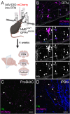
GPR4 is evident in the processes of Nmb-expressing RTN neurons but undetectable at terminals in the preBötC or lPBN. A, Illustration of the viral expression and immunostaining strategy for assessing GPR4 protein expression in processes and terminals of NMB neurons in the RTN area, lPBN, or PreBötC, generated in BioRender. B, HA staining in mCherry labeled neurons in the RTN area of an NmbCre/+; Gpr4HA/HA mouse injected with AAV-DIO-mCherry with high power images of areas bounded by dotted lines (i,ii); arrows denote staining in RTN cell bodies, arrowheads denote staining in RTN processes. C, mCherry labeled terminals in the preBötC area (bregma −7.48 mm). D, mCherry labeled terminals in the lPBN (denoted by CGRP staining). Scale bar, 25 µm.

Similar articles

Cited by

References

    1. An S, Tsai C, Goetzl EJ (1995) Cloning, sequencing and tissue distribution of two related G protein-coupled receptor candidates expressed prominently in human lung tissue. FEBS Lett 375:121–124. 10.1016/0014-5793(95)01196-L - DOI - PubMed
    1. Anthony TE, Dee N, Bernard A, Lerchner W, Heintz N, Anderson DJ (2014) Control of stress-induced persistent anxiety by an extra-amygdala septohypothalamic circuit. Cell 156:522–536. 10.1016/j.cell.2013.12.040 - DOI - PMC - PubMed
    1. Bailey JE, Argyropoulos SV, Kendrick AH, Nutt DJ (2005) Behavioral and cardiovascular effects of 7.5% CO2 in human volunteers. Depress Anxiety 21:18–25. 10.1002/da.20048 - DOI - PubMed
    1. Bailey JE, Dawson GR, Dourish CT, Nutt DJ (2011) Validating the inhalation of 7.5% CO2 in healthy volunteers as a human experimental medicine: a model of generalized anxiety disorder (GAD). J Psychopharmacol 25:1192–1198. 10.1177/0269881111408455 - DOI - PubMed
    1. Bakken TE, et al. (2021) Single-cell and single-nucleus RNA-seq uncovers shared and distinct axes of variation in dorsal LGN neurons in mice, non-human primates, and humans. Elife 10. 10.7554/eLife.64875 - DOI - PMC - PubMed

LinkOut - more resources