. 2024 Feb 27;31(1):26.
doi: 10.1186/s12929-024-01014-9.

Reduced interleukin-18 secretion by human monocytic cells in response to infections with hyper-virulent Streptococcus pyogenes

Affiliations

Reduced interleukin-18 secretion by human monocytic cells in response to infections with hyper-virulent Streptococcus pyogenes

Lea A Tölken et al. J Biomed Sci. .

Abstract

Background: Streptococcus pyogenes (group A streptococcus, GAS) causes a variety of diseases ranging from mild superficial infections of the throat and skin to severe invasive infections, such as necrotizing soft tissue infections (NSTIs). Tissue passage of GAS often results in mutations within the genes encoding for control of virulence (Cov)R/S two component system leading to a hyper-virulent phenotype. Dendritic cells (DCs) are innate immune sentinels specialized in antigen uptake and subsequent T cell priming. This study aimed to analyze cytokine release by DCs and other cells of monocytic origin in response to wild-type and natural covR/S mutant infections.

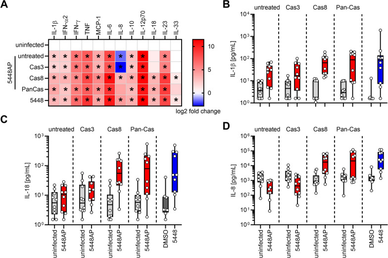
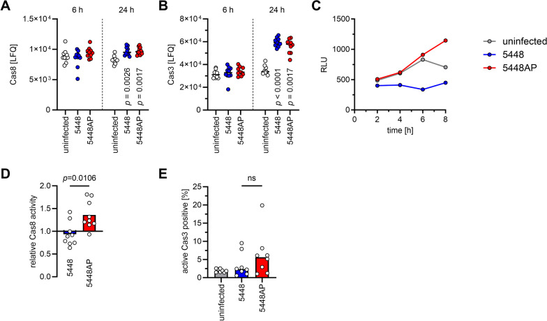
Methods: Human primary monocyte-derived (mo)DCs were used. DC maturation and release of pro-inflammatory cytokines in response to infections with wild-type and covR/S mutants were assessed via flow cytometry. Global proteome changes were assessed via mass spectrometry. As a proof-of-principle, cytokine release by human primary monocytes and macrophages was determined.

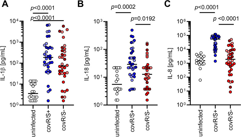
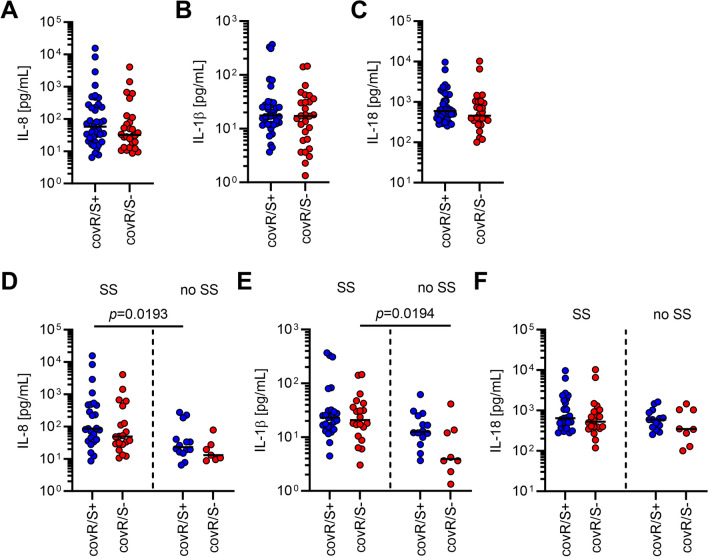
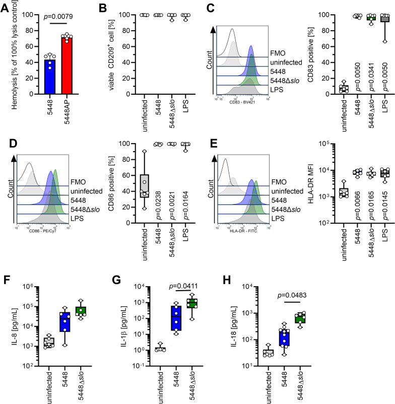
Results: In vitro infections of moDCs and other monocytic cells with natural GAS covR/S mutants resulted in reduced secretion of IL-8 and IL-18 as compared to wild-type infections. In contrast, moDC maturation remained unaffected. Inhibition of caspase-8 restored secretion of both molecules. Knock-out of streptolysin O in GAS strain with unaffected CovR/S even further elevated the IL-18 secretion by moDCs. Of 67 fully sequenced NSTI GAS isolates, 28 harbored mutations resulting in dysfunctional CovR/S. However, analyses of plasma IL-8 and IL-18 levels did not correlate with presence or absence of such mutations.

Conclusions: Our data demonstrate that strains, which harbor covR/S mutations, interfere with IL-18 and IL-8 responses in monocytic cells by utilizing the caspase-8 axis. Future experiments aim to identify the underlying mechanism and consequences for NSTI patients.

Keywords: CovR/S; Dendritic cells; Interleukin-18; Necrotizing soft tissue infection; Streptococcus pyogenes.


Conflict of interest statement

All authors declare no competing interests.

Figures

Fig. 1
Fig. 1
MoDCs infected with GAS strains harboring dysfunctional CovR/S secrete less IL-8 and IL-18. MoDCs were infected with four strains possessing functional CovR/S (5448, 5626, 2006, 8157) or four strains possessing non-functional CovR/S (5448AP, 2002, 5005, 8003) as assessed by sequence analyses and SpeB proteolytic assay. The concentrations of IL-1β (A), IL-18 (B), and IL-8 (C) were measured in supernatants of (un)infected moDCs. Each dot represents one independent experiment with cells from one donor (n ≥ 8). Horizontal lines denote median values. The level of significance was determined using Kruskal–Wallis test with Dunn’s post test. Different shades of color represent infections with different strains
Fig. 2
Fig. 2
Monocytic cells infected with 5448AP secrete less IL-18 and IL-8. MoDCs (A), monocytes (B), or monocyte-derived macrophages (C) were infected with 5448 or 5448AP and cytokine secretion was measured via a multiplex assay (n ≥ 8). The heatmaps represent the log2 fold change of cytokine concentration in relation to the uninfected controls (A–C). Original data are displayed in D–F and Additional file 1: Fig. S3. Original data of IL-1β (D), IL-18 (E), and IL-8 (F) concentration in supernatants of (un)infected moDCs, monocytes, and monocyte-derived macrophages. The data in (DF) are displayed as box plots. Each dot represents one independent experiment with cells from one donor (n ≥ 8). The level of significance was determined using Kruskal–Wallis test with Dunn’s posttest
Fig. 3
Fig. 3
5448AP does not impair DC maturation. MoDC were infected with 5448, 5448AP, or 5448Δemm1 for 1 h. Extracellular bacteria were killed by substituting the media with antibiotics for additional 23 h. MoDC phenotype (A, C–G) and viability (B) were evaluated via flow cytometry. Representative histograms for each marker are shown in (A). The maturation process was evaluated by assessing the expression of CD40 (B), frequencies of CD80+ (D) and CD83+ (E) cells as well as CD86 (F) and HLA-DR expression (G). H Principal component analysis of intracellular proteins 6 h and 24 h post infections with indicated strains. Each dot represents one donor (n = 10). The ellipses indicate the calculated 95% probability region for a bivariate normal distribution with an average center of groups. The data in (BG) are displayed as box plots. Each dot represents one independent experiment with cells from one donor (n = 10). The level of significance was determined using the Kruskal–Wallis test with Dunn’s posttest. FMO, fluorescence minus one; MFI, mean fluorescence intensity
Fig. 4
Fig. 4
Inhibition of caspase-8 restores IL-18 and IL-8 secretion in 5448AP infections. MoDCs were treated with caspase inhibitors (caspase-3: Cas3/7-Inhibitor I, Ac-DEVD-cho; caspase-8: z-IETD-fmk, pan-capase: z-VAD-fmk) and subsequently infected with 5448AP. Cytokine secretion by moDCs was measured via a multiplex assay. A The heatmap represents the log2 fold change of cytokine concentration in relation to the respective uninfected controls. Original data are displayed in B–D and Fig. S9. Original data of IL-1β (B), IL-18 (C), and IL-8 (D) concentration in supernatants of (un)infected moDCs. The data in (BD) are displayed as box plots. Each dot represents one independent experiment with cells from one donor (n = 10). A separate uninfected control with the respective inhibitor treatment was performed for each 5448AP infection. Untreated: indicates 5448AP infection without inhibitors. Each infection was compared to its respective uninfected controls. The level of significance was determined using Mann–Whitney U-test. Exact statistical analysis is displayed in Additional file 1: Table S6
Fig. 5
Fig. 5
5448AP-infected moDCs exhibit high caspase-8 activity. MoDCs were infected with 5448 or 5448AP. Relative abundance of caspase-8 (A) and caspase-3 (B) 6 h and 24 h post infection. Original calculations are displayed in Additional file 1: Table S4. C Caspase-8 activity of (un)infected moDCs as measured at indicated time points by luminescence. D Relative caspase-8 activity at 8 h post infection. E Frequencies of caspase-3+ moDCs 8 h post infection. Each dot represents one independent experiment with cells from one donor (n ≥ 8). Horizontal lines in (A, B) denote median values. Bars in (D, E) denote mean values. The level of significance was determined using Kruskal–Wallis test with Dunn’s posttest in (A, B, E) or Mann–Whitney U-test in (D). LFQ, label-free quantification intensities; RLU, relative light units
Fig. 6
Fig. 6
Cytokine/chemokine levels in plasma of NSTI patients. Levels of IL-8 (A), IL-1β (B), and IL-18 (C) were previously determined in plasma of NSTI patients [48] and reanalyzed. Comparison of IL-8 (D), IL-1β (E), and IL-18 (F) plasma concentrations between septic shock and no septic shock. Each dot represents data from one patient. Horizontal lines denote median values. The level of significance was determined using Kruskal–Wallis test with Dunn’s posttest. SS, septic shock
Fig. 7
Fig. 7
Knock-out of SLO in 5448 leads to elevated secretion of IL-1β and IL-18 by moDCs. A SLO hemolytic activity in supernatants of 5448 and 5448AP strains. MoDCs were infected with 5448 or 5448Δslo and moDC viability (B) and phenotype (C–E) were evaluated via flow cytometry. The maturation process was evaluated by assessing the frequencies of CD83+ (C) and CD86+ (D) cells as well as expression of HLA-DR (E). Representative histograms for each marker are shown in each respective left panel. The concentrations of IL-8 (F), IL-1β (G), and IL-18 (H) were measured in supernatants of (un)infected moDCs. The data in (BH) are displayed as box plots. Each dot represents one independent experiment with cells from one donor (n ≥ 6). The level of significance was determined using Kruskal–Wallis test with Dunn’s posttest. FMO, fluorescence minus one; MFI, mean fluorescence intensity

Similar articles

Cited by

References

    1. Akita K, Ohtsuki T, Nukada Y, Tanimoto T, Namba M, Okura T, Takakura-Yamamoto R, Torigoe K, Gu Y, Su MSS, Fujii M, Satoh-Itoh M, Yamamoto K, Kohno K, Ikeda M, Kurimoto M. Involvement of caspase-1 and caspase-3 in the production and processing of mature human interleukin 18 in monocytic THP1 cells*. J Biol Chem. 1997;272(42):26595–26603. doi: 10.1074/jbc.272.42.26595. - DOI - PubMed
    1. Anaya DA, McMahon K, Nathens AB, Sullivan SR, Foy H, Bulger E. Predictors of mortality and limb loss in necrotizing soft tissue infections. Arch Surg. 2005;140(2):151–157. doi: 10.1001/archsurg.140.2.151. - DOI - PubMed
    1. Anderson J, Imran S, Frost HR, Azzopardi KI, Jalali S, Novakovic B, Osowicki J, Steer AC, Licciardi PV, Pellicci DG. Immune signature of acute pharyngitis in a Streptococcus pyogenes human challenge trial. Nat Commun. 2022;13(1):769. doi: 10.1038/s41467-022-28335-3. - DOI - PMC - PubMed
    1. Antonopoulos C, El Sanadi C, Kaiser WJ, Mocarski ES, Dubyak GR. Proapoptotic chemotherapeutic drugs induce noncanonical processing and release of IL-1β via caspase-8 in dendritic cells. J Immunol. 2013;191(9):4789–4803. doi: 10.4049/jimmunol.1300645. - DOI - PMC - PubMed
    1. Ato M, Ikebe T, Kawabata H, Takemori T, Watanabe H. Incompetence of neutrophils to invasive Group A streptococcus is attributed to induction of plural virulence factors by dysfunction of a regulator. PLoS ONE. 2008;3(10):e3455. doi: 10.1371/journal.pone.0003455. - DOI - PMC - PubMed