. 2024 Feb 27;21(1):8.
doi: 10.1186/s12989-024-00569-7.

Differential pulmonary toxicity and autoantibody formation in genetically distinct mouse strains following combined exposure to silica and diesel exhaust particles

Affiliations

Differential pulmonary toxicity and autoantibody formation in genetically distinct mouse strains following combined exposure to silica and diesel exhaust particles

Lisa Mf Janssen et al. Part Fibre Toxicol. .

Abstract

Background: Inhalation of airborne particulate matter, such as silica and diesel exhaust particles, poses serious long-term respiratory and systemic health risks. Silica exposure can lead to silicosis and systemic autoimmune diseases, while DEP exposure is linked to asthma and cancer. Combined exposure to silica and DEP, common in mining, may have more severe effects. This study investigates the separate and combined effects of occupational-level silica and ambient-level DEP on lung injury, inflammation, and autoantibody formation in two genetically distinct mouse strains, thereby aiming at understanding the interplay between genetic susceptibility, particulate exposure, and disease outcomes. Silica and diesel exhaust particles were administered to mice via oropharyngeal aspiration. Assessments of lung injury and host response included in vivo lung micro-computed tomography, lung function tests, bronchoalveolar lavage fluid analysis including inflammatory cytokines and antinuclear antibodies, and histopathology with particle colocalization.

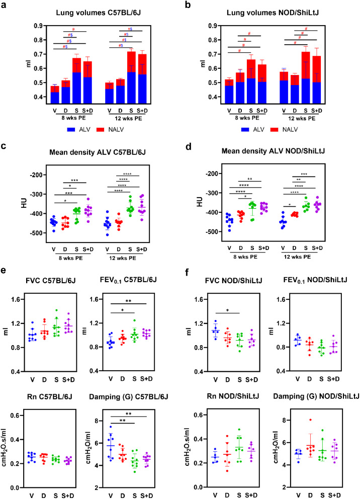
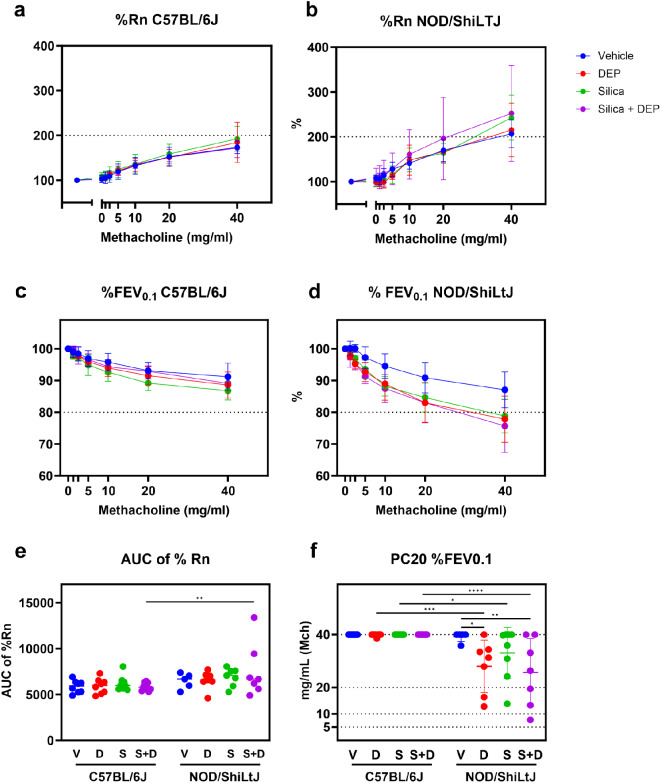
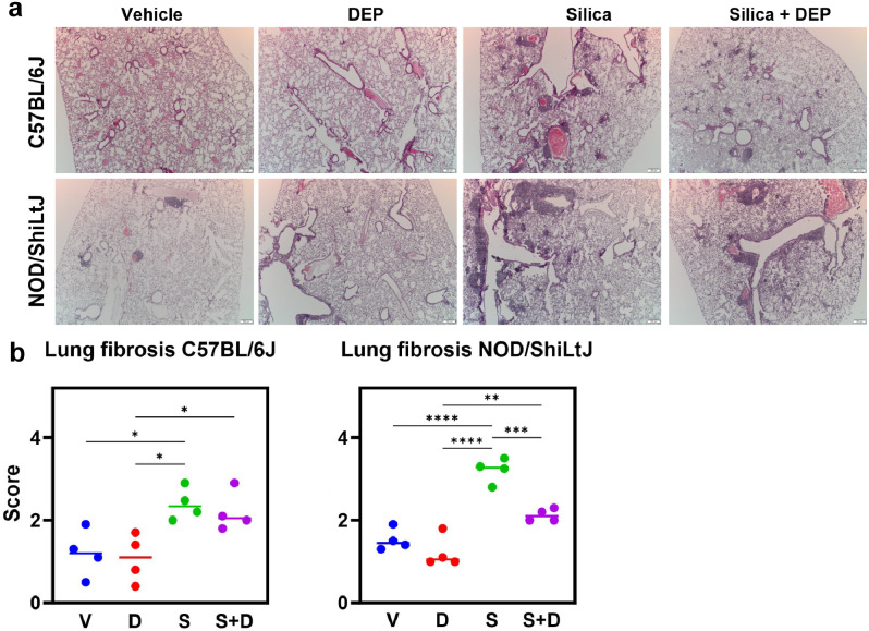
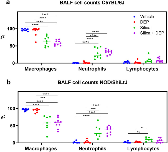
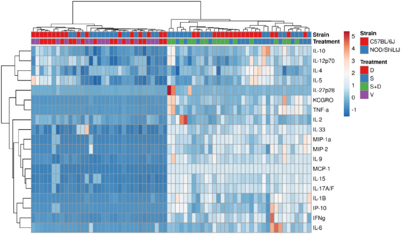
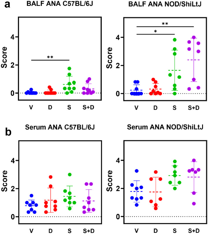
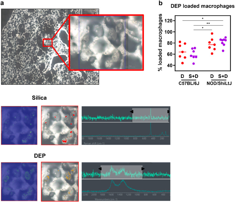
Results: The findings highlight the distinct effects of silica and diesel exhaust particles (DEP) on lung injury, inflammation, and autoantibody formation in C57BL/6J and NOD/ShiLtJ mice. Silica exposure elicited a well-established inflammatory response marked by inflammatory infiltrates, release of cytokines, and chemokines, alongside mild fibrosis, indicated by collagen deposition in the lungs of both C57BL/6J and NOD/ShilLtJ mice. Notably, these strains exhibited divergent responses in terms of respiratory function and lung volumes, as assessed through micro-computed tomography. Additionally, silica exposure induced airway hyperreactivity and elevated antinuclear antibody levels in bronchoalveolar lavage fluid, particularly prominent in NOD/ShiLtJ mice. Moreover, antinuclear antibodies correlated with extent of lung inflammation in NOD/ShiLTJ mice. Lung tissue analysis revealed DEP loaded macrophages and co-localization of silica and DEP particles. However, aside from contributing to airway hyperreactivity specifically in NOD/ShiLtJ mice, the ambient-level DEP did not significantly amplify the effects induced by silica. There was no evidence of synergistic or additive interaction between these specific doses of silica and DEP in inducing lung damage or inflammation in either of the mouse strains.

Conclusion: Mouse strain variations exerted a substantial influence on the development of silica induced lung alterations. Furthermore, the additional impact of ambient-level DEP on these silica-induced effects was minimal.

Keywords: Autoimmunity; Diesel exhaust particles; Lung inflammation; Mice; Silica; Silicosis.


Conflict of interest statement

The authors declare no competing interests.

Figures

Fig. 1
Fig. 1
(a) Stacked column plots representing non-aerated lung volumes (NALV) (ml) and aerated lung volumes (ALV) (ml) [resulting in total lung volumes (TLV) (ml)] in C57BL/6J and NOD/ShiLtJ mice. Data are presented as mean ± SD. Experimental groups were compared using repeated measures Two-Way ANOVA or mixed model in case of missing values with Tukey correction for multiple testing. Significant differences are represented as follows: $p < 0.05 between ALV, # p < 0.05 between NALV. N = 7–9 mice/group. (b) Mean aerated lung volume (HU) in DEP, silica, silica + DEP and vehicle exposed C57BL/6 and NOD/ShiLtJ mice. Data are presented as individual values with mean ± SD. *p < 0.05, **p < 0.01, ***p < 0.001 and ****p < 0.0001 by repeated measures Two-way ANOVA or mixed model in case of missing values with Tukey correction. N = 7–9 mice/group. (c) Forced vital capacity (FVC), Forced Expiratory Capacity in 0.1 s (FEV0.1) and Tissue Damping (G) in DEP, silica, silica + DEP and vehicle exposed C57BL/6 and NOD/ShiLtJ mice. Data are represented as individual values with mean ± SD. *p < 0.05, **p < 0.01, ***p < 0.001 and ****p < 0.0001 by One-way ANOVA with Tukey correction. N = 5–9 mice/group
Fig. 2
Fig. 2
Newtonian airway resistance (Rn) (% of baseline) (a&b) and FEV0.1 (% of baseline) (c&d) in response to increasing methacholine challenge in DEP, silica, silica + DEP and vehicle exposed C57BL/6 and NOD/ShiLtJ mice (e) Area under the curve of %Rn. (f) PC20 of %FEV0.1. Data are represented as mean ± SD. *p < 0.05, **p < 0.01, ***p < 0.001, ****p < 0.0001 with Two-way ANOVA with Tukey corrections for multiple testing
Fig. 3
Fig. 3
(a) Representative sections of H&E-stained lung tissue slides (5 μm) of vehicle-, DEP-, silica- and silica + DEP exposed C57BL/6J and NOD/ShiLtJ mice. (b) Pulmonary fibrosis scores of vehicle, DEP, silica and silica + DEP exposed C57BL/6J and NOD/ShiLtJ mice using the modified Ashcroft grading scale [29]. Data are represented as mean ± SD. *p < 0.05, **p < 0.01, ***p < 0.001, ****p < 0.0001 with One-way ANOVA. n = 4 mice/group
Fig. 4
Fig. 4
(a) % differential BAL fluid cell counts in C57Bl/6J mice, and (b) NOD/ShiLtJ mice. Data are represented as mean ± SD. *p < 0.05, **p < 0.01, ***p < 0.001, ****p < 0.0001 with One-way ANOVA and Tukey correction for multiple testing. n = 7–9/group
Fig. 5
Fig. 5
Heatmapping and co-clustering using Euclidean distance and Ward linkage of cytokine and chemokine values (pg/ml) in BAL fluid of vehicle, DEP, silica and silica + DEP exposed C57BL/6J and NOD/ShiLtJ mice. Values were normalized and unit-variance scaled
Fig. 6
Fig. 6
ANA scores based on indirect immunofluorescence assay using HEp2 slides. Scoring was performed by three independent reviewers and averaged for final scores. N = 6–9 mice/group. Data are represented as mean ± SD. *p < 0.05, **p < 0.01, ***p < 0.001, ****p < 0.0001 by One-way ANOVA with Tukey correction for multiple testing
Fig. 7
Fig. 7
Representative Raman microscopic images of unstained deparaffinized lung section of silica + DEP exposed C57BL/6J mouse; (a) Overview of scanned section showing localization of silica particles, indicated by red dots, and DEP particles indicated by yellow dots, obtained through spectrum identification using OMNIC™xi Software and automatic particle analyzer through library matching (library created using Min-U-Sil 5® and NIST2975 reference materials). (b) % of DEP loaded macrophages in DEP and silica + DEP exposed C57BL/6J and NOD/ShiLtJ mice. Data are represented as mean ± SD. *p < 0.05, **p < 0.01, ***p < 0.001, ****p < 0.0001 by One-way ANOVA with Tukey correction for multiple testing. N = 7–8 mice/group
Fig. 8
Fig. 8
Experimental design. Female C57BL/6J and NOD/ShiLtJ mice were obtained at 8 weeks old. Mice (n = 9 per group) were exposed four times over the course of 2 weeks, to either DEP (10 µg in 50 ul per dose), silica (1 mg in 50 µl per dose), both, or vehicle only (50 µl). Micro-CT scans were performed 8 weeks after the start of the experiment and on the day of harvest (12 weeks after start experiment). Lung function measurements using FlexiVent were performed on the day of harvest. Mice were sacrificed and harvested in week 12. BAL fluid was collected for differential cell counts, multiplex cytokine ELISA, and assessment of antinuclear antibodies. Serum was collected for assessment of antinuclear antibodies. Lungs were collected for histopathological assessment, based on formalin fixed paraffin embedded (PFFE) H&E and Sirius Red stained slides

Update of

Similar articles

Cited by

References

    1. Pollard KM. Silica, silicosis, and autoimmunity. Front Immunol. 2016;7:97. doi: 10.3389/fimmu.2016.00097. - DOI - PMC - PubMed
    1. Yassin A, Yebesi F, Tingle R. Occupational exposure to crystalline silica dust in the United States, 1988–2003. Environ Health Perspect. 2005;113(3):255–60. doi: 10.1289/ehp.7384. - DOI - PMC - PubMed
    1. Hoy RF. Artificial stone silicosis. Curr Opin Allergy Clin Immunol. 2021;21(2):114–20. doi: 10.1097/ACI.0000000000000715. - DOI - PubMed
    1. Leso V, Fontana L, Romano R, Gervetti P, Iavicoli I. Artificial Stone Associated Silicosis: a systematic review. Int J Env Res Pub He. 2019;16(4). - PMC - PubMed
    1. Parks CG, Miller FW, Pollard KM, Selmi C, Germolec D, Joyce K, et al. Expert panel workshop consensus statement on the role of the environment in the development of autoimmune disease. Int J Mol Sci. 2014;15(8):14269–97. doi: 10.3390/ijms150814269. - DOI - PMC - PubMed

Publication types