. 2024 Feb 26;14(1):4631.
doi: 10.1038/s41598-024-54463-5.

Cellular transfection using rapid decrease in hydrostatic pressure

Affiliations

Cellular transfection using rapid decrease in hydrostatic pressure

Shudi Huang et al. Sci Rep. .

Abstract

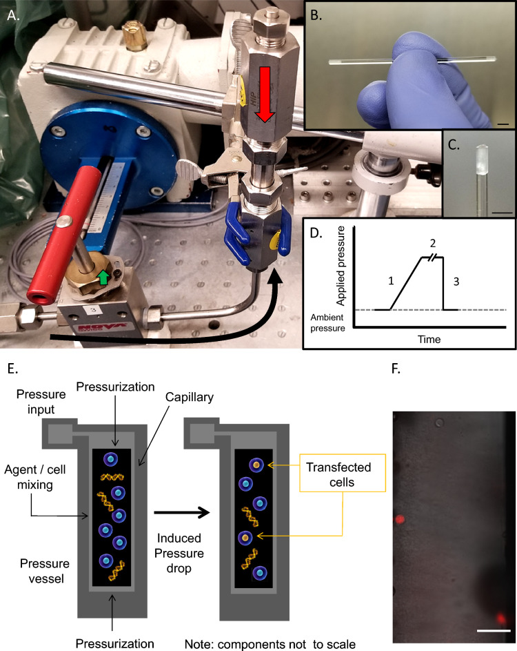
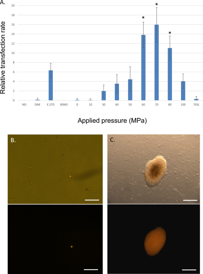
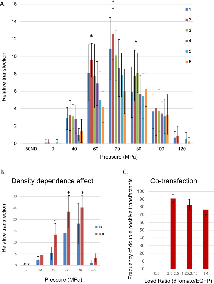
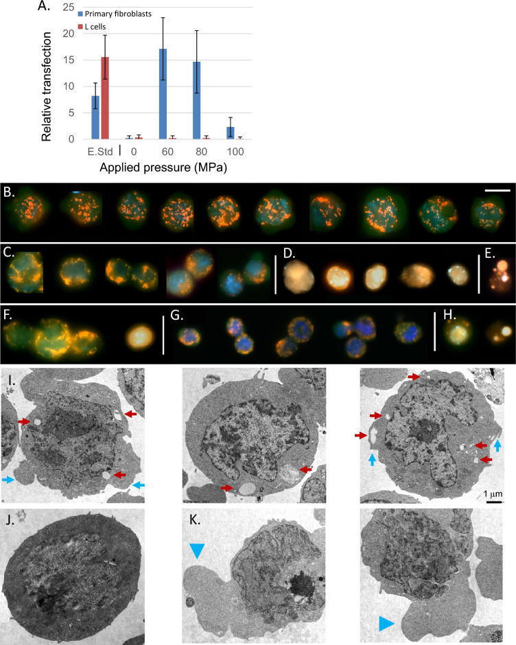
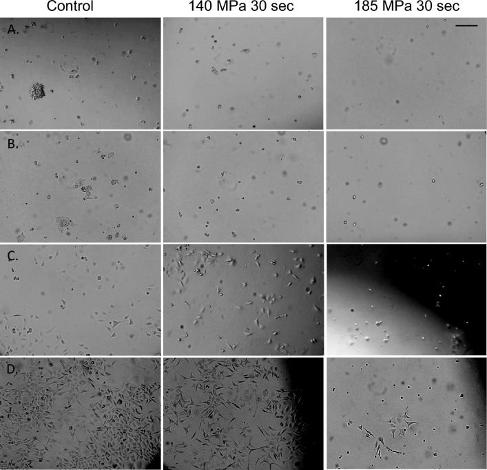
Of all methods exercised in modern molecular biology, modification of cellular properties through the introduction or removal of nucleic acids is one of the most fundamental. As such, several methods have arisen to promote this process; these include the condensation of nucleic acids with calcium, polyethylenimine or modified lipids, electroporation, viral production, biolistics, and microinjection. An ideal transfection method would be (1) low cost, (2) exhibit high levels of biological safety, (3) offer improved efficacy over existing methods, (4) lack requirements for ongoing consumables, (5) work efficiently at any scale, (6) work efficiently on cells that are difficult to transfect by other methods, and (7) be capable of utilizing the widest array of existing genetic resources to facilitate its utility in research, biotechnical and clinical settings. To address such issues, we describe here Pressure-jump-poration (PJP), a method using rapid depressurization to transfect even difficult to modify primary cell types such as embryonic stem cells. The results demonstrate that PJP can be used to introduce an array of genetic modifiers in a safe, sterile manner. Finally, PJP-induced transfection in primary versus transformed cells reveals a surprising dichotomy between these classes which may provide further insight into the process of cellular transformation.


Conflict of interest statement

The authors declare no competing interests.

Figures

Figure 1
Figure 1
Hydrostatic pressure application to mammalian cells. (A) High hydrostatic pressure generating device used for experimental pressures of 1–200 MPa. Pressure line with valve seat (black arrow) and pressure chamber (red arrow) are indicated. Total chamber capacity 10 mL. (B) Example of cell suspension isolated in borosilicate capillary. (C) Closeup of capillary petrolatum seal. Scale bars in figures (B) and (C) equal to 2 mm. (D) Schematic of the Pressure Jump-Poration. Phase 1: Sample is pressurized from ambient to the desired pressure at a rate of ~ 65 MPa/min; Phase 2: pressure is maintained at the desired static pressure for the indicated period (0.5–10 min); Phase 3: sample pressure is suddenly returned to ambient by opening valve. (E) Schematic of pressure-induced transfection of genetic material. (F) Detection of propidium iodine uptake in select cells at 60 MPa (borosilicate capillary) immediately following acute depressurization. Scale bar denotes 100 μm.
Figure 2
Figure 2
Efficacy of transfection of ES cells using pressure-jump-poration. (A) Transfection rate for the pressures indicated compared to standard electroporation conditions following transfection. NDN: (−) DNA control; DNE: (+) DNA, no electroporation; E.STD: (+) DNA, (+) electroporation; ND80: (−) DNA at P = 80 MPa; 0–100: (+) DNA, (+) pressure treatment; 70SL: (+) DNA, (+) pressure treatment, but with slow (3 min) pressure release. All other samples were held at the indicated pressure for 30 s followed by acute depressurization. For each pressure condition n = 9 independent experiments with three replicates within each experiment were performed, and n = 5 independent experiments with three replicates within each experiment for electroporation conditions. Results shown ± SD. (B) ES cells on gelatin at 24 h following pressure treatment at 60 MPa (no selection). Scale bar denotes 150 μm. (C) Clonal ES cell colony on fibroblast bed layer following pressure treatment at 60 MPa with subsequent puromycin selection for 5 days. All pressurization experiments were performed at a concentration of 4000 cells/μL with treated cells plated at 10,000 cells/well in a 6 well plate; electroporation standards were plated similarly. Scale bar denotes 200 μm. Results shown ± SD. *Denotes significant enhancement at p < 0.01 over electroporation. Transfection plasmid shown in (B) and (C) expresses dTomato/puromycin.
Figure 3
Figure 3
Expression of DNA constructs in mammalian cells. ES cells were transfected with plasmids containing several selection markers to assess expression competency. (AC) Fluorescent photomicrographs of pressure treated EYFP-expressing ES cells 24 h following treatment at the pressures indicated in the absence of selection. Pressurization experiments were performed at 4000 cells/μL with cells plated at 25,000 cells/well in a 24 well plate. Relative numbers of transfected, dTomato-expressing cells are indicated (red arrow). Transfected cells exhibit reduced levels of cytoplasmic citrine compared to non-transfected cells (green arrow). Scale bar denotes 100 μm. (DF) Beta-galactosidase expression in R1 transfected ES cells 48 h following pressure treatment. Pressurization experiments were performed at 4000 cells/μL with cells plated at 25,000 cells/well (D,E) or 10,000 cells/well (F) in a 24 well plate. Scale bar denotes 300 μm. (D) (+) DNA, (−) pressure treatment, (E) (−) DNA (+) 80 MPa pressure, (F) (+) DNA (+) pressure treatment. Beta-galactosidase positive cells are indicated (blue arrow), as are examples of dead cells following pressure treatment (box).
Figure 4
Figure 4
Characteristics of pressure-induced DNA transfection. All pressurization experiments were performed at a concentration of 4000 cells/μL. (A) Transfection response profiles of six ES cell lines to pressure treatment with treated cells plated in a 6 well plate at a density of 10,000 cells/well. *Denotes significant enhancement at p < 0.01 over values seen at 100 MPa. Lines: 1—R1, 2—Citrine, 3—Casp3KO, 4—RipK1KO, 5—RIPK3KO, 6—Casp8KO. (B) Initial plating density dependence on numbers of resulting transformants in pressure-treated ES cells. Cells were plated in a 24 well plate at 2000 or 10,000 cells/well as indicated. *Denotes significant enhancement at p < 0.01 over values seen at 2000 cells/well. (C) Co-transfection incidence in transfected R1 ES cells treated at 70 MPa with different reporter plasmid ratios at 48 h post transfection: 0 μg dTomato:5 μg EGFP; 2.5 μg dTomato:2.5 μg EGFP; 3.75 μg EGFP:1.25 μg dTomato; 4 μg EGFP:1 μg dTomato. Cells were plated at 25,000 cells/well in a 24 well plate. For each pressure condition, n = 3 independent experiments with three replicates within each experiment were performed. Results shown ± SD.
Figure 5
Figure 5
Properties of pressure-treated cells. (A) Differences in pressure-mediated transfection efficiency in immortalized versus primary cells. Shown are relative transfection efficiencies of primary fibroblasts (blue) vs. mouse L-cells (red) by electroporation and pressure-mediated transfection. Pressurization was performed at of 4000 cells/μL with cells plated at 10,000 cells/well in a 6 well plate; electroporation standards plated similarly. For each pressure condition, n = 3 independent experiments with three replicates within each experiment were performed. Results shown ± SD. (BH) Cellular features and morphology of pressure-treated R1 ES cells. Live citrine-expressing ES cells were treated with 1.8 μM (1 μg/mL) Hoechst 33342, 10 nM (5 ng/mL) TMRM and 0.75 μM (0.5 μg/mL) propidium iodide prior to pressure treatment. (B) Cells in the absence of pressure treatment; (C) Typical appearances of ES cells following 1 min of pressure treatment at 100 MPa (pressurization at 4000 cells/μL, cells were held for 1 h at 25,000 cells/well in the slide chamber). (D) A small subgroup of these cells become PI+ but retain cellular features. (E) A major portion of these PI+ cells go on to exhibit features of cellular destruction in the immediate (30 min) post-treatment period. (FH) ES cells following 5 min at 100 MPa. The majority of these cells demonstrate features shown in (F,G). (G) A portion of recovered cells exhibit features of reduced cellular volume. (H) The great majority of cell which become PI+ following treatment at 100 MPa for 5 min exhibit features of cellular degeneration. For (BH) scale bar indicate in (B) represents 10 μm. (IK) Electron photomicrographs of ES cells. (I) Following rapid depressurization at 80 MPa, a population of ES demonstrated the presence of intracellular voids proximal to the cell membrane (red arrows), frequently associated with protuberances of the cell membrane (blue arrows). ES cell held at ambient pressure is shown for comparison (J). By contrast ES cells subjected to slow pressure release at 80 MPa, (K) often demonstrated extensive extrusions (blue arrowheads). For figures (IK) scale bar represents distance of 1 μm.
Figure 6
Figure 6
Brightfield photomicrographs of primary and transformed cells after pressure treatment. Indicated below are images of (A) ES R1 cells 24 h post-treatment, (B) HEK293T cells 24 h post-treatment, (C) T24 cells 24 h post-treatment, and (D) L-cells 72 h post-treatment for each of the conditions indicated. Scale bar in (A) represents 100 μm for all images in (AD).

Similar articles

Cited by

References

    1. Hite B. H. The effect of pressure in the preservation of milk. Bull W. Va. Univ Agric. Experim Stn. 1–67 (1899).
    1. Patterson MF. Microbiology of pressure-treated foods. J. Appl. Microbiol. 2005 doi: 10.1111/j.1365-2672.2005.02564.x. - DOI - PubMed
    1. Chawla R, et al. High hydrostatic pressure technology in dairy processing: A review. J. Food Sci. Technol. Mysore. 2011 doi: 10.1007/s13197-010-0180-4. - DOI - PMC - PubMed
    1. Gollwitzer H, et al. High hydrostatic pressure for disinfection of bone grafts and biomaterials: An experimental study. Open Orthop. J. 2009 doi: 10.2174/1874325000903010001. - DOI - PMC - PubMed
    1. van de Sande MAJ, et al. Successful disinfection of femoral head bone graft using high hydrostatic pressure. Cell Tissue Bank. 2018 doi: 10.1007/s10561-017-9678-6. - DOI - PMC - PubMed