. 2024 Feb 26;15(1):1756.
doi: 10.1038/s41467-024-46092-3.

Transient disome complex formation in native polysomes during ongoing protein synthesis captured by cryo-EM

Affiliations

Transient disome complex formation in native polysomes during ongoing protein synthesis captured by cryo-EM

Timo Flügel et al. Nat Commun. .

Abstract

Structural studies of translating ribosomes traditionally rely on in vitro assembly and stalling of ribosomes in defined states. To comprehensively visualize bacterial translation, we reactivated ex vivo-derived E. coli polysomes in the PURE in vitro translation system and analyzed the actively elongating polysomes by cryo-EM. We find that 31% of 70S ribosomes assemble into disome complexes that represent eight distinct functional states including decoding and termination intermediates, and a pre-nucleophilic attack state. The functional diversity of disome complexes together with RNase digest experiments suggests that paused disome complexes transiently form during ongoing elongation. Structural analysis revealed five disome interfaces between leading and queueing ribosomes that undergo rearrangements as the leading ribosome traverses through the elongation cycle. Our findings reveal at the molecular level how bL9's CTD obstructs the factor binding site of queueing ribosomes to thwart harmful collisions and illustrate how translation dynamics reshape inter-ribosomal contacts.


Conflict of interest statement

The authors declare no competing interests.

Figures
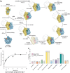
Fig. 1
Fig. 1. Visualization of actively translating E. coli polysomes.
a 14C-Val incorporation into nascent peptides over a 10-min period after incubation of 700 nM E. coli polysomes in the PURE system at 37 °C. Data (closed circles) represent mean values of triplicate experiments with error bars indicating the s. d. (n = 3). Data from duplicate experiments are shown individually (open cirles; t = 0 and t = 10 min). Data were fitted to Eqs. 1 and 2. b Translation cycle showing functional ribosome states isolated from ex vivo-derived polysomes reactivated in the PURE system. Shown are 30S (yellow), 50S (blue), initiation factors 1 (lavender), 2 (blue), and 3 (orange), translation factors (red), aminoacyl-tRNAs (A-tRNA, light violet), peptidyl-tRNAs (P-tRNA, green), and exit-tRNAs (E-tRNA, orange). For functional 70S state populations containing disome fractions, collided ribosomes are indicated as silhouettes. The EF-G bound translocation intermediate (Ti-POST: EF-G·GDP | P | E), not observed experimentally, is shown as transparent map simulated from PDB 7N2C. All maps were filtered to 5 Å. c Distribution of 70S functional states of all imaged 70S (filled bars) and of 70SL in disome complexes (dashed bars). Source data are provided as a Source Data file.
Fig. 2
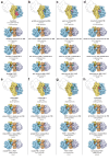
Fig. 2. 24 distinct functional disome complexes isolated from reactivated E. coli polysomes.
ah For each 70SL functional state (topmost of each column) three distinct functional 70SQ states are present: closed non-rotated PRE, rotated PRE-2, and POST. Shown are 50SL (blue), 30SL (yellow), 50SQ (lavender), 30SQ (peach), bL9L (turquoise), translation factors (red), A-tRNAs (light violet), P-tRNAs (green), and E-tRNAs (orange). Below each disome complex, functional state descriptions of 70SL and 70SQ (bold), particle numbers, and reconstruction resolutions are shown.
Fig. 3
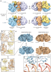
Fig. 3. Intermolecular interfaces between 70SL and 70SQ.
a Overview of the cryo-EM structure of the disome complex containing 70SL and 70SQ in closed non-rotated PRE states. Shown is a composite map generated from locally refined 70SL, 70SQ, and interface maps. Shown are 50SL (blue), 30SL (yellow), 50SQ (lavender), 30SQ (peach), bL9L (turquoise), EF-G (red), A-tRNAs (light violet), P-tRNAs (green), E-tRNAs (orange), and bS1Q (pink). Map corresponding to bS1Q is segmented and shown at lower threshold. Close-ups of the five interfaces between 70SL and 70SQ (shown is the interface map). Local resolution ranges of map regions shown in close-ups: (b) 4–6.3 Å, (c) 3.0–3.6 Å, (d) 3.3–3.8 Å, (e) 4–4.4 Å, (f) 2.7–3.5 Å. g Overlay of bL9L bound to 30SQ and EF-G (10 Å map simulated from an EF-G•GTP model (PDB: 7N2V).
Fig. 4
Fig. 4. The pre-nucleophilic attack state is enriched in 70SL.
Close-ups of the peptidyl transferase centers (PTC) of 70SQ (a) and 70SL (b). The cryo-EM densities reflect the weighted average of all tRNA species and amino acid occupants. a 70SQ bound to Val-A-tRNA (light violet), Phe-P-tRNA (green), and nascent peptide chain (blue) is in a post-nucleophilic attack state in which the nascent peptide has been transferred to the A-site tRNA. b 70SL bound to Ala-A-tRNA (light violet), Trp-P-tRNA (green), and nascent peptide chain (blue) represents a pre-nucleophilic attack state in which the nascent peptide has not yet been transferred to the A-site tRNA.
Fig. 5
Fig. 5. Disome interface dynamics during 70SL elongation.
a Intersubunit rotation of 70SL induces L1L stalk dissociation from 30SQ while bL9L CTD remains bound. Shown are 50SL (blue), 30SL (yellow), 50SQ (lavender), 30SQ (peach), bL9L (turquoise), A-tRNAs (light violet), P-tRNAs (green), E-tRNAs (orange), and mRNA (purple). bd non-rotated bS6L:uS4Q interface is dissolved upon 70SL intersubunit rotation and a new interface between uS11 and uS4Q forms. e Cryo-EM maps of rotated interface classes 1 (blue) and 2 (orange). f Overlay of rotated interface classes 1 and 2 with non-rotated disome (PDB: 8R3V) interface model showing an 8.7 Å upward movement of the bL9L CTD along h5/15. g Overlay of non-rotated interface classes 1 and 2 with the average non-rotated (PDB: 8R3V) disome interface model showing no significant conformational differences of the bL9L CTD.
Fig. 6
Fig. 6. Putative mechanism of functional disome formation and dissociation.
Two individual 70 S translate the same mRNA. bL9L switches between compact and stretched conformation, sampling the region upstream the mRNA exit. Upon translational pausing of 70SL, 70SL and 70SQ collide. The direct contact between 70SL and 70SQ stabilizes bL9L’s stretched conformation, induces binding of its CTD to 30SQ, and the establishment of four additional interfaces. bL9L CTD binding stalls 70SQ through obstruction of its factor binding site. Intersubunit rotation of 70SL induces L1L stalk dissociation from 30SQ while bL9L remains bound. Upon translocation events of 70SL, bL9L dissociates. 70SQ is no longer stalled and both 70SL and 70SQ resume translation as bL9L returns to the sampling mode. Shown are 50SL (blue), 30SL (yellow), 50SQ (lavender), 30SQ (peach), bL9L (turquoise), A-tRNAs (light violet), P-tRNAs (green), E-tRNAs (orange), and mRNA (purple).

Similar articles

Cited by

References

    1. Melnikov S, et al. One core, two shells: bacterial and eukaryotic ribosomes. Nat. Struct. Mol. Biol. 2012;19:560–567. - PubMed
    1. Jobe, A., Liu, Z., Gutierrez-Vargas, C. & Frank, J. New insights into ribosome structure and function. Cold Spring Harb. Perspect. Biol. 11, a032615 (2019). - PMC - PubMed
    1. Brandt F, et al. The native 3D organization of bacterial polysomes. Cell. 2009;136:261–271. - PubMed
    1. Xue L, et al. Visualizing translation dynamics at atomic detail inside a bacterial cell. Nature. 2022;610:205–211. - PMC - PubMed
    1. Juszkiewicz S, et al. ZNF598 is a quality control sensor of collided ribosomes. Mol. Cell. 2018;72:469–481.e7. - PMC - PubMed

MeSH terms