. 2024 Feb 26;14(1):7334.
doi: 10.1038/s41598-023-33629-7.

Sexual dimorphism during integrative endocrine and immune responses to ionizing radiation in mice

Affiliations

Sexual dimorphism during integrative endocrine and immune responses to ionizing radiation in mice

Marissa Burke et al. Sci Rep. .

Abstract

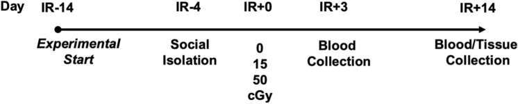
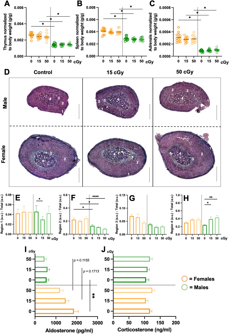
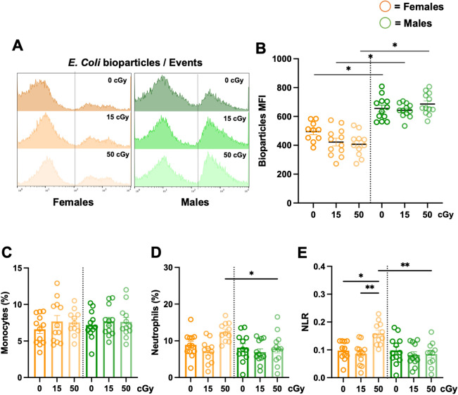
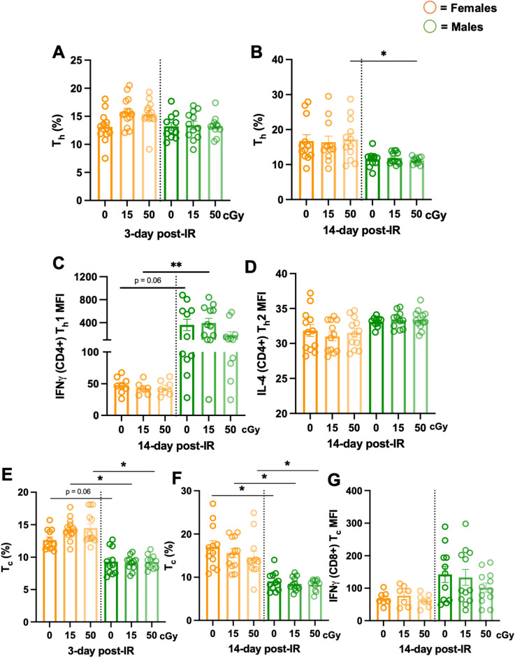
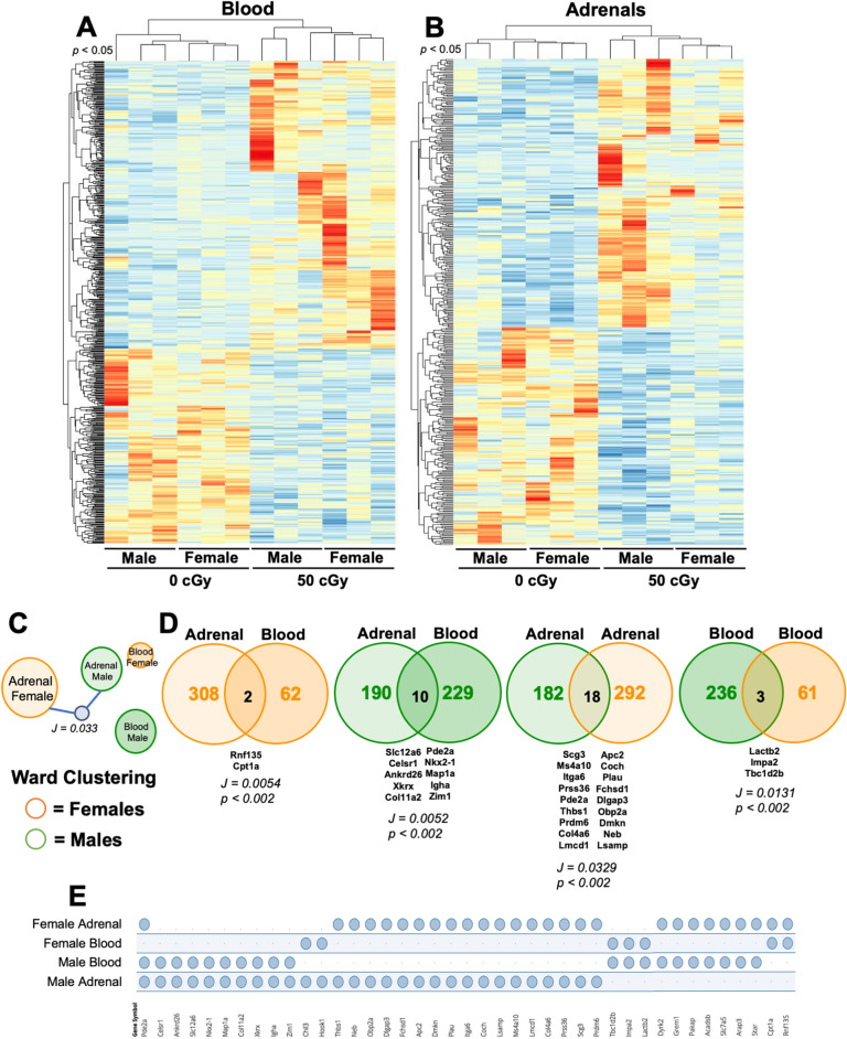
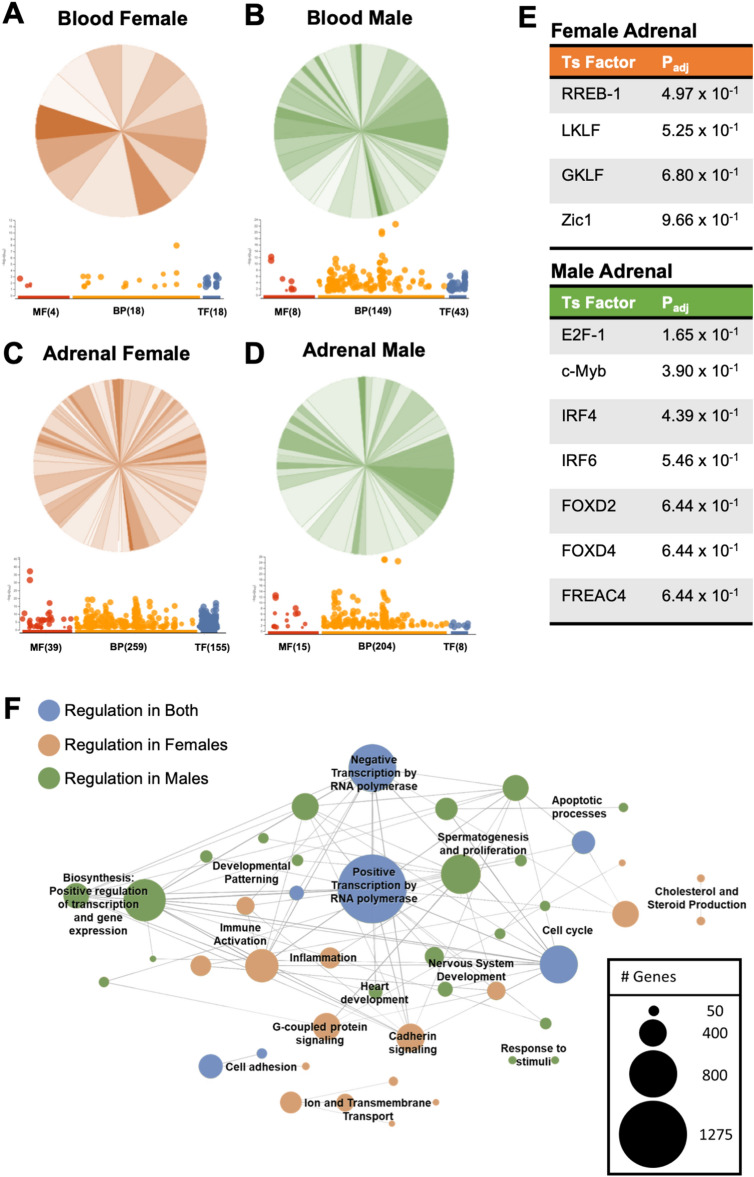
Exposure to cosmic ionizing radiation is an innate risk of the spaceflight environment that can cause DNA damage and altered cellular function. In astronauts, longitudinal monitoring of physiological systems and interactions between these systems are important to consider for mitigation strategies. In addition, assessments of sex-specific biological responses in the unique environment of spaceflight are vital to support future exploration missions that include both females and males. Here we assessed sex-specific, multi-system immune and endocrine responses to simulated cosmic radiation. For this, 24-week-old, male and female C57Bl/6J mice were exposed to simplified five-ion, space-relevant galactic cosmic ray (GCRsim) radiation at 15 and 50 cGy, to simulate predicted radiation exposures that would be experienced during lunar and Martian missions, respectively. Blood and adrenal tissues were collected at 3- and 14-days post-irradiation for analysis of immune and endocrine biosignatures and pathways. Sexually dimorphic adrenal gland weights and morphology, differential total RNA expression with corresponding gene ontology, and unique immune phenotypes were altered by GCRsim. In brief, this study offers new insights into sexually dimorphic immune and endocrine kinetics following simulated cosmic radiation exposure and highlights the necessity for personalized translational approaches for astronauts during exploration missions.


Conflict of interest statement

The authors declare no competing interests.

Figures

Figure 1
Figure 1
Representative experimental timeline. 24-week-old male and female C57Bl/6J were cage acclimated at 14-days prior to irradiation (IR-14) and socially isolated at IR-4. At IR + 0, mice were irradiated with 5-ion GCRsim ionizing radiation at 15 and 50 cGy, along with a unirradiated sham controls that were exposed to similar housing restraints and time as IR mice (0 cGy). Retro-orbital blood collection was performed on IR + 3 and abdominal aorta blood and tissues were collected on the time of euthanasia, IR + 14.
Figure 2
Figure 2
Sex-specific immune and endocrine system analysis, including adrenal gland architecture and function. Male and female mice were exposed to GCRsim (0, 15, and 50 cGy) and at 14-days post-irradiation mice were euthanized and immune thymus (A) and spleens (B) organs; and endocrine, adrenal glands (C) were isolated and weighed. All tissue weights were normalized to total body weights. 14-days post-irradiation glands were isolated, fixed, paraffin-embedded, sectioned (10 μm) and stained with hematoxylin and eosin for cellular architecture identification (D) and adrenal regional morphological size (EH). Three-days post-irradiation blood was isolated and enzyme-linked immunosorbent assays (ELISA) were performed for adrenal hormone characterization, including aldosterone (I) and corticosterone (J). Scale bars = 450 μm. Sectioned regions highlight: 1. Zona glomerulosa; 2. Zona fasciculata; 3. Vacuolated zona fasciculata; and 4. Medulla. Females (orange) and males (green) are displayed. Parametric statistical analysis was performed on (A) and (F). Nonparametric analyses were performed on (B, C, E, G, H, I, and J), as described in the methods section. Weight data represent ± SEM, p* < 0.05, n = 10–12 per group. H&E data represent ± SEM, p* < 0.05, p** < 0.01, p**** < 0.001, n = 2–3 per group. Adrenal region images were scaled up and quantified with metric measurements denoted as arbitrary units (a.u.). ELISA data represent ± SEM, p** < 0.01, n = 10 per group with technical replicates (n = 3) performed with each ELISA.
Figure 3
Figure 3
Sex-specific differences in phagocyte function, independent of phagocyte count. 24-week-old male and female C57Bl/6J mice were exposed to GCRsim (0, 15, and 50 cGy) and 3-days post-irradiation blood was collected. Red blood cells were lysed and leukocytes were stained and analyzed by flow cytometry. Phagocytic function was assessed using pH-sensitive E.coli bioparticles to determine the median fluorescence intensity (MFI) of phagocytosis (A, B). Cell count percentages (%) within total leukocytes (CD45+) were determined for monocytes (C), neutrophils (D), and neutrophil-to-lymphocyte ratio (NLR) (E). Parametric statistical analysis was performed on all data. Data represent ± SEM, p* < 0.05, p** < 0.01, n = 10–12 per group.
Figure 4
Figure 4
Longitudinal lymphocyte monitoring following irradiation displayed sex-specific effects. 24-week-old male and female C57Bl/6J mice were exposed to GCRsim (0, 15, and 50 cGy) and at 3- and 14-days post-irradiation, blood was collected. Red blood cells were lysed, leukocytes were stimulated for 16-h with a cell stimulation cocktail, stained, and analyzed by flow cytometry. Longitudinal monitoring of Thelper (Th, CD4+/CD3+) cell development percentages (%) post-irradiation in females (orange) and males (green) at day 3 (A) and day 14 (B) post-irradiation. Th cell function assessed Th1 (IFNγ) median fluorescence intensity (MFI) (C) and Th2 (IL-4) MFI production (D) from CD4+/CD3+ lymphocytes. Longitudinal monitoring of Tcytotoxic (Tc, CD8+/CD3+) cell development % post-irradiation in females and males at day 3 (E) and day 14 (F) post-irradiation. Tc cell function assessed (IFNγ) median fluorescence intensity (MFI) (G). Parametric statistical analysis was performed on (A) and (D). Nonparametric analyses were performed on (B, C, E, F, and G), as described in the methods section. Data represent ± SEM, p* < 0.05, p** < 0.01, n = 7–12 per group.
Figure 5
Figure 5
Sex- and dose-specific RNA sequencing profiles. Male and female mice were exposed to GCRsim (0 and 50 cGy) and at 14-days post-irradiation blood and adrenal glands were isolated and total RNA sequencing was performed. 50 cGy male and females were compared to 0 cGy sham controls. Heatmap of differentially expressed genes (DEG) in blood (A) and adrenal glands (B) from male and female mice exposed to 50 cGy versus 0 cGy controls (log2 fold change > 2 and p-value < 0.05). (C) Ward Clustering forceTree with Jaccard similarity child node (J = 0.033, p < 0.002) is displayed. (D) Venn diagrams of compared annotated datasets with overlapping Jaccard similarity score. (E) Jaccard similarity overlapping annotated genesets intersecting table displays overlapping genes across all tissue types and sexes. Clustering and geneset overlap (E) results generated using GeneWeaver and Venn diagrams (D) generated using Venny2.1. Data represent p < 0.05 (A, B) and p < 0.01 (C–E), n = 3 per group.
Figure 6
Figure 6
Adrenal and blood enriched gene ontology pathways are distinct and dimorphic. Male and female mice were exposed to GCRsim (0 and 50 cGy) and at 14-days post-irradiation blood and adrenal glands were isolated and total RNA sequencing was performed. 50 cGy male and females were compared to 0 cGy sham controls and enriched gene ontology PANTHER analysis converted gene to pathway (Venn Diagrams; shaded orange (female) and green (male) fractions all represent different PANTHER defined biological pathways and the number of genes involved in each pathway) and g:Profiler (dotplot; red denotes molecular function (MF), orange denotes biological pathways (BP), and blue denotes transcription factors (TF), and the number of genes identified in each) for annotated DEG (p < 0.01) in female blood (A) and adrenals (C), and male blood (B) and adrenals (D). Transcription factors analysis of female (orange label) and male (green label) adrenals, p < 0.01 (E). Overlapping blood and adrenal Consensus Pathways Analysis (CPA) for females (orange nodes) and males (green nodes) or both (blue nodes) and representative number of genes within each node. Significant pathways were identified as sharing at minimum one edge, three common genes, and a combined p-value < 0.01 (false discovery rate was not used for selection) (F). Data represent p < 0.01, n = 3 per group. PANTHER analysis orange circles denote biological pathways in female and green circles denote biological pathways in males.

Similar articles

Cited by

  • Taking the 3Rs to a higher level: replacement and reduction of animal testing in life sciences in space research.
    Vinken M, Grimm D, Baatout S, Baselet B, Beheshti A, Braun M, Carstens AC, Casaletto JA, Cools B, Costes SV, De Meulemeester P, Doruk B, Eyal S, Ferreira MJS, Miranda S, Hahn C, Helvacıoğlu Akyüz S, Herbert S, Krepkiy D, Lichterfeld Y, Liemersdorf C, Krüger M, Marchal S, Ritz J, Schmakeit T, Stenuit H, Tabury K, Trittel T, Wehland M, Zhang YS, Putt KS, Zhang ZY, Tagle DA. Vinken M, et al. Biotechnol Adv. 2025 Jul-Aug;81:108574. doi: 10.1016/j.biotechadv.2025.108574. Epub 2025 Apr 1. Biotechnol Adv. 2025. PMID: 40180136 Review.
  • To boldly go where no microRNAs have gone before: spaceflight impact on risk for small-for-gestational-age infants.
    Corti G, Kim J, Enguita FJ, Guarnieri JW, Grossman LI, Costes SV, Fuentealba M, Scott RT, Magrini A, Sanders LM, Singh K, Sen CK, Juran CM, Paul AM, Furman D, Calleja-Agius J, Mason CE, Galeano D, Bottini M, Beheshti A. Corti G, et al. Commun Biol. 2024 Oct 5;7(1):1268. doi: 10.1038/s42003-024-06944-6. Commun Biol. 2024. PMID: 39369042 Free PMC article.
  • A second space age spanning omics, platforms and medicine across orbits.
    Mason CE, Green J, Adamopoulos KI, Afshin EE, Baechle JJ, Basner M, Bailey SM, Bielski L, Borg J, Borg J, Broddrick JT, Burke M, Caicedo A, Castañeda V, Chatterjee S, Chin CR, Church G, Costes SV, De Vlaminck I, Desai RI, Dhir R, Diaz JE, Etlin SM, Feinstein Z, Furman D, Garcia-Medina JS, Garrett-Bakelman F, Giacomello S, Gupta A, Hassanin A, Houerbi N, Irby I, Javorsky E, Jirak P, Jones CW, Kamal KY, Kangas BD, Karouia F, Kim J, Kim JH, Kleinman AS, Lam T, Lawler JM, Lee JA, Limoli CL, Lucaci A, MacKay M, McDonald JT, Melnick AM, Meydan C, Mieczkowski J, Muratani M, Najjar D, Othman MA, Overbey EG, Paar V, Park J, Paul AM, Perdyan A, Proszynski J, Reynolds RJ, Ronca AE, Rubins K, Ryon KA, Sanders LM, Glowe PS, Shevde Y, Schmidt MA, Scott RT, Shirah B, Sienkiewicz K, Sierra MA, Siew K, Theriot CA, Tierney BT, Venkateswaran K, Hirschberg JW, Walsh SB, Walter C, Winer DA, Yu M, Zea L, Mateus J, Beheshti A. Mason CE, et al. Nature. 2024 Aug;632(8027):995-1008. doi: 10.1038/s41586-024-07586-8. Epub 2024 Jun 11. Nature. 2024. PMID: 38862027 Review.
  • Thymus ad astra, or spaceflight-induced thymic involution.
    Muramatsu W, Maryanovich M, Akiyama T, Karagiannis GS. Muramatsu W, et al. Front Immunol. 2025 Jan 24;15:1534444. doi: 10.3389/fimmu.2024.1534444. eCollection 2024. Front Immunol. 2025. PMID: 39926601 Free PMC article. Review.

References

    1. Simonsen LC, Slaba TC, Guida P, Rusek A. NASA's first ground-based galactic cosmic ray simulator: Enabling a new era in space radiobiology research. PLoS Biol. 2020;18:e3000669. doi: 10.1371/journal.pbio.3000669. - DOI - PMC - PubMed
    1. Crucian BE, Choukèr A, Simpson RJ, Mehta S, Marshall G, Smith SM, Zwart SR, Heer M, Ponomarev S, Whitmire A, Frippiat JP, Douglas GL, Lorenzi H, Buchheim JI, Makedonas G, Ginsburg GS, Ott CM, Pierson DL, Krieger SS, Baecker N, Sams C. Immune system dysregulation during spaceflight: potential countermeasures for deep space exploration missions. Front. Immunol. 2018;9:1437. doi: 10.3389/fimmu.2018.01437. - DOI - PMC - PubMed
    1. Pecaut MJ, Mao XW, Bellinger DL, Jonscher KR, Stodieck LS, Ferguson VL, Bateman TA, Mohney RP, Gridley DS. Is spaceflight-induced immune dysfunction linked to systemic changes in metabolism? PLoS ONE. 2017;12:e0174174. doi: 10.1371/journal.pone.0174174. - DOI - PMC - PubMed
    1. Cruz-Topete D, Cidlowski JA. One hormone, two actions: Anti- and pro-inflammatory effects of glucocorticoids. NeuroImmuno Modul. 2015;22:20–32. doi: 10.1159/000362724. - DOI - PMC - PubMed
    1. Rummell JA, Sawin CF, Buderer MC, Mauldin DG, Michel EL. Physiological response to exercise after space flight–Apollo 14 through Apollo 17. Aviat. Space Environ. Med. 1975;46:679–683. - PubMed