. 2024 Jul;20(7):1505-1522.
doi: 10.1080/15548627.2024.2323785. Epub 2024 Mar 6.

Sodium butyrate ameliorates high glucose-suppressed neuronal mitophagy by restoring PRKN expression via inhibiting the RELA-HDAC8 complex

Affiliations

Sodium butyrate ameliorates high glucose-suppressed neuronal mitophagy by restoring PRKN expression via inhibiting the RELA-HDAC8 complex

Ji Hyeon Cho et al. Autophagy. 2024 Jul.

Abstract

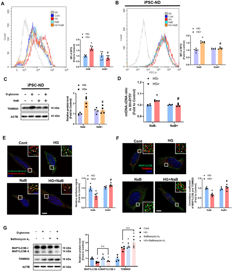
Damaged mitochondria accumulation in diabetes is one of the main features that contribute to increased incidence of cognitive impairment by inducing apoptosis. Butyrate is a major metabolite produced by microbiota that has neuroprotective effects by regulating mitochondrial function. However, detailed mechanisms underlying how butyrate can regulate neuronal mitophagy remain unclear. Here, we examined the regulatory effects of sodium butyrate (NaB) on high glucose-induced mitophagy dysregulation, neuronal apoptosis, and cognitive impairment and its underlying mechanisms in human-induced pluripotent stem cell-derived neurons, SH-SY5Ys, and streptozotocin (STZ)-induced diabetic mice. In our results, diabetic mice showed gut-microbiota dysbiosis, especially a decreased number of butyrate-producing bacteria and reduced NaB plasma concentration. NaB ameliorated high glucose-induced neuronal mitochondrial dysfunction by recovering PRKN/Parkin-mediated mitophagy. High glucose-induced reactive oxygen species (ROS) and -inhibited PRKAA/AMPKα stimulated the RELA/p65-HDAC8 complex, which downregulated PRKN protein expression by binding to the PRKN promoter region. NaB restored PRKN expression by blocking RELA nuclear translocation and directly inhibiting HDAC8 in the nucleus. In addition, HDAC8 overexpression inhibited the positive effect of NaB on high glucose-induced mitophagy dysfunction and neuronal apoptosis. Oral administration of NaB improved cognitive impairment in diabetic mice by restoring mitophagy in the hippocampus. Taken together, NaB ameliorates neuronal mitophagy through PRKN restoration by inhibiting RELA-HDAC8 complexes, suggesting that NaB is an important substance for protecting neuronal apoptosis in diabetes-associated cognitive impairment.

Keywords: autophagy; diabetes; gut-brain axis; mitochondria; neuronal apoptosis; short-chain fatty acids.


Conflict of interest statement

No potential conflict of interest was reported by the author(s).

Figures

Figure 1.
Figure 1.
Among SCFAs, NaB ameliorated high glucose-induced mitochondrial dysfunction, and neuronal cell death. (A–D) SH-SY5Ys and iPSC-NDs were pretreated with NaA (500 µM), NaB (500 µM), NaP (500 µM) for 30 min prior to high glucose (HG; 25 mM) treatment for 24 h. (A and B) The mean fluorescence intensities of MitoSOX were measured by flow cytometric analysis. n = 4 and n = 5, respectively. (C and D) The mean fluorescence intensities of TMRE were measured by flow cytometric analysis. n = 4,n = 5, respectively. (E and F) SH-SY5Ys and iPSC-NDs were pretreated with NaA (500 µM), NaB (500 µM), NaP (500 µM) for 30 min prior to HG (25 mM) treatment for 72 h. (E) The protein levels of cleaved-CASP3 and ACTB were detected by western blotting. n = 4. (F) The percentage of apoptotic cells (ANXA5 and PI positive) were measured by flow cytometric analysis. n = 5. (G–J) Mice were injected with either vehicle (0.05 M citrate buffer) or STZ (75 mg/kg/day) for the first 3 days of 8 weeks of diabetes induction. (G) β diversity between vehicle (red) and STZ (blue); weighted UniFrac distance, bray – Curtis. (H) The taxonomic abundance of annotation at the genus level. n = 4. (I) The concentration of NaB in the plasma of mice, measured by GC – MS. n = 4. (J) Representative immunohistochemistry images of the hippocampus stained with cleaved-CASP3 and DAPI. n = 4. Scale bars: 20 μm. All data are representative. Quantitative data are represented as mean ± SEM. *p < 0.05 versus vehicle-injected mice or control; #p < 0.05 versus HG.
Figure 2.
Figure 2.
NaB recovered neuronal mitophagy suppressed by high glucose. (A–F) SH-SY5Ys and iPSC-NDs were pretreated with NaB for 30 min prior to HG treatment for 24 h. (A and B) The mean fluorescence intensities of MitoTracker green (MTG) were measured by flow cytometric analysis. n = 5. (C) The protein levels of TOMM20 and ACTB were detected by western blotting. n = 4. (D) The DNA levels of mtDNA and nDNA were analyzed by real-time PCR. ACTB was used as a loading control. (E) SH-SY5Y cells were transfected with mt-keima 24 h prior to NaB treatment for 30 min and visualized. n = 4. Scale bars: 10 μm. (F) Immunofluorescence staining of TOMM20, MAP1LC3B, and DAPI were visualized. n = 4. Scale bars: 10 μm. (G) SH-SY5Ys were treated with HG for 24 h. Bafilomycin A1 (10 nM) was applied for 2 h prior to harvest. The protein levels of TOMM20, MAP1LC3B – I, MAP1LC3B – II, and ACTB were detected by western blotting. All data are representative. Quantitative data are represented as mean ± SEM. *p < 0.05 versus control; #p < 0.05 versus HG.
Figure 3.
Figure 3.
NaB recovered PRKN expression decreased by high glucose. (A) Mice were injected with either vehicle (0.05 M citrate buffer) or STZ (75 mg/kg/day) for the first 3 days of 8 weeks of diabetes induction. The protein level of PINK1, BNIP3, BNIP3L/NIX, PRKN, OPTN/optineurin, CALCOCO2/NDP52, and ACTB in hippocampal were subjected to western blotting. n = 4. (B) SH-SY5Ys were exposed to HG for 24 h. The protein levels of PINK1, BNIP3, BNIP3L, PRKN, OPTN, CALCOCO2, and ACTB were subjected to western blotting. n = 4. (C – G) SH-SY5Ys and iPSC-NDs were pretreated with NaB for 30 min prior to HG treatment for 24 h. (C) The mRNA expression levels of PINK1, BNIP3, BNIP3L, PRKN, OPTN, CALCOCO2 were investigated. n = 4. (D) The mRNA expression levels of PRKN were investigated n = 4. (E) Protein levels of the PRKN and ACTB were determined by western blotting. n = 4. (F) Cells were transfected with mt-keima-red or mt-keima-red-PRKN 24 h prior to NaB treatment for 30 min and visualized. n = 4. Scale bars: 10 μm. (G) Immunofluorescence staining of MFN1, UB, and DAPI were visualized. n = 4. Scale bars: 10 μm. All data are representative. Quantitative data are represented as mean ± SEM. *p < 0.05 versus vehicle-injected mice or control; #p < 0.05 versus HG.
Figure 4.
Figure 4.
SLC5A8 is involved in the inhibitory effect of NaB of PRKN expression on suppressing high glucose activated RELA and HDAC8. (A and B) SH-SY5Ys were pretreated with NaB or PTX (200 nM), or Ibuprofen (500 µM) for 30 min before HG treatment for 24 h. (A) The mean fluorescence intensities of MTG were measured by flow cytometric analysis. n = 4. (B) The protein levels of PRKN, TOMM20, and ACTB were detected by western blotting. n = 4. (C) SH-SY5Ys were pretreated with NAC (2 mM) or NaB for 30 min prior to HG treatment for 24 h. Immunofluorescence staining of RELA and DAPI were visualized. n = 4. Scale bars: 10 μm. (D) SH-SY5Ys were transfected with RELA siRNA or NT siRNA for 24 h and exposed to HG for 24 h. The protein levels of PRKN and ACTB were detected by western blotting. n = 4. (E) SH-SY5Ys were pretreated with TC-H 106 (10 µm) + PCI 34051 (10 µm), or TMP269 (5 µm) for 30 min before HG treatment for 24 h. The protein levels of PRKN, TOMM20, and ACTB were detected by western blotting. n = 4. (F and G) SH-SY5Ys were exposed to HG for 24 h. (F) The mRNA expression levels of HDAC1, HDAC2, HDAC3, HDAC8 were investigated. n = 4. (G) The protein levels of HDAC1, HDAC2, HDAC3, HDAC8 were detected by western blotting. n = 4. (H and I) SH-SY5Ys were transfected with HDAC1, HDAC2, HDAC3, HDAC8 siRNA or NT siRNA for 24 h, and exposed to HG for 24 h. (H) The mRNA expression of PRKN was detected by PCR. n = 4. (I) The protein levels of PRKN and ACTB were detected by western blotting. n = 4. All data are representative. Quantitative data are represented as mean ± SEM. *p < 0.05 versus control; #p < 0.05 versus HG.
Figure 5.
Figure 5.
PRKAA-mediated HDAC8 activity and ROS-induced RELA stimulate complex that suppresses transcription of PRKN under high glucose conditions. (A) SH-SY5Ys were treated with HG for 1, 3, and 6 h. The protein levels of p-PRKAA, PRKAA, p-HDAC8, HDAC8 and ACTB were detected by western blotting. n = 4. (B) SH-SY5Ys were pretreated with AICAR (1 mM) for 30 min prior to treatment of HG for 6 h. The protein levels of p-HDAC8, HDAC8 and ACTB were detected by western blotting. n = 4. (C – E) NaB were pretreated 30 min prior to HG treatment for 24 h. (C) HDAC8 activity in the nucleus were measured with HDAC8 activity assay kit. n = 4. (D) Immunofluorescence staining of HDAC8, RELA, and DAPI were visualized in SH-SY5Ys. n = 4. Scale bars: 10 μm. (E) Interaction between RELA and HDAC8 (RELA+HDAC8, red) was assessed by PLA assay. n = 4. Scale bars: 10 μm. (F – I) SH-SY5Y cells were were pretreated with NaB 30 min prior to HG treatment for 6 h. (F, G, I) DNA was immunoprecipitated with RELA, HDAC8, and acetyl-histone H3 antibody, respectively. The immunoprecipitation and input samples were amplified with primers of GAPDH and PRKN gene. n = 3. (H) Fold enrichment of PRNK region was assayed by chromatin accessibility assay kit. n = 4. All data are representative. Quantitative data are represented as mean ± SEM. *p < 0.05 versus control; #p < 0.05 versus HG.
Figure 6.
Figure 6.
Overexpressed HDAC8 blocked the protective effect of NaB under high glucose conditions. (A – C) SH-SY5Ys were transfected with pcDNA3.1-GFP or pcDNA3.1-HDAC8-GFP 24 h prior to NaB treatment for 30 min and exposed to HG for 24 h. (A) The mean fluorescence intensities of MitoSOX were measured by flow cytometric analysis. n = 4. (B) The mean fluorescence intensities of TMRE were measured by flow cytometric analysis, respectively. n = 4. (C) tThe protein levels of PRKN, TOMM20 and ACTB were detected by western blotting. n = 3. (D and E) SH-SY5Ys were transfected with pcDNA3.1-GFP or pcDNA3.1-HDAC8-GFP prior to NaB treatment for 30 min and exposed to HG for 72 h. (D) The protein levels of cleaved-CASP3 and ACTB were detected by western blotting. n = 3. (E) LDH from cell supernatant were measured by LDH cytotoxicity assay kit. n = 4. All data are representative. Quantitative data are represented as mean ± SEM. *p < 0.05 versus pcDNA3.1-GFP; #p < 0.05 versus HG+pcDNA3.1-GFP.
Figure 7.
Figure 7.
Oral administration of NaB recovered cognitive impairment in diabetic mice by preventing apoptosis through mitophagy restoration. (A – H) Mice were injected with either vehicle (0.05 M citrate buffer) or STZ (75 mg/kg/day) for the first 3 days of 8 weeks of diabetes induction. Mice injected with either vehicle or STZ were orally administered vehicle or NaB (0.5 g/kg/day) once daily for 8 weeks after diabetes induction. (A) The protein levels of the hippocampal PRKN and ACTB were determined by western blotting. n = 4. (B) Representative immunohistochemistry images of the hippocampus stained with TOMM20, MAP1LC3B, and DAPI. n = 4. Scale bars: 20 μm. (C) Representative immunohistochemistry images of the hippocampus stained with TOMM20, 4-HNE, and DAPI. n = 4. Scale bars: 20 μm. (D) Representative immunohistochemistry images of the hippocampus stained with TOMM20, 8-oxo-dG, and DAPI. n = 4. Scale bars: 20 μm. (E) The protein levels of the hippocampal cleaved-CASP3 and ACTB were determined by western blotting. n = 4. (F – H) The mice were subjected to Y-maze test, NOR test, and open field test, respectively. n = 5. (I) The schematic model for the protective effect of NaB against HG-suppressed PRKN-dependent mitophagy leading to cognitive impairment. All data are representative. Quantitative data are represented as mean ± SEM. *p < 0.05 versus vehicle-injected mice; #p < 0.05 versus STZ-injected mice.

References

    1. Onphachanh X, Lee HJ, Lim JR, et al. Enhancement of high glucose-induced PINK1 expression by melatonin stimulates neuronal cell survival: involvement of MT2/Akt/NF-κB pathway. J Pineal Res. 2017;63(2):63. doi: 10.1111/jpi.12427 - DOI - PubMed
    1. Wilson DM 3rd, Cookson MR, Van Den Bosch L, et al. Hallmarks of neurodegenerative diseases. Cell. 2023;186(4):693–714. doi: 10.1016/j.cell.2022.12.032 - DOI - PubMed
    1. Chae CW, Yoon JH, Lim JR, et al. TRIM16-mediated lysophagy suppresses high-glucose-accumulated neuronal Aβ. Autophagy. 2023;19(10):2752–2768. doi: 10.1080/15548627.2023.2229659 - DOI - PMC - PubMed
    1. Li Q, Zhao Y, Guo H, et al. Impaired lipophagy induced-microglial lipid droplets accumulation contributes to the buildup of TREM1 in diabetes-associated cognitive impairment. Autophagy. 2023;19(10):2639–2656. doi: 10.1080/15548627.2023.2213984 - DOI - PMC - PubMed
    1. Palikaras K, Lionaki E, Tavernarakis N.. Mechanisms of mitophagy in cellular homeostasis, physiology and pathology. Nat Cell Biol. 2018;20(9):1013–1022. doi: 10.1038/s41556-018-0176-2 - DOI - PubMed

Publication types

MeSH terms