

Prevalence of adenomyosis features in women scheduled for assisted reproductive treatment, using the Morphological Uterus Sonographic Assessment group definitions
- PMID: 38410091
- PMCID: PMC11103150
- DOI: 10.1111/aogs.14812
Prevalence of adenomyosis features in women scheduled for assisted reproductive treatment, using the Morphological Uterus Sonographic Assessment group definitions
Abstract
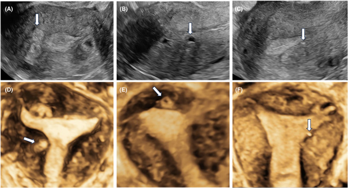
Introduction: Studies that use standardized ultrasonographic criteria to diagnose adenomyosis in subfertile women are needed. These would improve the understanding of the disease burden and enable further studies on its impact on fertility and assisted reproductive treatment (ART) outcome. The aim of this study was to determine the prevalence of different features of adenomyosis in women scheduled for their first ART, diagnosed at two (2D) and three-dimensional (3D) transvaginal ultrasonography (TVUS) using the revised Morphological Uterus Sonographic Assessment (MUSA) group definitions.
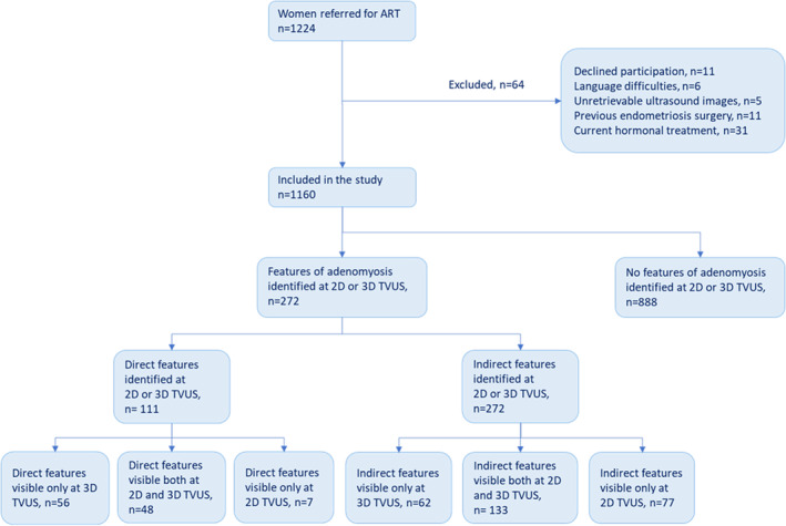
Material and methods: This was a prospective, observational cross-sectional study of subfertile women aged 25 to ≤39 years, that were referred to a university hospital for their first ART between December 2018 and May 2021. Of 1224 eligible women, 1160 women fulfilled the inclusion criteria and consented to participate in the study. All women underwent a systematic 2D and 3D TVUS examination. The primary outcome was the presence of direct and indirect features of adenomyosis, as proposed by the MUSA group. Secondary outcomes were to describe the ultrasonographic characteristics of the different features, as well as any difference in the diagnostics at 2D or 3D TVUS and any association with clinical characteristics such as endometriosis.
Results: At least one direct or indirect feature of adenomyosis was observed in 272 (23.4%, 95% confidence interval [CI] 21.0-25.9) women. Direct features that are pathognomonic for the disease were observed in 111 (9.6%, 95% CI, 7.9-11.3) women. Direct features were visible only at 3D TVUS in 56 (4.8%, 95% CI 3.6-6.1) women, that is, 56/111 (50.5%) of women with at least one direct adenomyosis feature. Direct features were more common in women with endometriosis (OR 2.8, 95% CI 1.8-4.3).
Conclusions: We found than one in 10 women scheduled for ART had direct features of adenomyosis at ultrasound examination. The present study suggests that the use of 3D TVUS is an important complement to 2D in the diagnostics of adenomyosis. Our results may further improve the counseling of women scheduled for ART and enables future studies on the impact of different features of adenomyosis on subfertility, ART results and obstetric outcomes.
Keywords: adenomyosis; assisted reproduction; infertility; ultrasound.
© 2024 The Authors. Acta Obstetricia et Gynecologica Scandinavica published by John Wiley & Sons Ltd on behalf of Nordic Federation of Societies of Obstetrics and Gynecology (NFOG).
Conflict of interest statement
The authors have stated explicitly that there are no conflicts of interest in connection with this article.
Figures

Similar articles
-
Correlation of adenomyosis features to live birth rates after the first IVF/ICSI treatment, when using the revised Morphological Uterus Sonographic Assessment group definitions.Acta Obstet Gynecol Scand. 2024 Dec;103(12):2540-2553. doi: 10.1111/aogs.14986. Epub 2024 Oct 9. Acta Obstet Gynecol Scand. 2024. PMID: 39382305 Free PMC article.
-
Association of 2D and 3D transvaginal ultrasound findings with adenomyosis in symptomatic women of reproductive age: a prospective study.Clinics (Sao Paulo). 2021 Aug 16;76:e2981. doi: 10.6061/clinics/2021/e2981. eCollection 2021. Clinics (Sao Paulo). 2021. PMID: 34406269 Free PMC article.
-
Transvaginal sonographic features of diffuse adenomyosis in 18-30-year-old nulligravid women without endometriosis: association with symptoms.Ultrasound Obstet Gynecol. 2015 Dec;46(6):730-6. doi: 10.1002/uog.14834. Ultrasound Obstet Gynecol. 2015. PMID: 25728241
-
Transvaginal Ultrasound for the Diagnosis of Adenomyosis: Systematic Review and Meta-Analysis.J Minim Invasive Gynecol. 2018 Feb;25(2):257-264. doi: 10.1016/j.jmig.2017.08.653. Epub 2017 Aug 30. J Minim Invasive Gynecol. 2018. PMID: 28864044
-
The First Lugano Workshop on the role of adenomyosis in ART.Reprod Biomed Online. 2025 Jan;50(1):104444. doi: 10.1016/j.rbmo.2024.104444. Epub 2024 Sep 12. Reprod Biomed Online. 2025. PMID: 39672080 Review.
Cited by
-
How Reproducible Are the Ultrasound Features of Adenomyosis Defined by the Revised MUSA Consensus?J Clin Med. 2025 Jan 13;14(2):456. doi: 10.3390/jcm14020456. J Clin Med. 2025. PMID: 39860462 Free PMC article.
-
The impact of adenomyosis on intrauterine insemination success in unexplained infertile women: a retrospective cross-sectional study.BMC Pregnancy Childbirth. 2025 Jun 3;25(1):650. doi: 10.1186/s12884-025-07769-9. BMC Pregnancy Childbirth. 2025. PMID: 40462001 Free PMC article.
-
Correlation of adenomyosis features to live birth rates after the first IVF/ICSI treatment, when using the revised Morphological Uterus Sonographic Assessment group definitions.Acta Obstet Gynecol Scand. 2024 Dec;103(12):2540-2553. doi: 10.1111/aogs.14986. Epub 2024 Oct 9. Acta Obstet Gynecol Scand. 2024. PMID: 39382305 Free PMC article.
References
-
- Bird CC, McElin TW, Manalo‐Estrella P. The elusive adenomyosis of the uterus—revisited. Am J Obstet Gynecol. 1972;112:583‐593. - PubMed
-
- Tomassetti C, Meuleman C, Timmerman D, D'Hooghe T. Adenomyosis and subfertility: evidence of association and causation. Semin Reprod Med. 2013;31:101‐108. - PubMed
-
- Younes G, Tulandi T. Effects of adenomyosis on in vitro fertilization treatment outcomes: a meta‐analysis. Fertil Steril. 2017;108:483‐490.e3. - PubMed
-
- Vercellini P, Consonni D, Dridi D, Bracco B, Frattaruolo MP, Somigliana E. Uterine adenomyosis and in vitro fertilization outcome: a systematic review and meta‐analysis. Hum Reprod. 2014;29:964‐977. - PubMed
Publication types
MeSH terms
Grants and funding
LinkOut - more resources
Full Text Sources
Medical
