

Primary extra hepatic hydatid cyst of the kidney: A case report
- PMID: 38410691
- PMCID: PMC10896061
- DOI: 10.1177/2050313X241233188
Primary extra hepatic hydatid cyst of the kidney: A case report
Abstract
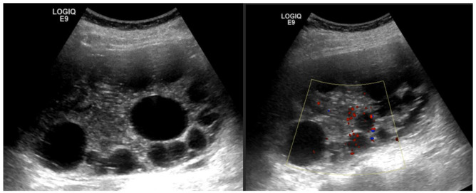
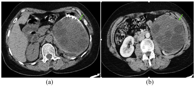
Human echinococcosis (hydatid disease) is highly endemic in Morocco and reemerging as a significant public health problem in this country. It is a cyclozoonotic infection introduced by Echinococcus granulosus, that can invade any organ in the human body. The liver is the mostly affected organ, which is the primary site of human infestation, followed by lungs. Renal involvement by hydatid disease is commonly secondary, occurring owing to primary cyst rupture or disseminated disease. Primary renal localization, often presented as solitary cysts with no visceral involvement, is uncommon even in endemic zone. Herein, a rare cause of renal mass due to an isolated hydatid cyst in a 56-year-old woman who presented with ambiguous left flank pain for the last 10 years. Findings imaging did not reveal any other localizations of hydatid disease. For this scolicidal effect and to prevent peritoneal seeding, the patient was placed on albendazole 400 mg. Three weeks later, the surgery was performed successfully with a left total nephrectomy.
Keywords: Renal mass; imaging; primary hydatid cyst.
© The Author(s) 2024.
Conflict of interest statement
The authors declared no potential conflicts of interest with respect to the research, authorship, and/or publication of this article.
Figures

Similar articles
-
Traumatic rupture of a solitary splenic hydatid cyst: A case report.Trauma Case Rep. 2015 Apr 23;1(1-2):1-3. doi: 10.1016/j.tcr.2015.02.001. eCollection 2015 Feb. Trauma Case Rep. 2015. PMID: 30101166 Free PMC article.
-
Peritoneal hydatidosis secondary to an asymptomatic liver hydatid cyst rupture: A case report.Int J Surg Case Rep. 2024 Oct;123:110220. doi: 10.1016/j.ijscr.2024.110220. Epub 2024 Aug 28. Int J Surg Case Rep. 2024. PMID: 39216290 Free PMC article.
-
Rare isolated primary peritoneal hydatid cysts: A case report from Syria.Qatar Med J. 2017 Apr 21;2016(2):13. doi: 10.5339/qmj.2016.13. eCollection 2016. Qatar Med J. 2017. PMID: 28567367 Free PMC article.
-
Laparoscopic management of renal hydatid cyst.JSLS. 2014 Apr-Jun;18(2):361-6. doi: 10.4293/108680813X13753907291396. JSLS. 2014. PMID: 24960508 Free PMC article. Review.
-
Albendazole and Treatment of Hydatid Cyst: Review of the Literature.Infect Disord Drug Targets. 2019;19(2):101-104. doi: 10.2174/1871526518666180629134511. Infect Disord Drug Targets. 2019. PMID: 29956639 Review.
Cited by
-
Isolated renal hydatid cyst: A rare presentation of echinococcosis.Urol Case Rep. 2025 Jul 16;62:103125. doi: 10.1016/j.eucr.2025.103125. eCollection 2025 Sep. Urol Case Rep. 2025. PMID: 40703091 Free PMC article.
References
-
- El Hadj Sidi C, Mgarrech I, Alimi F, et al.. Four pathways for hydatid pulmonary embolism. J Card Surg 2020; 35(8): 1877–1884. - PubMed
Publication types
LinkOut - more resources
Full Text Sources
