. 2024 Mar 19;98(3):e0198223.
doi: 10.1128/jvi.01982-23. Epub 2024 Feb 27.

NLRP1 restricts porcine deltacoronavirus infection via IL-11 inhibiting the phosphorylation of the ERK signaling pathway

Affiliations

NLRP1 restricts porcine deltacoronavirus infection via IL-11 inhibiting the phosphorylation of the ERK signaling pathway

Haojie He et al. J Virol. .

Abstract

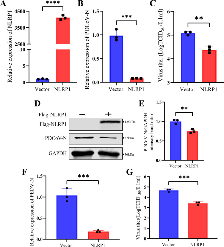
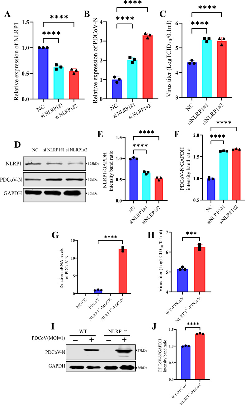
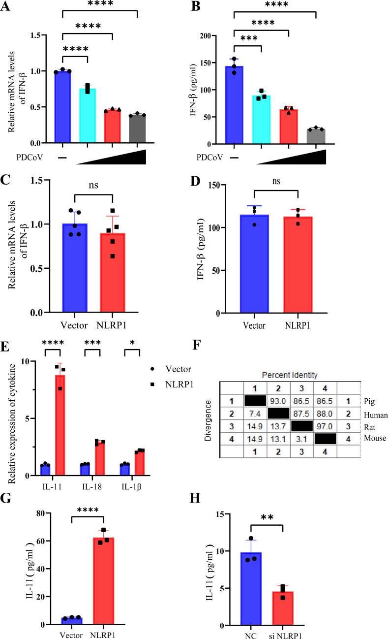
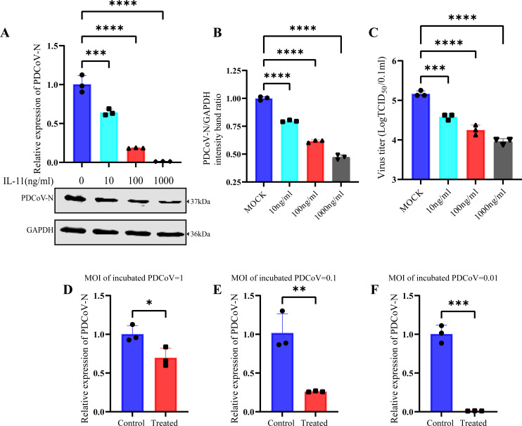
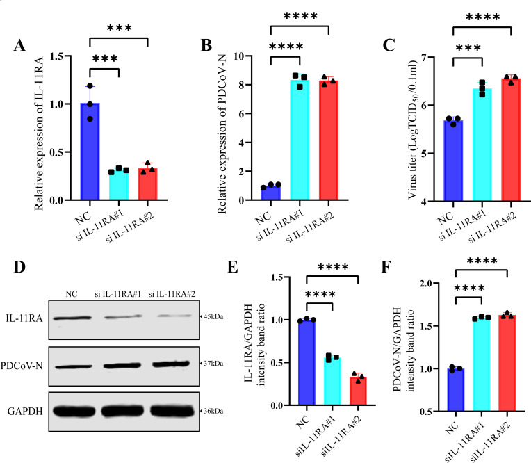
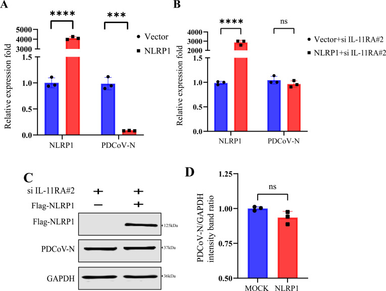
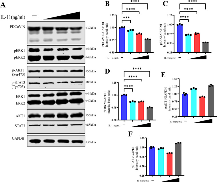
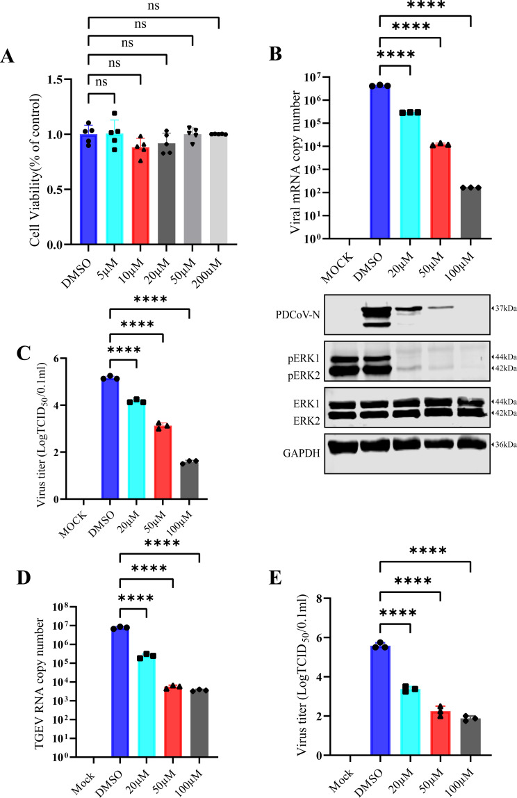
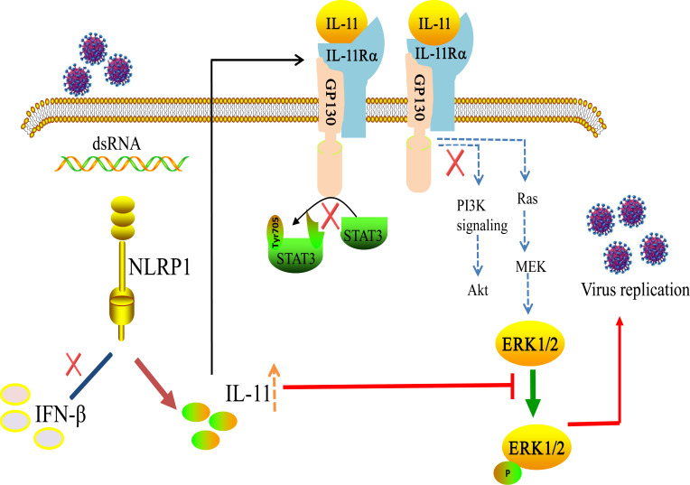
Continuously emerging highly pathogenic coronaviruses remain a major threat to human and animal health. Porcine deltacoronavirus (PDCoV) is a newly emerging enterotropic swine coronavirus that causes large-scale outbreaks of severe diarrhea disease in piglets. Unlike other porcine coronaviruses, PDCoV has a wide range of species tissue tropism, including primary human cells, which poses a significant risk of cross-species transmission. Nucleotide-binding oligomerization domain-like receptor (NLR) family pyrin domain-containing 1 (NLRP1) has a key role in linking host innate immunity to microbes and the regulation of inflammatory pathways. We now report a role for NLRP1 in the control of PDCoV infection. Overexpression of NLRP1 remarkably suppressed PDCoV infection, whereas knockout of NLRP1 led to a significant increase in PDCoV replication. A mechanistic study revealed that NLRP1 suppressed PDCoV replication in cells by upregulating IL-11 expression, which in turn inhibited the phosphorylation of the ERK signaling pathway. Furthermore, the ERK phosphorylation inhibitor U0126 effectively hindered PDCoV replication in pigs. Together, our results demonstrated that NLRP1 exerted an anti-PDCoV effect by IL-11-mediated inhibition of the phosphorylation of the ERK signaling pathway, providing a novel antiviral signal axis of NLRP1-IL-11-ERK. This study expands our understanding of the regulatory network of NLRP1 in the host defense against virus infection and provides a new insight into the treatment of coronaviruses and the development of corresponding drugs.IMPORTANCECoronavirus, which mainly infects gastrointestinal and respiratory epithelial cells in vivo, poses a huge threat to both humans and animals. Although porcine deltacoronavirus (PDCoV) is known to primarily cause fatal diarrhea in piglets, reports detected in plasma samples from Haitian children emphasize the potential risk of animal-to-human spillover. Finding effective therapeutics against coronaviruses is crucial for controlling viral infection. Nucleotide-binding oligomerization-like receptor (NLR) family pyrin domain-containing 1 (NLRP1), a key regulatory factor in the innate immune system, is highly expressed in epithelial cells and associated with the pathogenesis of viruses. We demonstrate here that NLRP1 inhibits the infection of the intestinal coronavirus PDCoV through IL-11-mediated phosphorylation inhibition of the ERK signaling pathway. Furthermore, the ERK phosphorylation inhibitor can control the infection of PDCoV in pigs. Our study emphasizes the importance of NLRP1 as an immune regulatory factor and may open up new avenues for the treatment of coronavirus infection.

Keywords: ERK signaling; anti-coronavirus; interleukin-11; nucleotide-binding oligomerization domain-like receptor family pyrin domain-containing 1; porcine deltacoronavirus.


Conflict of interest statement

The authors declare no conflict of interest.

Figures

Fig 1
Fig 1
pNLRP1 inhibits the replication of PDCoV. IPI-2I cells were transfected with Flag-tagged NLRP1 (0 or 1 µg) for 24 h, followed by infection with PDCoV at an MOI of 0.1 for 24 h. (A) The overexpression of NLRP1 was verified by RT-qPCR. (B) The relative expression of PDCoV was determined by RT-qPCR. (C) The PDCoV TCID50 in the supernatants was titrated on swine testis cells. (D) The protein expression of NLRP1 and PDCoV-N was detected by Western blotting. (E) The relative intensities of PDCoV-N were normalized to glyceraldehyde-3-phosphate dehydrogenase (GAPDH). (F) IPI-2I cells were transfected with Flag-tagged NLRP1 (0 or 1 µg) for 24 h, followed by infection with PEDV at an MOI of 1 for 24 h. The relative expression of PEDV was determined by RT-qPCR. (G) The PEDV TCID50 in the supernatants was titrated on Vero-E6 cells. The means and SD of the results from three independent experiments are shown. **P < 0.01; ***P < 0.001; and ****P < 0.0001.
Fig 2
Fig 2
Knockdown or knockout of NLRP1 enhances PDCoV replication. IPI-2I cells were transfected with siNLRP1#1, siNLRP1#2, or NC at 50 nM for 24 h and subsequently infected with PDCoV at an MOI of 0.1 for 24 h. (A and B) mRNA levels of NLRP1 and PDCoV-N in NLRP1 knockdown cells. (C) The PDCoV TCID50 in the supernatants was titrated on swine testis (ST) cells. (D–F) Expression of NLRP1 and PDCoV-N was detected by Western blotting, and the relative intensities of NLRP1 and PDCoV-N were normalized to that of glyceraldehyde-3-phosphate dehydrogenase (GAPDH). (G and I) WT IPI-2I cells and NLRP1−/− IPI-2I cells were infected with PDCoV at an MOI of 0.1 for 24 h, and then the relative mRNA levels and protein levels of PDCoV-N were detected by RT-qPCR (G) and Western blotting (I). (H) The PDCoV TCID50 in the supernatants of WT or NLRP1−/− IPI-2I cell was titrated on ST cells. (J) The relative intensity of PDCoV-N was normalized to that of GAPDH. The means and SD of the results from three independent experiments are shown. ***P < 0.001 and ****P < 0.0001.
Fig 3
Fig 3
NLRP1 does not rely on the IFN pathway to inhibit PDCoV replication. (A and B) The mRNA levels of IFN-β were detected by RT-qPCR (A) and the protein levels of IFN-β were detected by ELISA (B) in IPI-2I cells either mock infected or infected with PDCoV at MOI of 0.01, 0.1, and 1. (C–E) The mRNA expression of IFN-β (C) and cytokines (E) was measured by RT-qPCR, and the protein expression of IFN-β was measured by ELISA in NLRP1-overexpression cells. (F) Multiple sequence alignment between the pig, human, rat, and mouse. (G and H) Protein levels of IL-11 in the supernatant of NLRP1-overexpression cells (G) or NLRP1-knockdown cells (H) were analyzed by ELISA. The means and SD of the results from three independent experiments are shown. ns, no significant difference. *P < 0.05; **P < 0.01; ***P < 0.001; and ****P < 0.0001.
Fig 4
Fig 4
The treatment of IL-11 recombinant protein inhibits the replication of PDCoV. After incubation with 0.1 MOI of PDCoV for 2 h at 37°C, IPI-2I cells were washed three times with phosphate-buffered saline (PBS) to remove unbound virus and then treated with different concentrations of IL-11 protein (0, 10, 100, or 1,000 ng/mL) for 24 h. (A) The relative viral copy number of PDCoV and the protein expression level of PDCoV-N were measured by RT-qPCR and Western blotting, respectively. (B) The relative intensity of PDCoV-N was normalized to that of glyceraldehyde-3-phosphate dehydrogenase (GAPDH). (C) The PDCoV TCID50 in the supernatants of IPI-2I cells was titrated on swine testis cells. (D–F) The relative viral RNA copy number of PDCoV in IL-11-treated cells. After incubation with 0.01 (F), 0.1 (E), or 1 (D) MOI of PDCoV for 2 h at 37°C, IPI-2I cells were washed three times with PBS to remove unbound virus and then treated with 100 ng/mL of IL-11 protein for 24 h. The means and SD of the results from three independent experiments are shown. ns, no significant difference. *P < 0.05; **P < 0.01; ***P < 0.001; and ****P < 0.0001.
Fig 5
Fig 5
Knockdown of IL-11RA enhances PDCoV infection. IPI-2I cells were transfected with IL-11RA#1, IL-11RA#2, or NC at 50 nM for 24 h and subsequently infected with PDCoV at an MOI of 0.1 for 24 h. (A and B) mRNA levels of IL-11RA and PDCoV-N in IL-11RA knockdown cells. (C) The PDCoV TCID50 in the supernatants was titrated on swine testis cells. (D–F) Expression of IL-11RA and PDCoV-N was detected by Western blotting, and the relative intensities of NLRP1 and PDCoV-N were normalized to that of glyceraldehyde-3-phosphate dehydrogenase (GAPDH). The means and SD of the results from three independent experiments are shown. ***P < 0.001 and ****P < 0.0001.
Fig 6
Fig 6
NLRP1 relies on the IL-11 pathway to inhibit PDCoV replication. After transfection with 50 nM of siIL-11RA#2 or NC for 12 h, IPI-2I cells were transfected with 1 µg of Flag-tagged NLRP1 for 24 h and then infected with PDCoV at an MOI of 0.1 for 24 h. (A and B) The relative expression of NLRP1 and PDCoV-N in NC (A) or IL-11-knockdown cells (B). (C) The protein expression of PDCoV-N was detected by Western blotting. (D) The relative intensity of PDCoV-N was normalized to that of glyceraldehyde-3-phosphate dehydrogenase (GAPDH). The means and SD of the results from three independent experiments are shown. ns, no significant difference. ***P < 0.001 and ****P < 0.0001.
Fig 7
Fig 7
The treatment of IL-11 inhibits the phosphorylation of the ERK signaling pathway. After infection with PDCoV at an MOI of 1, IPI-2I cells were treated with different doses (0, 10, 100, and 500) μg/mL of recombinant IL-11 protein. (A) The total proteins and phosphorylation proteins’ expression of ERK1, ERK2, AKT1, and STAT3 were measured by Western blotting. (B–F) The relative intensities of PDCoV-N (B), pERK1 (C), pERK2 (D), pAKT1 (E), and pSTAT3 (F) were normalized to that of glyceraldehyde-3-phosphate dehydrogenase (GAPDH). The means and SD of the results from three independent experiments are shown. ***P < 0.001 and ****P < 0.0001.
Fig 8
Fig 8
The phosphorylation inhibitor of ERK1/2 (U0126) significantly inhibits the replication of PDCoV in cells. (A) Cell viability of IPI-2I treated with different concentrations (0, 5, 10, 20, 50, and 200 µM) of U0126. (B) The viral copy number and the expression of PDCoV-N were tested by RT-qPCR and Western blotting in U0126-treated cells. (C) The PDCoV TCID50 in the supernatants was titrated on swine testis (ST) cells. (D) IPI-2I cells were treated with different concentrations (0, 20, 50, and 100 µM) of U0126, followed by infection with TGEV at an MOI of 0.1 for 24 h. The viral copy number of TGEV was determined by RT-qPCR. (E) The TGEV TCID50 in the supernatants was titrated on ST cells. The means and SD of the results from three independent experiments are shown. ns, no significant difference. ***P < 0.001 and ****P < 0.0001.
Fig 9
Fig 9
U0126 suppresses PDCoV replication in piglets. SPF pigs were infected with PDCoV (n = 3) or infected with PDCoV and treated with U0126 (15 mg kg−1, n = 3), or mock infected with Dulbecco’s modified Eagle’s medium (n = 3). (A) Daily virus shedding in feces from the different groups was measured by RT-qPCR detection of viral genomes in fecal swabs. (B) Gross lesions in piglets inoculated with the PDCoV group or U0126-treated group. The intestinal lesions were examined on the day of death or after euthanasia at the final time points. The necropsy images show transparent intestines observed in piglets inoculated with the PDCoV group but not in mock-inoculated piglets. (C) Histopathological examination of the jejunum from the PDCoV-infected piglets. Jejunum was taken from each group and then processed for hematoxylin and eosin staining. Representative images are shown. (D) PDCoV viral RNA in the jejunum was measured by RT-qPCR. (E) Immunohistochemistry of jejunum from the piglets. The black arrows point to the virus in the tissue.
Fig 10
Fig 10
The mechanism of NLRP1 inhibits PDCoV infection. NLRP1 suppressed the PDCoV replication by promoting the secretion of IL-11 to inhibit the phosphorylation of the ERK signaling pathway.

Similar articles

References

    1. Dong BQ, Liu W, Fan XH, Vijaykrishna D, Tang XC, Gao F, Li LF, Li GJ, Zhang JX, Yang LQ, Poon LLM, Zhang SY, Peiris JSM, Smith GJD, Chen H, Guan Y. 2007. Detection of a novel and highly divergent coronavirus from Asian leopard cats and Chinese ferret badgers in Southern China. J Virol 81:6920–6926. doi:10.1128/JVI.00299-07 - DOI - PMC - PubMed
    1. Woo PCY, Lau SKP, Lam CSF, Lai KKY, Huang Y, Lee P, Luk GSM, Dyrting KC, Chan K-H, Yuen K-Y. 2009. Comparative analysis of complete genome sequences of three avian coronaviruses reveals a novel group 3c coronavirus. J Virol 83:908–917. doi:10.1128/JVI.01977-08 - DOI - PMC - PubMed
    1. Woo PCY, Lau SKP, Lam CSF, Lau CCY, Tsang AKL, Lau JHN, Bai R, Teng JLL, Tsang CCC, Wang M, Zheng B-J, Chan K-H, Yuen K-Y. 2012. Discovery of seven novel mammalian and avian coronaviruses in the genus deltacoronavirus supports bat coronaviruses as the gene source of alphacoronavirus and betacoronavirus and avian coronaviruses as the gene source of gammacoronavirus and deltacoronavirus. J Virol 86:3995–4008. doi:10.1128/JVI.06540-11 - DOI - PMC - PubMed
    1. Wang L, Byrum B, Zhang Y. 2014. Detection and genetic characterization of deltacoronavirus in pigs, Ohio, USA, 2014. Emerg Infect Dis 20:1227–1230. doi:10.3201/eid2007.140296 - DOI - PMC - PubMed
    1. Ma Y, Zhang Y, Liang X, Lou F, Oglesbee M, Krakowka S, Li J. 2015. Origin, evolution, and virulence of porcine deltacoronaviruses in the United States. mBio 6:e00064. doi:10.1128/mBio.00064-15 - DOI - PMC - PubMed

Supplementary concepts

LinkOut - more resources