

Patterns of subregional cerebellar atrophy across epilepsy syndromes: An ENIGMA-Epilepsy study
- PMID: 38411286
- PMCID: PMC11120093
- DOI: 10.1111/epi.17881
Patterns of subregional cerebellar atrophy across epilepsy syndromes: An ENIGMA-Epilepsy study
Erratum in
-
Correction to "patterns of subregional cerebellar atrophy across epilepsy syndromes: An ENIGMA-Epilepsy study".Epilepsia. 2024 Aug;65(8):2501. doi: 10.1111/epi.18053. Epub 2024 Jun 22. Epilepsia. 2024. PMID: 39119782 No abstract available.
Abstract
Objective: The intricate neuroanatomical structure of the cerebellum is of longstanding interest in epilepsy, but has been poorly characterized within the current corticocentric models of this disease. We quantified cross-sectional regional cerebellar lobule volumes using structural magnetic resonance imaging in 1602 adults with epilepsy and 1022 healthy controls across 22 sites from the global ENIGMA-Epilepsy working group.
Methods: A state-of-the-art deep learning-based approach was employed that parcellates the cerebellum into 28 neuroanatomical subregions. Linear mixed models compared total and regional cerebellar volume in (1) all epilepsies, (2) temporal lobe epilepsy with hippocampal sclerosis (TLE-HS), (3) nonlesional temporal lobe epilepsy, (4) genetic generalized epilepsy, and (5) extratemporal focal epilepsy (ETLE). Relationships were examined for cerebellar volume versus age at seizure onset, duration of epilepsy, phenytoin treatment, and cerebral cortical thickness.
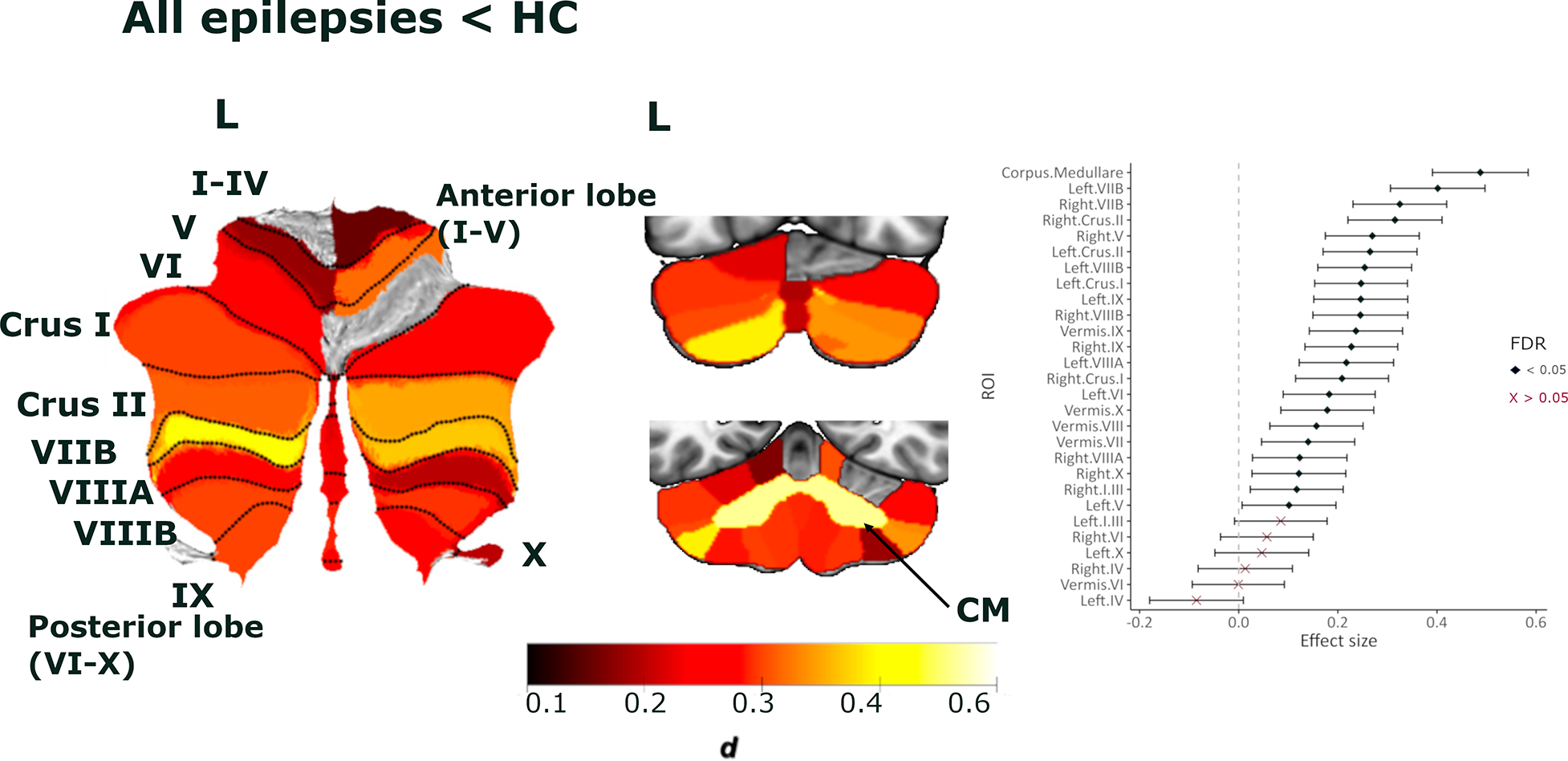
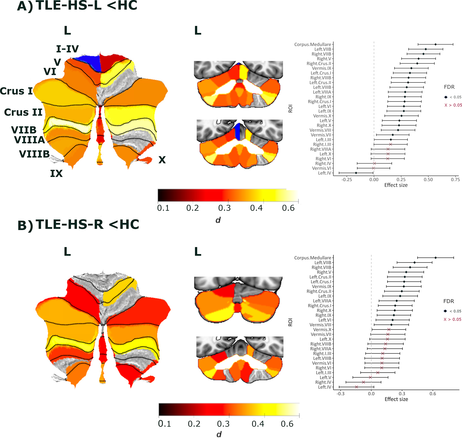
Results: Across all epilepsies, reduced total cerebellar volume was observed (d = .42). Maximum volume loss was observed in the corpus medullare (dmax = .49) and posterior lobe gray matter regions, including bilateral lobules VIIB (dmax = .47), crus I/II (dmax = .39), VIIIA (dmax = .45), and VIIIB (dmax = .40). Earlier age at seizure onset ( = .05) and longer epilepsy duration ( = .06) correlated with reduced volume in these regions. Findings were most pronounced in TLE-HS and ETLE, with distinct neuroanatomical profiles observed in the posterior lobe. Phenytoin treatment was associated with reduced posterior lobe volume. Cerebellum volume correlated with cerebral cortical thinning more strongly in the epilepsy cohort than in controls.
Significance: We provide robust evidence of deep cerebellar and posterior lobe subregional gray matter volume loss in patients with chronic epilepsy. Volume loss was maximal for posterior subregions implicated in nonmotor functions, relative to motor regions of both the anterior and posterior lobe. Associations between cerebral and cerebellar changes, and variability of neuroanatomical profiles across epilepsy syndromes argue for more precise incorporation of cerebellar subregional damage into neurobiological models of epilepsy.
Keywords: MRI; anterior lobe; cerebellum; epilepsy; posterior lobe.
© 2024 The Authors. Epilepsia published by Wiley Periodicals LLC on behalf of International League Against Epilepsy.
Conflict of interest statement
CONFLICT OF INTEREST STATEMENT
L.Vivash. reports research funding from Biogen Australia, Life Molecular Imaging and Eisai.
T.J. O’Brien has received consulting fees from Eisai, UCB, Supernus, Biogen, ES Therapeutics, Epidarex, LivaNova, Kinoxis Therapeutics. He participates on the Data Safety Monitoring Board for ES Therapeutics, Kinoxis Therapeutics. He has served as President (past) for Epilepsy Society of Australia, and is the current chair for Australian Epilepsy Clinical Trials Network (AECTN) and the American Epilepsy Society (Translational Research Committee).
B. Bender is the cofounder of AIRAmed GmbH, a company that offers brain segmentation.
P. Martin. has received honorary as an advisory board member from Biogen unrelated to the submitted work.
P. Striano received speaker fees and advisory boards for Biomarin, Zogenyx, GW Pharmaceuticals; research funding by ENECTA BV, GW Pharmaceuticals, Kolfarma srl., Eisai.
P.M. Thompson received a research grant from Biogen, Inc., and was a paid consultant for Kairos Venture Capital, Inc., USA, for projects unrelated to this work.
C.L. Yasuda has received personal payments from Torrent, Zodiac and UCB.
S.M Sisodiya has received research grants from UCB Pharma and Jazz Pharmaceuticals, speakers fees from UCB, Eisai and Zogenix; honoraria or other fees from Eisai, Jazz Pharma, Stoke Therapeutics, UCB and Zogenix. (payments to institution)
The remaining authors have no conflicts of interest.
Figures


Update of
-
Patterns of subregional cerebellar atrophy across epilepsy syndromes: An ENIGMA-Epilepsy study.bioRxiv [Preprint]. 2023 Oct 23:2023.10.21.562994. doi: 10.1101/2023.10.21.562994. bioRxiv. 2023. Update in: Epilepsia. 2024 Apr;65(4):1072-1091. doi: 10.1111/epi.17881. PMID: 37961570 Free PMC article. Updated. Preprint.
References
MeSH terms
Substances
Grants and funding
- R01 AG058854/AG/NIA NIH HHS/United States
- P41 EB015922/EB/NIBIB NIH HHS/United States
- HCRW_/HCRW_/United Kingdom
- RF1 NS033310/GF/NIH HHS/United States
- R01 NS127524/NS/NINDS NIH HHS/United States
- R01 NS122827/NS/NINDS NIH HHS/United States
- R01 MH116147/MH/NIMH NIH HHS/United States
- R01 NS106957/NS/NINDS NIH HHS/United States
- P41EB015922/GF/NIH HHS/United States
- R01 NS120976/NS/NINDS NIH HHS/United States
- R01 NS106957/GF/NIH HHS/United States
- MR/M00841X/1/MRC_/Medical Research Council/United Kingdom
- R01 NS033310/NS/NINDS NIH HHS/United States
- R01 NS1127524/GF/NIH HHS/United States
- R01AG058854/GF/NIH HHS/United States
- R01MH116147/GF/NIH HHS/United States
- MR/S00355X/1/MRC_/Medical Research Council/United Kingdom
- R01 NS124585/NS/NINDS NIH HHS/United States
- G0802012/MRC_/Medical Research Council/United Kingdom
- 180365/SNSF_/Swiss National Science Foundation/Switzerland
