. 2024 Feb 27;15(1):57.
doi: 10.1186/s13244-024-01625-8.

T2WI-based MRI radiomics for the prediction of preoperative extranodal extension and prognosis in resectable rectal cancer

Affiliations

T2WI-based MRI radiomics for the prediction of preoperative extranodal extension and prognosis in resectable rectal cancer

Hang Li et al. Insights Imaging. .

Abstract

Objective: To investigate whether T2-weighted imaging (T2WI)-based intratumoral and peritumoral radiomics can predict extranodal extension (ENE) and prognosis in patients with resectable rectal cancer.

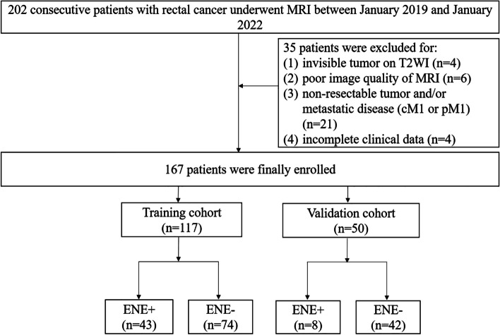
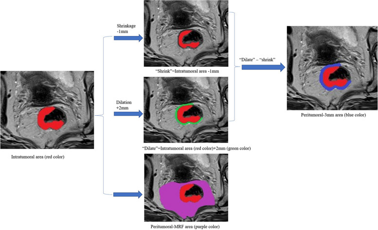
Methods: One hundred sixty-seven patients with resectable rectal cancer including T3T4N + cases were prospectively included. Radiomics features were extracted from intratumoral, peritumoral 3 mm, and peritumoral-mesorectal fat on T2WI images. Least absolute shrinkage and selection operator regression were used for feature selection. A radiomics signature score (Radscore) was built with logistic regression analysis. The area under the receiver operating characteristic curve (AUC) was used to evaluate the performance of each Radscore. A clinical-radiomics nomogram was constructed by the most predictive radiomics signature and clinical risk factors. A prognostic model was constructed by Cox regression analysis to identify 3-year recurrence-free survival (RFS).

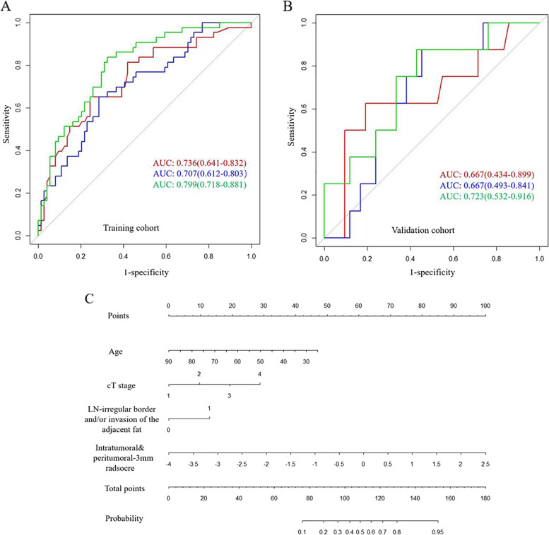
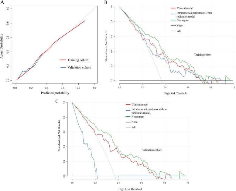
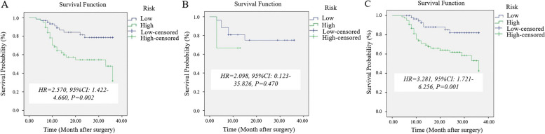
Results: Age, cT stage, and lymph node-irregular border and/or adjacent fat invasion were identified as independent clinical risk factors to construct a clinical model. The nomogram incorporating intratumoral and peritumoral 3 mm Radscore and independent clinical risk factors achieved a better AUC than the clinical model in the training (0.799 vs. 0.736) and validation cohorts (0.723 vs. 0.667). Nomogram-based ENE (hazard ratio [HR] = 2.625, 95% CI = 1.233-5.586, p = 0.012) and extramural vascular invasion (EMVI) (HR = 2.523, 95% CI = 1.247-5.106, p = 0.010) were independent risk factors for predicting 3-year RFS. The prognostic model constructed by these two indicators showed good performance for predicting 3-year RFS in the training (AUC = 0.761) and validation cohorts (AUC = 0.710).

Conclusion: The nomogram incorporating intratumoral and peritumoral 3 mm Radscore and clinical risk factors could predict preoperative ENE. Combining nomogram-based ENE and MRI-reported EMVI may be useful in predicting 3-year RFS.

Critical relevance statement: A clinical-radiomics nomogram could help preoperative predict ENE, and a prognostic model constructed by the nomogram-based ENE and MRI-reported EMVI could predict 3-year RFS in patients with resectable rectal cancer.

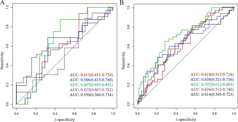
Key points: • Intratumoral and peritumoral 3 mm Radscore showed the most capability for predicting ENE. • Clinical-radiomics nomogram achieved the best predictive performance for predicting ENE. • Combining clinical-radiomics based-ENE and EMVI showed good performance for 3-year RFS.

Keywords: Lymph node; Magnetic resonance imaging; Nomograms; Rectal Neoplasms.


Conflict of interest statement

XZ is an employee of GE Healthcare. The remaining authors declare that they have no competing interests.

Figures

Fig. 1
Fig. 1
Flowchart of patient selection
Fig. 2
Fig. 2
Tumor segmentation process on MRI. First, we manually segmented the whole tumor on axial T2WI images and labeled it as the intratumoral area. Second, “shrink” was defined as the tumor border automatically shrinking by 1 mm on the inside. “Dilate” was defined as automated dilation of the tumor border by 2 mm on the outside. “Dilate-shrink” resulted in a ring with a thickness of 3 mm. Thus, the peritumoral 3 mm area was obtained, including the most peripheral portion of the tumor and the surrounding tissues. Moreover, the peritumoral-mesorectal fat (MRF) area was obtained by drawing along the mesorectal fascia
Fig. 3
Fig. 3
Receiver operating characteristic curves of intratumoral radiomics score (red line), intratumoral&peritumoral-MRF radiomics score (blue line), intratumoral and peritumoral-3-mm radiomics score (green line), peritumoral-3mm radiomics score (purple line), and peritumoral-MRF radiomics score (black line) for predicting extranodal extension in the training cohort (A) and validation cohort (B)
Fig. 4
Fig. 4
The performance and validation of the final selected model to predict extranodal extension (ENE). ROC of clinical model (red line), intratumoral and peritumoral-3-mm radiomics model (blue line), and nomogram (green line)  for predicting ENE in the training cohort (A) and validation cohort (B). C The predictive nomogram of ENE
Fig. 5
Fig. 5
Fit and usefulness evaluation of the clinical-radiomics nomogram. Calibration curve of the clinical-radiomics nomogram for predicting extranodal extension (ENE) in the training cohort (red line) and validation cohort (blue line) (A); decision curve analysis (DCA) of the nomogram for assessing its clinical usefulness; this indicates that a nomogram to predict ENE gains more benefit than the “treat all,” “treat none,” radiomics model and the clinical model when the threshold probability ranges from 0.18 to 0.73 in the training cohort (B) and from 0.10 to 0.74 in the validation cohort (C)
Fig. 6
Fig. 6
Kaplan–Meier survival curves of the nomogram-based extranodal extension (ENE) for 3-year recurrence-free survival in patients with rectal cancer in the entire cohort (A), at T1-T2 stage (B), and T3a/b-T4a stage (C)

Similar articles

Cited by

References

    1. Sung H, Ferlay J, Siegel RL, et al. Global Cancer Statistics 2020: GLOBOCAN estimates of incidence and mortality worldwide for 36 Cancers in 185 countries. CA Cancer J Clin. 2021;71:209–249. doi: 10.3322/caac.21660. - DOI - PubMed
    1. Siegel RL, Miller KD, Goding Sauer A, et al. Colorectal cancer statistics. CA Cancer J Clin. 2020;70:145–164. doi: 10.3322/caac.21601. - DOI - PubMed
    1. Bhattacharya P, Mukherjee R. Lymph node extracapsular extension as a marker of aggressive phenotype: classification, prognosis and associated molecular biomarkers. Eur J Surg Oncol. 2021;47:721–731. doi: 10.1016/j.ejso.2020.09.005. - DOI - PubMed
    1. Wind J, Lagarde SM, Ten Kate FJ, et al. A systematic review on the significance of extracapsular lymph node involvement in gastrointestinal malignancies. Eur J Surg Oncol. 2007;33:401–408. doi: 10.1016/j.ejso.2006.11.001. - DOI - PubMed
    1. Alakus H, Hölscher AH, Grass G, et al. Extracapsular lymph node spread: a new prognostic factor in gastric cancer. Cancer. 2010;116:309–315. doi: 10.1002/cncr.24764. - DOI - PubMed

LinkOut - more resources