Review
. 2024 Feb 27;15(1):61.
doi: 10.1186/s13244-024-01607-w.

Rotator cuff tear patterns: MRI appearance and its surgical relevance

Affiliations
Review

Rotator cuff tear patterns: MRI appearance and its surgical relevance

Alexeys Perez Yubran et al. Insights Imaging. .

Abstract

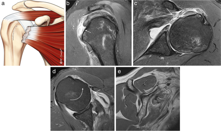
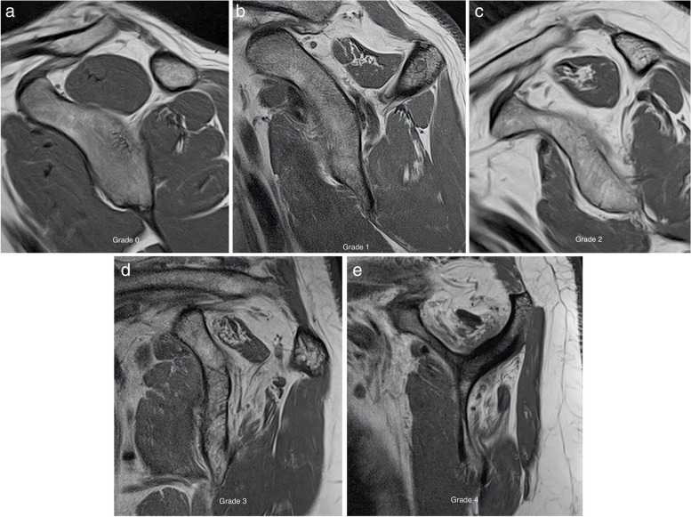
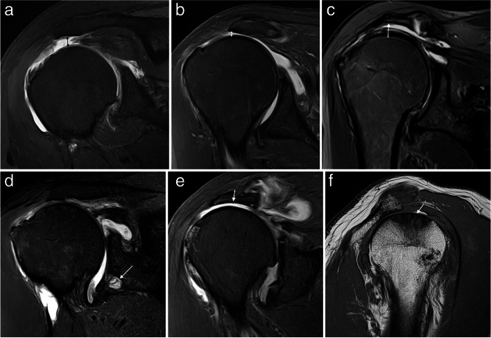
A new perspective on rotator cuff anatomy has allowed a better understanding of the patterns of the different rotator cuff tears. It is essential for radiologists to be aware of these different patterns of tears and to understand how they might influence treatment and surgical approach. Our objective is to review the arthroscopy correlated magnetic resonance imaging appearance of the different types of rotator cuff tears based on current anatomical concepts.Critical relevance statement Knowledge of the characteristics of rotator cuff tears improves our communication with the surgeon and can also make it easier for the radiologist to prepare a report that guides therapeutic conduct and serves as a prognosis for the patient.Key points• There is no universally accepted classification for RC tears.• New patterns such as delamination or myotendinous junction tears have been defined.• The most difficult feature to assess in full thickness tears on MRI is the pattern.• Fatty infiltration of the RC tendons is crucial in the prognosis and outcome.• The radiological report is an effective way of communication with the surgeon.

Keywords: Delaminated; Massive tears; Outcome; Rotator cuff; Surgery.


Conflict of interest statement

The authors declare that they have no competing interests.

Figures

Fig. 1
Fig. 1
Supraspinatus components. a Drawing showing the bellies and tendons of supraspinatus. b, c Axial fat-suppressed PD-weighted MRI showing the anterior belly with its long tendon and insertion (arrows) and the posterior belly with a broader tendon (dashed arrows)
Fig. 2
Fig. 2
Normal supraspinatus and infraspinatus insertions. The SS inserts on a small triangular area in the humeral head, extending from the bicipital groove anteromedially to the top of the greater tuberosity. The IS footprint instead has a broader insertion and covers the posterior border of the SS superiorly and inserts onto the anterolateral area and the middle facet of the greater tuberosity
Fig. 3
Fig. 3
Normal supraspinatus and infraspinatus ultrastructure. a Drawing shows the different layers of RC. b, c Oblique-sagittal PD-weighted MRI arthrography. Layer 1, superficial fibers of the coracohumeral ligament; layer 2 are dense parallel collagen fibers from SS and IS tendons or bursal fibers and layer 3 are smaller collagen fibers of less uniform orientation or articular fibers. These are very difficult to differentiate even with high field MRI. Layer 4 is the rotator cable and layer 5, superior capsule
Fig. 4
Fig. 4
Normal rotator cable. a Drawing showing the cable and crescent. b Axial fat-suppressed PD-weighted MRI reveals the normal rotator cable (arrow) and the crescent (c) lateral to the cable. b Arthroscopic correlation view from the posterior portal showing the crescent and cable
Fig. 5
Fig. 5
Normal subscapularis. a Oblique coronal fat-suppressed PD-weighted MRI showing the comma shape of the SSC insertion (white outline). b Oblique sagittal fat-suppressed PD-weighted FS MRI demonstrate the normal appearance of the tendon (arrows)
Fig. 6
Fig. 6
Partial articular side tear of the supraspinatus tendon. a Drawing showing the disruption of the articular layer of the SS. b Oblique coronal fat-suppressed PD-weighted MRI arthrography demonstrates a discontinuity and irregularity of the articular side of the tendon (arrow) affecting more than 50% of tendon thickness. c Arthroscopic view from the posterior portal confirming the tear with fraying of the articular side of the tendon (black arrow). b biceps tendon
Fig. 7
Fig. 7
Partial articular-sided tear of the supraspinatus tendon (two different patients). a ABER fat-suppressed PD-weighted arthrographic MRI demonstrate the partial articular tear (arrow). b ABER T1-weighted MRI arthrography reveals an articular tear with horizontal intratendinous extension. Note how the contrast is filling the tear (arrow)
Fig. 8
Fig. 8
Partial bursal side tear of the supraspinatus tendon. a Illustration shows where the bursal side tears are located. b Oblique coronal fat-suppressed PD-weighted MRI reveals a discontinuity of the SS bursal side (arrow) greater than 50% of the tendon thickness. c Correlation with its arthroscopic view from a lateral portal where the tear is confirmed (black arrow)
Fig. 9
Fig. 9
Partial intrasubstance tear of the supraspinatus tendon. a Illustration of the morphology of intrasubstance tears. b Oblique coronal fat-suppressed PD-weighted arthrographic MRI showing a tear contained within the thickening of the tendon (arrow)
Fig. 10
Fig. 10
Full-thickness tear of the supraspinatus tendon. a Illustration of a full thickness tendon tear. b Oblique coronal fat-suppressed PD-weighted MRI showing a SS tendon tear that communicates the articular side (white arrow) and bursal side (black arrow). c Arthroscopic view from the lateral portal showing a complete tear and the communication between the articular and extra-articular spaces
Fig. 11
Fig. 11
Full thickness tear patterns. ac Illustrations of the different full thickness tear patterns: crescent-shaped, U-shaped, and L-shaped. df Axial fat-suppressed PD-weighted MRI arthrography showing the different patterns (arrows)
Fig. 12
Fig. 12
Full thickness tear extension. a Oblique sagittal fat-suppressed PD-weighted MR arthrography showing a C1 tear consisting in a small full thickness tear or pinhole (arrow). b Oblique coronal fat-suppressed PD-weighted MR demonstrates a C2 tear with a small retraction, less than 2 cm (double-headed arrow). c Oblique coronal and sagittal shows a C3 tear which is a large full thickness tear with a retraction usually between 3 and 4 cm (double-headed arrow) affecting only one tendon. d Oblique sagittal fat-suppressed PD-weighted MRI showing a C4 tear consisting in a complete tear of two or more tendons. In this case, there is a tear of both supraspinatus and infraspinatus (arrows). e Arthroscopic view of a C1 tear from the posterior portal (arrow). fh Arthroscopic views of C2, C3, and C4 tears from a lateral portal
Fig. 13
Fig. 13
Supraspinatus full thickness tears different retraction stages. a, b Oblique coronal fat-suppressed PD-weighted MRI arthrography showing a complete full thickness tear with a small retraction (arrow in a) and a full thickness tear with fibers retracted over the humeral head vertex (arrow in b). c Oblique coronal fat-suppressed PD-weighted MRI shows tendon fibers retracted over the superior glenoid margin (arrow)
Fig. 14
Fig. 14
Delaminated tear classification. Illustrations showing the different types of delaminated tears. a Complete tear with articular layer more medially retracted than the superior layer. b Complete tear with bursal layer more medially retracted than the articular layer. c Complete tear with both layers equally retracted. d Partial thickness articular side tear with horizontal splitting extension. e Partial thickness bursal side tear with intratendinous splitting tear. f Intrasubstance tear with intratendinous extension
Fig. 15
Fig. 15
Delaminated tears of supraspinatus tendon. af Oblique coronal fat-suppressed PD-weighted MRI images. Full thickness delaminated tears. Articular layer more retracted than bursal layer (arrow in a). Bursal layer more retracted than articular layer (arrow in b) and bursal and articular layers equally retracted (arrow in c). Partial thickness delaminated tears. Articular side tear with intratendinous extension and retraction (arrow in d). Bursal side tear (arrow in e) with intratendinous extension (dashed arrow). Intrasubstance tear with horizontal intratendinous extension (arrow in f)
Fig. 16
Fig. 16
Full thickness delaminated tear of the supraspinatus tendon with articular layer flipped medially into the articular space. Oblique coronal fat-suppressed PD-weighted MRI reveals a complete SS tendon tear (dashed arrow) and note how the articular layer is flipped downwards into the articular space (arrow)
Fig. 17
Fig. 17
Flipped supraspinatus full-thickness tear “Fosbury flop tear”. a Draw illustrating the flipped SS tendon. b, c Oblique coronal fat-suppressed PD-weighted MRI demonstrate a full thickness tear of supraspinatus (arrow in b) with tendon stump flipped upon itself (arrow in c)
Fig. 18
Fig. 18
Supraspinatus myotendinous junction tears. a, b Axial fat-suppressed PD-weighted MRI. a Shows edema of the anterior muscle belly of the SS and a partial tear of the myotendinous junction (arrow). b Reveals a full thickness retracted tear of the myotendinous junction of the anterior belly of the SS (arrow)
Fig. 19
Fig. 19
Anterior tears (subscapularis). ac Illustrations of the different types (1 to 3) of the SSC tendon tears. df Oblique sagittal fat-suppressed PD-weighted MRI arthrography showing a partial tear of the upper third of the SSC tendon (arrow in d), a complete tear of the upper third of the tendon (arrow in e) and a complete tear of the upper and middle portions of the tendon (arrow in f) and intact lower third (dashed arrow)
Fig. 20
Fig. 20
Anterior tears (subscapularis). a Illustration of type 4 and 5 tears of the SSC. bc Axial and oblique sagittal fat-suppressed PD-weighted MRI showing a subscapularis full thickness tear (arrow) with retraction of the tendon (dashed arrow), note the humeral head is centered on the glenoid. de Axial fat-suppressed PD-weighted and oblique sagittal PD-weighted MRI images showing a type 5 tear (arrow) with anterior subluxation of the humeral head (curved arrow) and fatty infiltration Goutallier type 3
Fig. 21
Fig. 21
Traumatic subscapularis tears. a, b Axial fat-suppressed PD-weighted MRI images showing an acute SSC full thickness tear with retraction of the tendon (arrow in a) and an acute bony avulsion of the insertion of SSC tendon in the lesser tuberosity (arrow)
Fig. 22
Fig. 22
Bridging sign. Oblique coronal fat-suppressed PD-weighted MRI showing the torn cranial portion of the SSC tendon with superomedial displacement (arrow) due to retraction of the coracohumeral and superior glenohumeral ligament complex (asterisk)
Fig. 23
Fig. 23
Fatty infiltration. ae Oblique sagittal T1-weighted MRI showing the different grades of fatty atrophy following Goutallier modify classification for SS and IS tendons. Grade 0 = normal muscle, grade 1 = some fatty streaks, grade 2 = less than 50% fatty muscle atrophy (more muscle than fat), grade 3 = 50% fatty muscle atrophy, (equal muscle and fat), and grade 4 = more than 50% muscle atrophy (more fat than muscle)
Fig. 24
Fig. 24
Hamada rotator cuff arthropathy evolutive stages. ac Oblique coronal fat-suppressed PD-weighted MRI. Stage 1 showing an acromiohumeral interval (AHI) > 5 mm (double-headed arrow in a). Stage 2 the AHI is < 5 mm (double-headed arrow in b). Stage 3 is same as stage 2 but with the acetabularization of the acromion undersurface (arrow in c). de Oblique coronal fat-suppressed PD-weighted MRI. Stage 4a is similar to stage 2 but with degenerative changes in the glenohumeral joint (arrow in d). Stage 4B adds acromion acetabularization (arrow in e). f Sagittal oblique T1 MRI. Stage 5, there are osteonecrotic changes in the humeral head vertex (arrow in f)

References

    1. Kuhn JE, Dunn WR, Ma B, et al. Interobserver agreement in the classification of rotator cuff tears. Am J Sports Med. 2007;35:437–441. doi: 10.1177/0363546506298108. - DOI - PubMed
    1. Calvo E, Rebollon GC, Morcillo D, Arce G. Rotator Cuff Across the Life Span ISAKOS Consensus Book. New York: Springer; 2019. Diagnosis and Classification of Rotator Cuff Tears.
    1. Oñate M, Bureau NJ (2019) Supraspinatus myotendinous junction injuries: MRI Findings and prevalence. AJR Am J Roentgenol 212(1):W1–W9 - PubMed
    1. Choo HJ, Lee SJ, Kim JH, et al. Delaminated tears of the rotator cuff: prevalence, characteristics, and diagnostic accuracy using indirect MR arthrography. AJR Am J Roentgenol. 2015;204:360–366. doi: 10.2214/AJR.14.12555. - DOI - PubMed
    1. Davidson J, Burkhart SS. The geometric classification of rotator cuff tears: a system linking tear pattern to treatment and prognosis. Arthroscopy. 2010;26:417–424. doi: 10.1016/j.arthro.2009.07.009. - DOI - PubMed

LinkOut - more resources