Review
. 2024 Feb 27;15(1):66.
doi: 10.1186/s13244-024-01622-x.

Liver imaging and pregnancy: what to expect when your patient is expecting

Affiliations
Review

Liver imaging and pregnancy: what to expect when your patient is expecting

Giorgia Porrello et al. Insights Imaging. .

Abstract

Liver diseases in pregnancy can be specific to gestation or only coincidental. In the latter case, the diagnosis can be difficult. Rapid diagnosis of maternal-fetal emergencies and situations requiring specialized interventions are crucial to preserve the maternal liver and guarantee materno-fetal survival. While detailed questioning of the patient and a clinical examination are highly important, imaging is often essential to reach a diagnosis of these liver diseases and lesions. Three groups of liver diseases may be observed during pregnancy: (1) diseases related to pregnancy: intrahepatic cholestasis of pregnancy, pre-eclampsia, eclampsia, hemolysis, elevated liver enzymes and low platelets (HELLP) syndrome, and acute fatty liver of pregnancy; (2) liver diseases that are more frequent during or exacerbated by pregnancy: acute herpes simplex hepatitis, Budd-Chiari syndrome, hemorrhagic hereditary telangiectasia, hepatocellular adenoma, portal vein thrombosis, and cholelithiasis; (3) coincidental conditions, including acute hepatitis, incidental focal liver lesions, metabolic dysfunction-associated steatotic liver disease, cirrhosis, hepatocellular carcinoma, liver abscesses and parasitosis, and liver transplantation. Specific knowledge of the main imaging findings is required to reach an early diagnosis, for adequate follow-up, and to avoid adverse consequences in both the mother and the fetus.Critical relevance statement Pregnancy-related liver diseases are the most important cause of liver dysfunction in pregnant patients and, in pregnancy, even common liver conditions can have an unexpected turn. Fear of radiations should never delay necessary imaging studies in pregnancy.Key points• Pregnancy-related liver diseases are the most frequent cause of liver dysfunction during gestation.• Fear of radiation should never delay necessary imaging studies.• Liver imaging is important to assess liver emergencies and for the diagnosis and follow-up of any other liver diseases.• Common liver conditions and lesions may take an unexpected turn during pregnancy.• Pregnancy-specific diseases such as pre-eclampsia and HELLP syndrome must be rapidly identified. However, imaging should never delay delivery when it is considered to be urgent for maternal-fetal survival.

Keywords: Complications; Focal liver lesions; Liver disorders; Liver transplant; Pregnancy.


Conflict of interest statement

GP, JB, A.A., G.B., MDB, and VV declare that they have nothing to disclose. RC is a member of the Insights into Imaging Editorial Board and is the Insights into Imaging Social Media Editor. He has not taken part in the review or selection process of this article.

Figures

Fig. 1
Fig. 1
Intrahepatic cholestasis of pregnancy in a 36-year-old woman. a Transverse abdominal US scan shows multiple gallstones (short arrow) into the gallbladder. Contextual transverse scan of the pancreas (b) reveals the presence of a blurred hypoechoic lesion on the pancreas that (arrow), together with laboratory evidence of raised amylase and lipase, led to the diagnosis of acute pancreatitis
Fig. 2
Fig. 2
Hepatic rupture in a 27-year-old woman around childbirth. The patient complained of severe right upper quadrant and scapular pain. Alanine aminotransferase and aspartate aminotransferase were augmented, prothrombin time was 30%. Transverse abdominal US scan (a) shows the presence of hypoechoic areas (*), free fluid around the liver (stars), and striated thickening of the gallbladder walls (arrow), raising suspicion for pre-eclampsia. Axial (b) and coronal (c) CT scan on portal venous phase (b) demonstrate hepatic lacerations (arrowhead) with surrounding hemoperitoneum and a large peritoneal hematoma (*). Patient underwent emergency cesarean section and liver transplant
Fig. 3
Fig. 3
MRI evidence of necrosis due to HELLP in a 32-year-old patient. Necrosis appears as multiple slightly hypointense bands on T1w sequences (a), corresponding to areas of T2 hyperintensity (b). Ascites is also seen (*)
Fig. 4
Fig. 4
HELLP Syndrome natural history in a 31-year-old woman. Three subsequent scans, all in coronal plane reconstruction, portal venous phase. The first one (a) was held at the end of third trimester, when symptoms first appeared. Liver infarctions (*), ascites (star), and a little subcapsular hematoma (arrow) are seen, typical signs of HELLP syndrome. Immediate delivery was promptly performed after the radiologist’s diagnosis. Control CT scans held after 10 (b) and 40 days (c) show progressive disappearance of subcapsular hematoma and ascites, with calcifications all along the infarcted areas
Fig. 5
Fig. 5
Acute fatty liver of pregnancy (AFLP) in a 32-year-old primiparous woman. US performed in the first month of the pregnancy showed no signs of hepatic steatosis (a), while US scan performed at the beginning of third trimester (b) demonstrates a markedly increased hepatic echogenicity that, combined with clinical examination, is consistent with acute fatty liver
Fig. 6
Fig. 6
Ultrasound (US) surveillance during pregnancy in a 29-year-old woman with chronic Budd-Chiari syndrome. Pre-pregnancy gadobenate dimeglumine contrast-enhanced MR images (a, b) obtained during portal venous phase (a) and hepatobiliary phase (b) show the occlusion of the hepatic veins (a—arrows) and multiple benign regenerative nodules with peripheral hyperintensity on hepatobiliary phase (b—arrows). Note the presence of a trans-jugular intrahepatic portosystemic shunt (TIPS) connecting the superior vena cava and the portal system. US examination performed during pregnancy (c, d) allowed to evaluate the patency of the TIPS (c), to monitoring the lesion size (d) and to eliminate the presence of ascites
Fig. 7
Fig. 7
Hereditary hemorrhagic telangiectasia discovered in a 33-year-old pregnant woman. The patient had an episode of hemoptysis that led to a contrast-enhanced CT. Liver is slightly heterogeneous on unenhanced image (a). Enhanced CT shows the presence of a heterogeneously enhancing liver on hepatic arterial phase (b) with portal phase homogenization (c). Coronal plane MIP reconstruction on arterial phase (d) shows the presence of enlarged intra- and extrahepatic artery (> 6 mm—thick arrow) and early enhancement of left hepatic vein (thin arrow)
Fig. 8
Fig. 8
Hepatocellular adenoma evolution during pregnancy in a 29-year-old woman, 39th week of pregnancy, presenting with sudden and strong acute pain in right upper abdomen, associated drop of hemoglobin levels (Hb = 10 g/dL). Axial CT scan on delayed phase (a) shows a tumor (*) measuring more than 5 cm, with active bleeding (arrow) and hemoperitoneum (arrowhead). Due to patient instability, arterial embolization combining gelatin sponge injection and selective distal coiling was performed (b) followed by fetal extraction. Six years after the event, axial scan, T1w MRI (c) shows shrinkage of the lesion
Fig. 9
Fig. 9
Pre-eclampsia (PE) outcomes in a 24-year-old primiparous woman. This patient did not perform imaging studies during pregnancy, but presented with persistent right upper quadrant pain around delivery, increased AST/ALT and arterial blood hypertension; hence, PE was suspected. Axial, portal venous phase CT scan performed after childbirth demonstrates portal vein thrombosis (arrow) and parenchymal infarction (dotted lines)
Fig. 10
Fig. 10
Focal liver lesion management in a 27-year-old pregnant woman. During routine US, a liver mass is seen (first picture, upper row), showing vascularization and arterial flow on Doppler US (first picture, lower row). MR study was requested and demonstrates a T1 hypointense lesion, slightly hyperintense on T2. This lesion was not deemed malignant therefore contrast-enhanced MRI was withheld until childbirth and showed avid enhancement on hepatic arterial phase (HAP) with homogenization on delayed phase (DP) and a central scar (arrow), typical of focal nodular hyperplasia
Fig. 11
Fig. 11
During a routine US (a), hyperechoic hepatic lesions (arrows) were seen in a 37-year-old pregnant woman. These lesions showed no vascularization on Doppler US nor stiffness, and the liver had no signs of chronic liver disease, so they were classified as hemangiomas. Months later, one lesion slightly grew, and an unenhanced MR was performed (b), showing high T2 signal with low T1 signal (upper row) and no diffusion restriction (lower row), thus confirming the diagnosis of hemangiomas
Fig. 12
Fig. 12
Cirrhotic decompensation during an unplanned pregnancy in a 31-year-old patient with hepatitis B virus-related cirrhosis. Surveillance abdominal US scan during the second trimester shows the presence of pleural effusion (*) and mild ascites (*). The therapy of the patient was therefore changed accordingly, and the gestation was otherwise uneventful
Fig. 13
Fig. 13
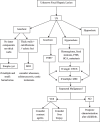
Diagnostic algorithm presenting the management and differential diagnoses to consider in an asymptomatic pregnant woman with focal liver lesions. abbreviations: US = Ultrasonography; MRI = Magnetic Resonance Imaging; CEUS = Contrast Enhanced US; FNH = Focal Nodular Hyperplasia; HCA = Hepatocellular Adenoma
Fig. 14
Fig. 14
Diagnostic Algorithm for liver urgencies and emergencies in pregnancy. abbreviations: RUQ = Right Upper Quadrant; ICP = Intrahepatic Cholestasis of Pregnancy; AFLP = Acute Fatty Liver of Pregnancy; HELLP = Elevated Liver Enzymes, Low Platelet; US = Ultrasonography; MRI = Magnetic Resonance Imaging; MRCP= Magnetic Resonance Cholangiopancreatography; CT = Computed Tomography; MRI = Magnetic Resonance Imaging; AST = Aspartate transaminase; ALT = alanine aminotransferase (ALT); NAFLD = Non-Alcoholic Fatty Liver Disease

Similar articles

Cited by

References

    1. Feldt-Rasmussen U, Mathiesen ER. Endocrine disorders in pregnancy: physiological and hormonal aspects of pregnancy. Best Pract Res Clin Endocrinol Metab. 2011;25:875–884. doi: 10.1016/j.beem.2011.07.004. - DOI - PubMed
    1. Tkachenko O, Shchekochikhin D, Schrier RW. Hormones and Hemodynamics in Pregnancy. Int J Endocrinol Metab. 2014;12(2):e14098. doi: 10.5812/ijem.14098. - DOI - PMC - PubMed
    1. Mandic-Markovic VD, Mikovic ZM, Djukic MK, et al. Doppler parameters of the maternal hepatic artery blood flow in normal pregnancy: maternal hepatic artery blood flow in normal pregnancy. Eur J Obstet Gynecol Reprod Biol. 2014;181:275–279. doi: 10.1016/j.ejogrb.2014.08.010. - DOI - PubMed
    1. Costantine MM. Physiologic and pharmacokinetic changes in pregnancy. Front Pharmacol. 2014;5:65. doi: 10.3389/fphar.2014.00065. - DOI - PMC - PubMed
    1. Bissonnette J, Durand F, De Raucourt E, et al. Pregnancy and vascular liver disease. J Clin Exp Hepatol. 2015;5:41–50. doi: 10.1016/j.jceh.2014.12.007. - DOI - PMC - PubMed

LinkOut - more resources