. 2024 Mar 19;98(3):e0172023.
doi: 10.1128/jvi.01720-23. Epub 2024 Feb 27.

A VRC13-like bNAb response is associated with complex escape pathways in HIV-1 envelope

Affiliations

A VRC13-like bNAb response is associated with complex escape pathways in HIV-1 envelope

Vinita R Joshi et al. J Virol. .

Abstract

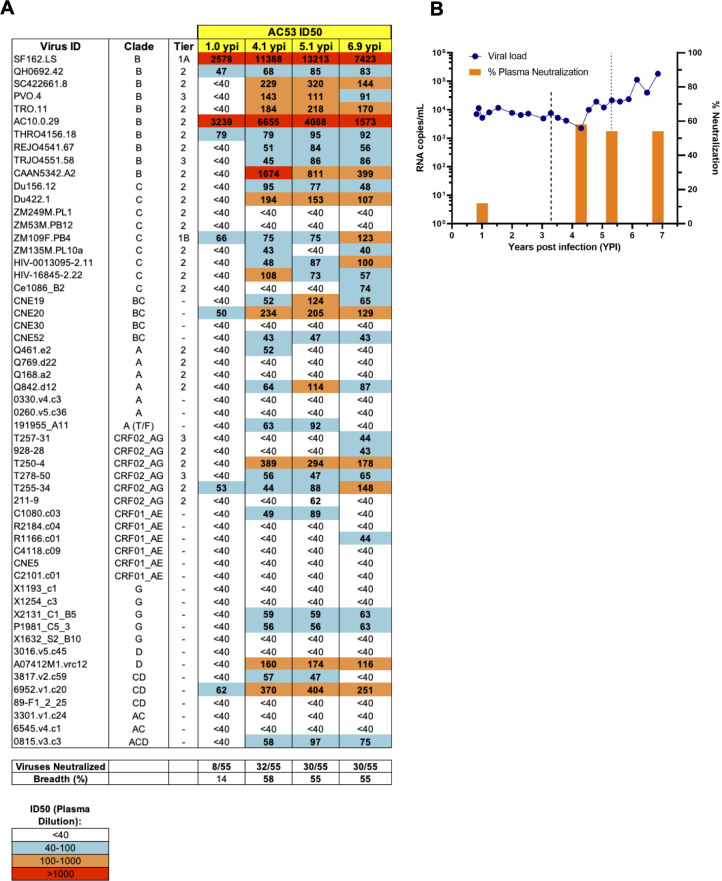
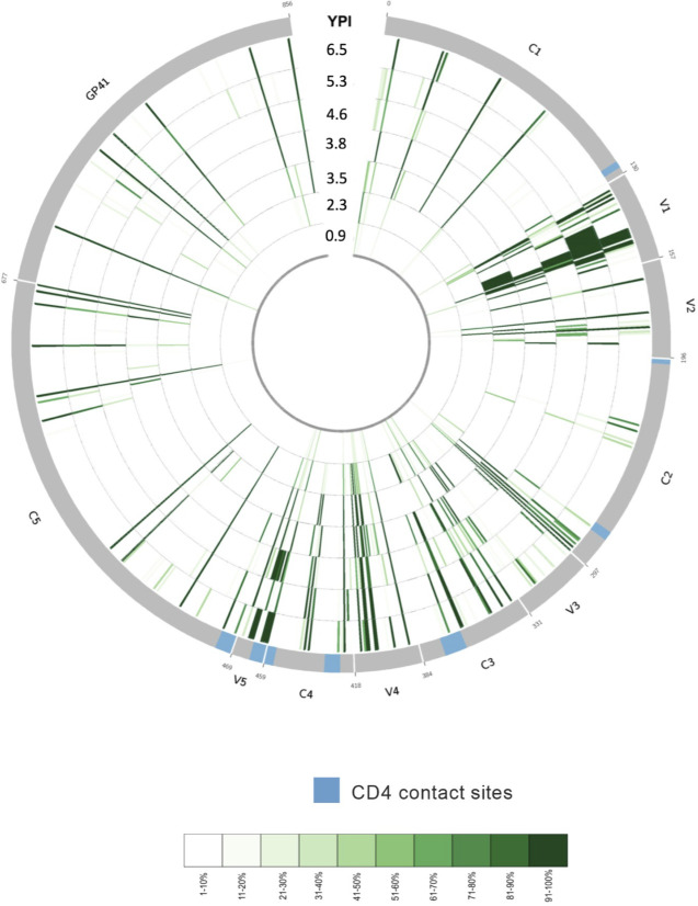
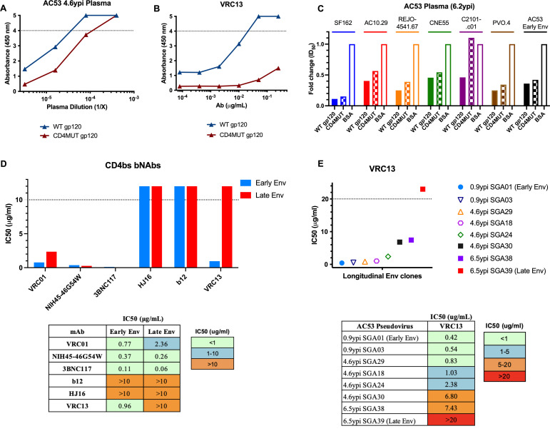
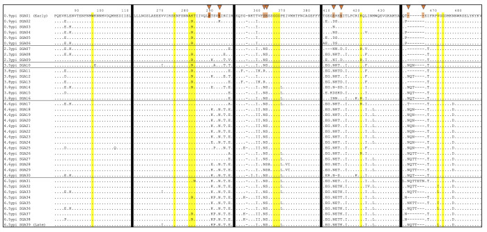
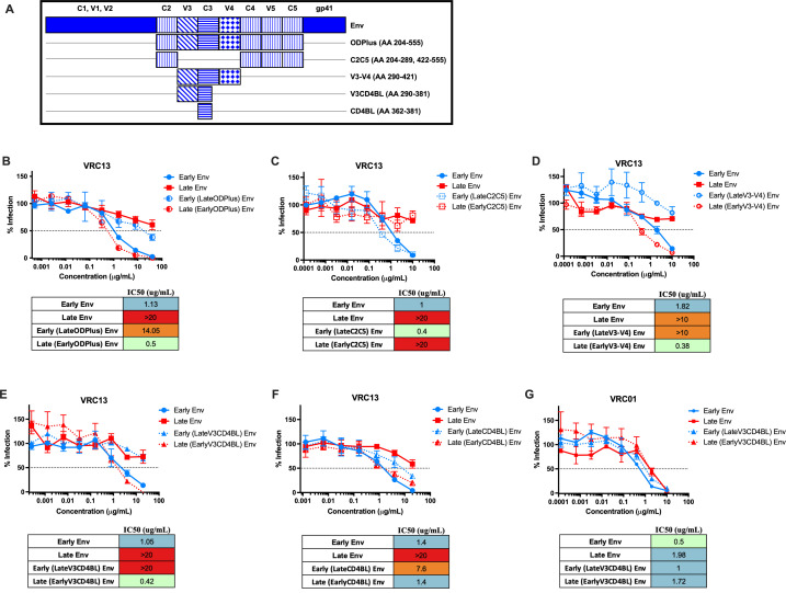
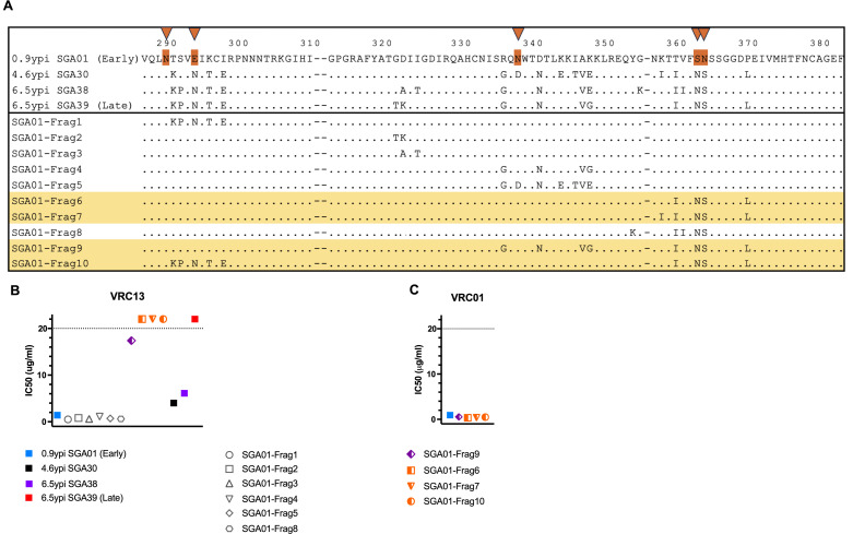
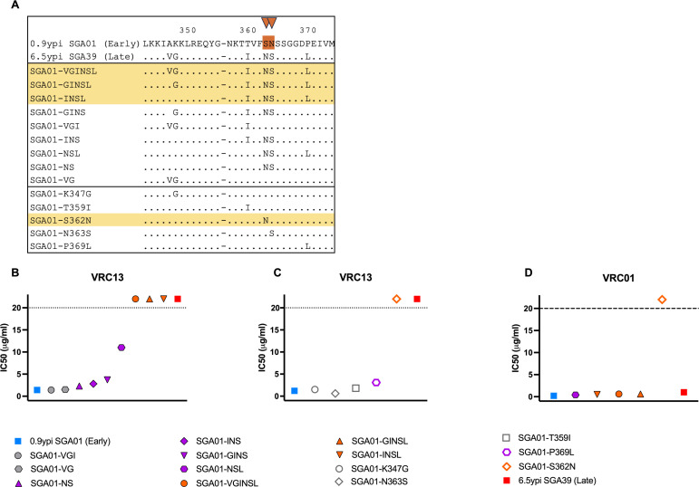
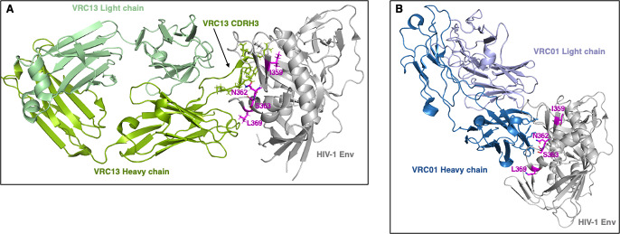
The rational design of HIV-1 immunogens to trigger the development of broadly neutralizing antibodies (bNAbs) requires understanding the viral evolutionary pathways influencing this process. An acute HIV-1-infected individual exhibiting >50% plasma neutralization breadth developed neutralizing antibody specificities against the CD4-binding site (CD4bs) and V1V2 regions of Env gp120. Comparison of pseudoviruses derived from early and late autologous env sequences demonstrated the development of >2 log resistance to VRC13 but not to other CD4bs-specific bNAbs. Mapping studies indicated that the V3 and CD4-binding loops of Env gp120 contributed significantly to developing resistance to the autologous neutralizing response and that the CD4-binding loop (CD4BL) specifically was responsible for the developing resistance to VRC13. Tracking viral evolution during the development of this cross-neutralizing CD4bs response identified amino acid substitutions arising at only 4 of 11 known VRC13 contact sites (K282, T283, K421, and V471). However, each of these mutations was external to the V3 and CD4BL regions conferring resistance to VRC13 and was transient in nature. Rather, complete resistance to VRC13 was achieved through the cooperative expression of a cluster of single amino acid changes within and immediately adjacent to the CD4BL, including a T359I substitution, exchange of a potential N-linked glycosylation (PNLG) site to residue S362 from N363, and a P369L substitution. Collectively, our data characterize complex HIV-1 env evolution in an individual developing resistance to a VRC13-like neutralizing antibody response and identify novel VRC13-associated escape mutations that may be important to inducing VRC13-like bNAbs for lineage-based immunogens.IMPORTANCEThe pursuit of eliciting broadly neutralizing antibodies (bNAbs) through vaccination and their use as therapeutics remains a significant focus in the effort to eradicate HIV-1. Key to our understanding of this approach is a more extensive understanding of bNAb contact sites and susceptible escape mutations in HIV-1 envelope (env). We identified a broad neutralizer exhibiting VRC13-like responses, a non-germline restricted class of CD4-binding site antibody distinct from the well-studied VRC01-class. Through longitudinal envelope sequencing and Env-pseudotyped neutralization assays, we characterized a complex escape pathway requiring the cooperative evolution of four amino acid changes to confer complete resistance to VRC13. This suggests that VRC13-class bNAbs may be refractory to rapid escape and attractive for therapeutic applications. Furthermore, the identification of longitudinal viral changes concomitant with the development of neutralization breadth may help identify the viral intermediates needed for the maturation of VRC13-like responses and the design of lineage-based immunogens.

Keywords: CD4bs; HIV; VRC13; bNAb; escape; evolution.


Conflict of interest statement

The authors declare no conflict of interest.

Figures

Fig 1
Fig 1
Development of neutralization breadth in donor AC53 over 6.9 years post HIV-1 infection. (A) ID50 values of AC53 plasma from 1.0, 4.1, 5.1, and 6.9 ypi tested against a standardized 55-pseudovirus panel. ID50 values indicate plasma dilutions that reduced maximal viral infection by 50% and are color-coded according to the legend shown at the bottom. (B) Plasma viremia is shown as HIV-1 RNA copies/mL (left y-axis, blue line), and plasma neutralization breadth is shown as % of viruses neutralized (right y-axis, orange bars) during untreated infection. The dashed lines at 3.5 and 5.3 ypi indicate cross-neutralizing CD4bs and N160-directed V1V2 specificities in AC53 plasma, respectively, as per Mikell et al. (67).
Fig 2
Fig 2
Env evolution in donor AC53 over 6.9 years post HIV-1 infection. Circos ideogram indicating Env amino acid sequence evolution from 0.9 ypi (innermost circle) to 6.5 ypi (outermost circle). Env sequences were derived using bulk PCR amplification from patient plasma and sequenced by next-generation sequencing. Green tick marks denote the presence of amino acid substitutions differing from the baseline (0.9 ypi) consensus amino acid residue at any given position and time point. A light green tick mark denotes the presence of a low-frequency amino acid variant, and a dark green tick mark denotes the presence of a high-frequency amino acid variant as depicted in the color-coded legend. The 0.9 ypi time point depicts numerous amino acid sites in the early viral population exhibiting low diversity, while later time points radiating outward illustrate increased sequence divergence from baseline as well as numerous residues evolving to fixed amino acid variants. Known CD4-contact sites are indicated in blue on the outer gray circle.
Fig 3
Fig 3
Env clones from donor AC53 exhibit resistance to a VRC13-like antibody response. The presence of CD4bs-directed antibodies determined by ELISA using (A) plasma from donor AC53 or (B) bNAb VRC13. AC53 plasma and VRC13 were tested for the presence of binding antibodies against an HIV-1 YU2 gp120 (WT gp120) or a CD4bs mutant containing four mutations (A281R, G366R, D368R, and P369R; termed CD4MUT gp120). (C) Heterologous neutralization of AC53 plasma (6.2 ypi) depleted with either WT gp120 or CD4MUT gp120 plotted as fold change in ID50 values, compared to depletion with BSA (control). IC50 values determined by TZM-bl neutralization assays of (D) a panel of CD4-binding site bNAbs against pseudoviruses expressing AC53 Early Env (SGA01 0.9 ypi) and AC53 Late Env (SGA39 6.5 ypi), with accompanying numerical values, and (E) VRC13 against pseudoviruses expressing various longitudinal AC53 SGAs over 6.5 years post infection, with accompanying numerical values. All experiments were conducted in triplicate. The dotted line in (A) and (B) indicates the maximum detectable absorbance value. The dotted line in (D) and (E) indicates the maximum tested antibody concentration of 10 µg/mL and 20 µg/mL, respectively. Values above these maximums are arbitrarily graphically represented and are not quantifiable.
Fig 4
Fig 4
Longitudinal Env evolution in donor AC53 flanking known VRC13 contact sites. Amino acid sequence alignment of Env clones isolated by SGA from donor AC53. Amino acid numbering corresponds to the HIV-1 HXB2 reference strain. Each row reflects a single Env SGA clone, denoted by date post infection isolated and SGA clone number. Time points are separated by horizontal black lines. Residues highlighted in yellow indicate 11 known VRC13 contact sites. Vertical black lines denote alignment breaks between windows containing known VRC13 contact sites. Orange triangles at the top denote PNLG sites where sequence evolution was observed. Identical amino acids are shown as dots and deletions are shown as dashes. “0.9 ypi SGA01” and “6.5 ypi SGA39” denote the “Early Env” and “Late Env” pseudoviruses used in neutralization assays.
Fig 5
Fig 5
Mutations within the V3 and CD4-binding loops are responsible for escape from VRC13. Env chimeras generated between donor AC53 Early Env and Late Env SGA clones are indicated in the schematic in (A). TZM-bl neutralization assays of Env chimeras testing the (B) ODPlus, (C) C2C5, (D) V3–V4, (E) V3CD4BL, and (F) CD4BL regions against VRC13 are shown. (G) TZM-bl neutralization assays of the V3CD4BL chimeras against VRC01 are shown. The dotted line on the y-axis indicates 50% infection. All assays were conducted in triplicate. Data are shown as mean values; error bars represent SEM.
Fig 6
Fig 6
AC53-specific mutations and their contribution to VRC13 neutralization in donor AC53. Env variants generated based on amino acid substitutions between donor AC53 Early Env SGA01 and Late Env SGA39 are indicated in the schematic in (A), with selected substitutions incorporated into the Early Env SGA01 backbone. Orange triangles at top denote PNLG sites where sequence evolution was observed. Graphical representation of IC50 values of chimeras indicated in (A) determined by TZM-bl neutralization assays with (B) VRC13 and (C) VRC01 are shown. Env chimeras that demonstrated resistance to VRC13 neutralization are highlighted yellow in (A). The dotted line in (B) and (C) indicates the maximum tested Ab concentration of 20 µg/mL. Values above these maximums are arbitrarily graphically represented and are not quantifiable. (B–C) Recombinant Envs are color coded to denote no resistance (gray), partial resistance (purple), or complete resistance (orange) to VRC13. Amino acid numbering corresponds to the HIV-1 HXB2 reference strain.
Fig 7
Fig 7
Novel VRC13 escape mutations and their contribution to autologous neutralization in donor AC53. (A) Alignment of env chimeras expressing combinations of mutations implicated in resistance to VRC13 incorporated into the Early Env SGA01 backbone. (A). Graphical representation of IC50 values of chimeras indicated in (A) as determined by TZM-bl neutralization assays with (B, C) VRC13 and (D) VRC01. Env chimeras that demonstrated complete escape from VRC13 neutralization are highlighted yellow in (A). The dotted line in (B–D) indicates the maximum tested Ab concentration of 20 µg/mL. Values above these maximums are arbitrarily graphically represented and are not quantifiable. (B–D) Recombinant Envs are color coded to denote no resistance (gray), partial resistance (purple), or complete resistance (orange) to VRC13. Amino acid numbering corresponds to the HIV-1 HXB2 reference strain.
Fig 8
Fig 8
Structural modeling of potential novel VRC13 escape mutations. The four potential VRC13 escape changes T359I, S362N, N363S, and P369L were modeled on previously published HIV-1 Env structures bound to either (A) VRC13 (PDB ID 4YDJ) or (B) VRC01 (PDB ID 3NGB). HIV-1 Env is denoted in gray, and (A) VRC13 heavy chain and light chains are shown in dark green and pale green, respectively. (B) VRC01 heavy chain and light chains are shown in dark blue and pale violet, respectively. Residue changes are highlighted in magenta and labeled. All images were created using the PyMOL Molecular Graphics System, Version 2.0 Schrödinger, LLC, using PDB 4YDJ and 3NGB as template (49, 55).

Similar articles

References

    1. Moore JP, Cao Y, Ho DD, Koup RA. 1994. Development of the anti-gp120 antibody response during seroconversion to human immunodeficiency virus type 1. J Virol 68:5142–5155. doi:10.1128/JVI.68.8.5142-5155.1994 - DOI - PMC - PubMed
    1. Wei X, Decker JM, Wang S, Hui H, Kappes JC, Wu X, Salazar-Gonzalez JF, Salazar MG, Kilby JM, Saag MS, Komarova NL, Nowak MA, Hahn BH, Kwong PD, Shaw GM. 2003. Antibody neutralization and escape by HIV-1. Nature 422:307–312. doi:10.1038/nature01470 - DOI - PubMed
    1. Cuevas JM, Geller R, Garijo R, López-Aldeguer J, Sanjuán R. 2015. Extremely high mutation rate of HIV-1 in vivo. PLoS Biol 13:e1002251. doi:10.1371/journal.pbio.1002251 - DOI - PMC - PubMed
    1. Richman DD, Wrin T, Little SJ, Petropoulos CJ. 2003. Rapid evolution of the neutralizing antibody response to HIV type 1 infection. Proc Natl Acad Sci U S A 100:4144–4149. doi:10.1073/pnas.0630530100 - DOI - PMC - PubMed
    1. Li B, Decker JM, Johnson RW, Bibollet-Ruche F, Wei X, Mulenga J, Allen S, Hunter E, Hahn BH, Shaw GM, Blackwell JL, Derdeyn CA. 2006. Evidence for potent autologous neutralizing antibody titers and compact envelopes in early infection with subtype C human immunodeficiency virus type 1. J Virol 80:5211–5218. doi:10.1128/JVI.00201-06 - DOI - PMC - PubMed