. 2024 Feb 26;4(2):100715.
doi: 10.1016/j.crmeth.2024.100715.

PXPermute reveals staining importance in multichannel imaging flow cytometry

Affiliations

PXPermute reveals staining importance in multichannel imaging flow cytometry

Sayedali Shetab Boushehri et al. Cell Rep Methods. .

Abstract

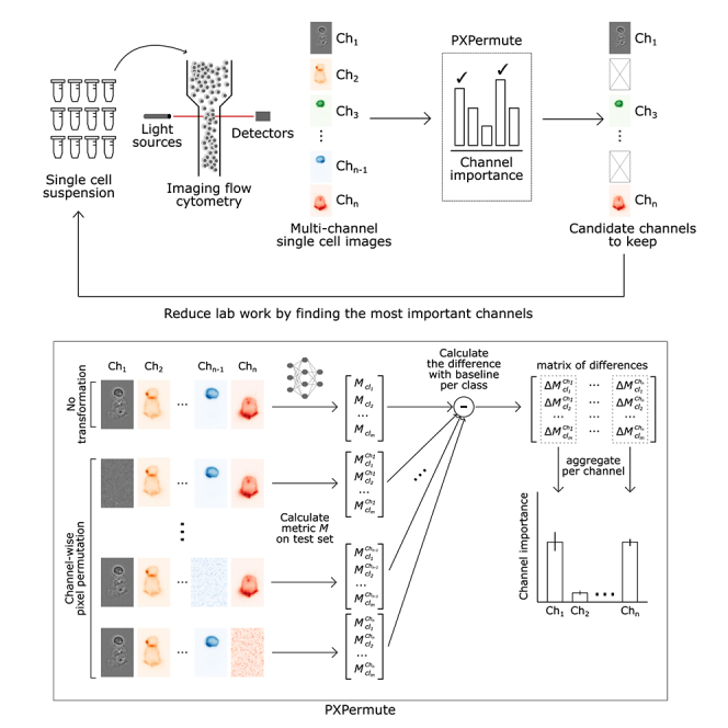
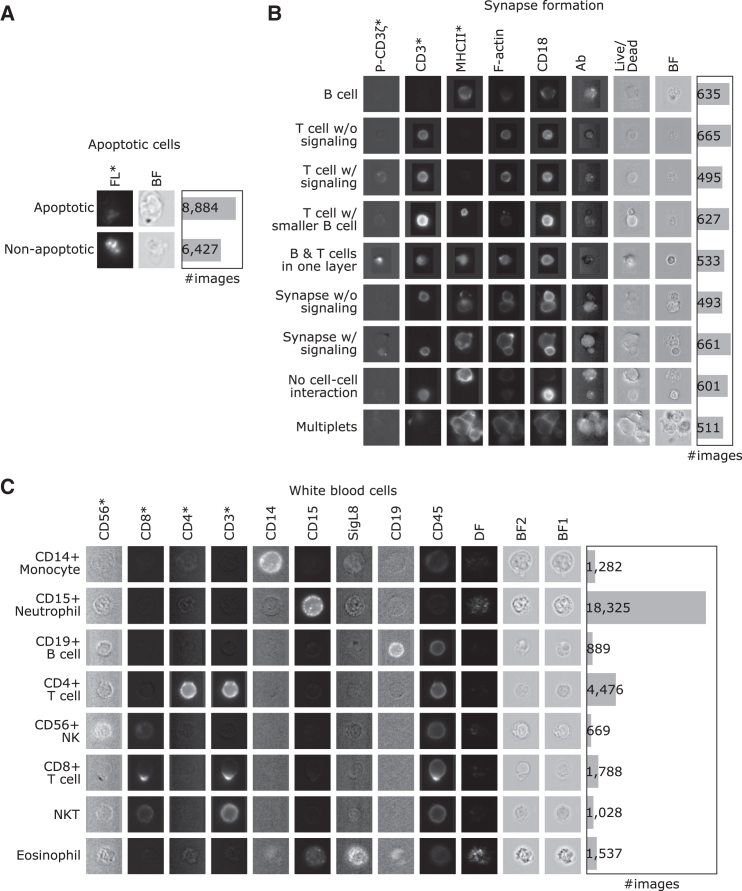
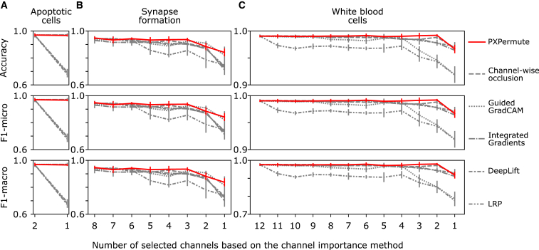
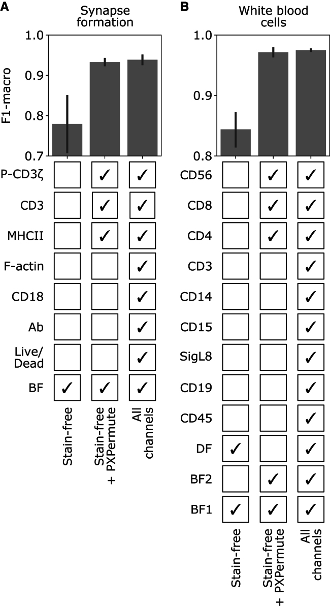
Imaging flow cytometry (IFC) allows rapid acquisition of numerous single-cell images per second, capturing information from multiple fluorescent channels. However, the traditional process of staining cells with fluorescently labeled conjugated antibodies for IFC analysis is time consuming, expensive, and potentially harmful to cell viability. To streamline experimental workflows and reduce costs, it is crucial to identify the most relevant channels for downstream analysis. In this study, we introduce PXPermute, a user-friendly and powerful method for assessing the significance of IFC channels, particularly for cell profiling. Our approach evaluates channel importance by permuting pixel values within each channel and analyzing the resulting impact on machine learning or deep learning models. Through rigorous evaluation of three multichannel IFC image datasets, we demonstrate PXPermute's potential in accurately identifying the most informative channels, aligning with established biological knowledge. PXPermute can assist biologists with systematic channel analysis, experimental design optimization, and biomarker identification.

Keywords: CP: Imaging; CP: Systems biology; cell profiling; channel importance; computer vision; deep learning; image flow cytometry; interpretable artificial intelligence; machine learning; staining importance.


Conflict of interest statement

Declaration of interests The authors declare no competing interests.

Figures

None
Graphical abstract
Figure 1
Figure 1
PXPermute allows identifying the most important fluorescent (FL) channels in a multichannel imaging flow cytometry experiment and thus reduces lab work and expenses (A) Schematic of a PXPermute embedded end-to-end analysis. In the first part, biologists image thousands of single cells using imaging flow cytometry in different FL and bright-field (BF) channels. The second part, PXPermute, will select the most important channels based on the task. (B) Schematic of PXPermute, a simple yet powerful method to find the most important channels. In the first step, a performance metric Mcl (such as accuracy or F1-score) is calculated per class cl. Then, each channel Ch is shuffled, and the performance per class cl is calculated as MclCh. Finally, the difference between the original performance and the permuted one is calculated. These differences are averaged and yield the channel importance.
Figure 2
Figure 2
Three datasets with various numbers of images and channels are used to evaluate PXPermute Rows indicate classes, and columns indicate the channels. Channels marked with an asterisk (∗) indicate that those channels have been identified as the most important channels in previous works. (A) Apoptotic cells: a dataset containing 15,311 images with one stain-free, BF, and an FL channel. (B) Synapse formation: a dataset containing 5,221 images with one stain-free channel (BF) and seven FL channels, namely antibody (Ab), CD18, F-actin, MHC class II, CD3, P-CD3ζ and live/dead. (C) White blood cells: a dataset containing 29,994 images with three stain-free channels, two BFs (BF1 and BF2), a dark-field (DF), and nine stained channels including CD15, SigL8, CD14, CD19, CD3, CD45, CD4, CD56, and CD8.
Figure 3
Figure 3
PXPermute robustly identifies the most important channels Each dataset’s channel importance is normalized between zero and one for PXPermute and five other methods, including channel-wise occlusion, Guided GradCAM, integrated gradients, DeepLift, and layerwise relevance propagation (LRP). The error bars are based on a 5-fold cross-validation scheme, representing the mean with a 95% confidence interval. Channels marked with an asterisk (∗) indicate that those channels have been identified as the most important channels in previous works. Note that PXPermute channel rankings align better with the findings from previous studies than any other method.
Figure 4
Figure 4
PXPermute outperforms other methods in identifying the correct channel ranking based on a remove-and-retrain procedure The remove-and-retrain based on the channel ranking is performed on the apoptotic cell (2 channels), synapse formation (8 channels), and white blood cell (12 channels) datasets. The error bars represent the mean with a 95% confidence interval. In each case, the channels are sorted in ascending order according to their predicted importance score, from the least important channel to the most important. Then, the channels are iteratively removed from the dataset, from the least important to the most important channel. After each removal, the classification model was retrained on the dataset containing the subset of channels to perform the same classification task as before. Methods with better rankings would stay higher throughout the plot. PXPermute performed better than other methods in finding the optimal channel rankings.
Figure 5
Figure 5
PXPermute finds an optimal channel selection that performs similarly to using all the channels For the synapse formation (A) and white blood cell (B) datasets, the model was trained on the stain-free channels (lower bound), stain-free + top 3 channels identified by PXPermute, and all channels (upper bound). The error bars represent the mean with a 95% confidence interval. PXPermute rankings lead to fewer stainings (3 out of 7 in A, 3 out of 8 in B) without a significant loss in performance.

References

    1. Rane A.S., Rutkauskaite J., deMello A., Stavrakis S. High-Throughput Multi-parametric Imaging Flow Cytometry. Chem. 2017;3:588–602. doi: 10.1016/j.chempr.2017.08.005. - DOI
    1. Barteneva N.S., Vorobjev I.A. Imaging flow cytometry. J. Immunol. Methods. 2016 - PubMed
    1. Doan M., Vorobjev I., Rees P., Filby A., Wolkenhauer O., Goldfeld A.E., Lieberman J., Barteneva N., Carpenter A.E., Hennig H. Diagnostic Potential of Imaging Flow Cytometry. Trends Biotechnol. 2018;36:649–652. doi: 10.1016/j.tibtech.2017.12.008. - DOI - PubMed
    1. Barteneva N.S., Fasler-Kan E., Vorobjev I.A. Imaging Flow Cytometry: Coping with Heterogeneity in Biological Systems. J. Histochem. Cytochem. 2012;60:723–733. doi: 10.1369/0022155412453052. - DOI - PMC - PubMed
    1. Blasi T., Hennig H., Summers H.D., Theis F.J., Cerveira J., Patterson J.O., Davies D., Filby A., Carpenter A.E., Rees P. Label-free cell cycle analysis for high-throughput imaging flow cytometry. Nat. Commun. 2016;7 doi: 10.1038/ncomms10256. - DOI - PMC - PubMed

MeSH terms