. 2024 Mar 7;111(3):509-528.
doi: 10.1016/j.ajhg.2024.01.013. Epub 2024 Feb 26.

Haploinsufficiency of ZFHX3, encoding a key player in neuronal development, causes syndromic intellectual disability

Collaborators, Affiliations

Haploinsufficiency of ZFHX3, encoding a key player in neuronal development, causes syndromic intellectual disability

María Del Rocío Pérez Baca et al. Am J Hum Genet. .

Abstract

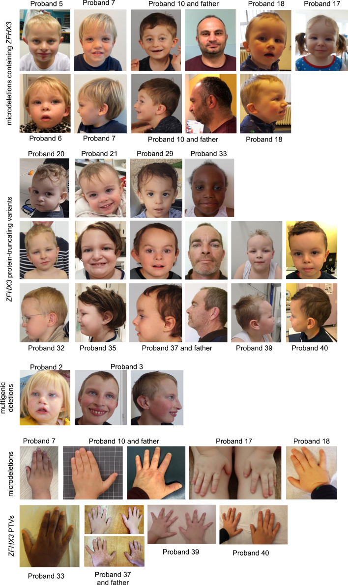
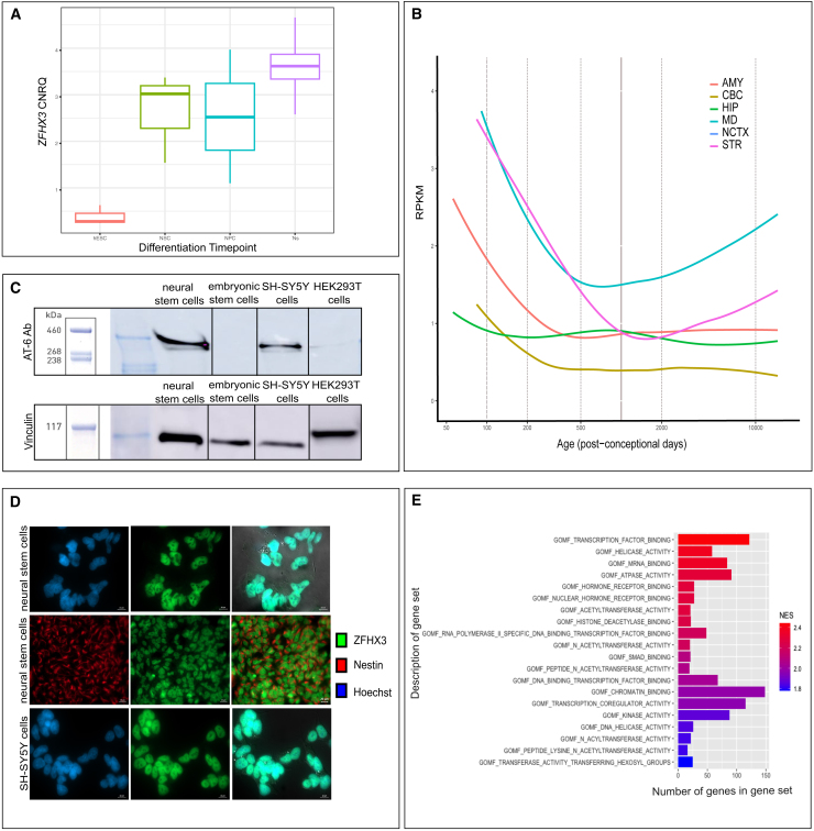
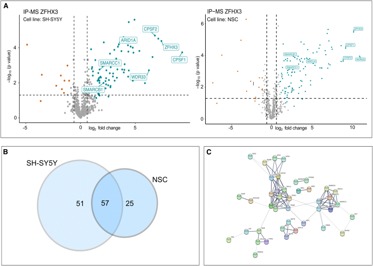
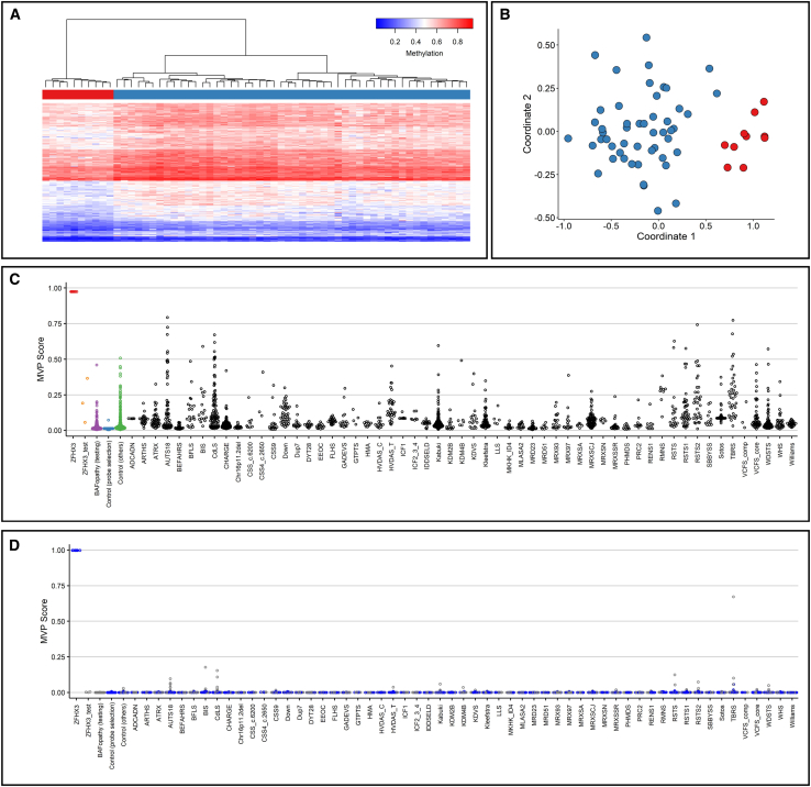
Neurodevelopmental disorders (NDDs) result from impaired development and functioning of the brain. Here, we identify loss-of-function (LoF) variation in ZFHX3 as a cause for syndromic intellectual disability (ID). ZFHX3 is a zinc-finger homeodomain transcription factor involved in various biological processes, including cell differentiation and tumorigenesis. We describe 42 individuals with protein-truncating variants (PTVs) or (partial) deletions of ZFHX3, exhibiting variable intellectual disability and autism spectrum disorder, recurrent facial features, relative short stature, brachydactyly, and, rarely, cleft palate. ZFHX3 LoF associates with a specific methylation profile in whole blood extracted DNA. Nuclear abundance of ZFHX3 increases during human brain development and neuronal differentiation. ZFHX3 was found to interact with the chromatin remodeling BRG1/Brm-associated factor complex and the cleavage and polyadenylation complex, suggesting a function in chromatin remodeling and mRNA processing. Furthermore, ChIP-seq for ZFHX3 revealed that it predominantly binds promoters of genes involved in nervous system development. We conclude that loss-of-function variants in ZFHX3 are a cause of syndromic ID associating with a specific DNA methylation profile.

Keywords: ZFHX3; chromatin remodeling complex; mRNA polyadenylation and cleavage complex; neurodevelopmental disorder.


Conflict of interest statement

Declaration of interests L.R. is an employee of GeneDx, LLC. X.W. is a co-founder and employee of AiLife Diagnostics.

Figures

None
Graphical abstract
Figure 1
Figure 1
Overview of ZFHX3 aberrations in 42 probands (A) (Micro)deletions identified in probands 1–18 (#) are represented by the red bars, all affecting ZFHX3 (gray box). RefSeq coding and non-coding genes are indicated respectively in blue and green. Genomic positions are according to hg38; ZFHX3 is located on the reverse strand. (B) Nonsense and frameshift variants in ZFHX3 are identified in probands 19–42. Gene and protein structure of the canonical transcript (GenBank: NM_006885.4; GenBank: NP_008816.3) are shown in the top and middle part, respectively. The majority of PTVs are located in exon 9. Top: coding exons are indicated as dark gray boxes, while UTR regions are indicated with a light gray bar. Middle: LoF variants are present in the ZFHX3 cohort and located in different domains of ZFHX3. Bottom: Frameshift and nonsense variants present in gnomAD (v3.1.2) are enriched at the C-terminus, i.e., 25 of the 44 reported variants are located in exon 10. (C) Clinical presentation of individuals with ZFHX3 aberration. Left: de-identified face mask of images from 17 individuals with either a microdeletion or truncating variant affecting ZFHX3 (left) and matched healthy individuals (right). Note a high and broad forehead, laterally sparse eyebrows, upslanted palpebral fissures, a low-hanging columella, flat philtrum, a relatively short midface, and a thin upper lip. Right: score distribution plots of the ZFHX3 cohort (green) and the control cohort (orange) and the receiver operating characteristic (ROC) curve to determine the capacity of Face2Gene to identify individuals in the ZFHX3 cohort against controls.
Figure 2
Figure 2
Front and side profile pictures, as well as hand and digits pictures of individuals with a deletion or PTV affecting ZFHX3 Rows 1–2: frontal and profile photographs of probands with a microdeletion containing ZFHX3. Rows 3–5: frontal and profile photographs of probands with a ZFHX3 protein-truncating variant (PTV). Row 6: frontal and profile photographs of probands with a multigenic deletion containing ZFHX3. Rows 7–8: photographs showing the hands of probands with a microdeletion (row 7) or PTV (row 8) affecting ZFHX3.
Figure 3
Figure 3
ZFHX3 is increasingly abundant during neural differentiation and localizes to the nucleus (A) ZFHX3 levels during in vitro neural differentiation. RNA was sampled at the following stages: hESCs, NSCs, neural progenitor cells (NPC), and direct differentiated neurons (Ns). An increasing expression is observed upon differentiation. The median expression value is indicated by the middle horizontal line. The whiskers indicate the minimum and maximum values. CNRQ, calibrated normalized relative quantity. (B) ZFHX3 levels in developing human brain. ZFHX3 expression in the developing human brain (normalized RPKM data) showing higher expression during early prenatal development followed by decreased expression upon brain maturation. Data obtained from BrainSpan: http://www.brainspan.org. Abbreviations: AMY, amygdala; CBC, cerebellum; HIP, hippocampus; MD: medial dorsal nucleus of the thalamus; NCTX: neocortex; STR: striatum. The dashed vertical lines indicate the age post-conception to the corresponding expression level. (C) Endogenous ZFHX3 (404 kDa) accumulation in NSCs, hESCs, SH-SY5Y, and HEK293T as detected with western blot using the AT-6 antibody (top) and a vinculin antibody as a loading control (bottom). (D) ZFHX3 is localized in the nucleus of SH-SY5Y cells and NSCs. Indirect immunofluorescence staining for ZFHX3 (SH-SY5Y and NSCs) and nestin (cytoplasmic neuronal marker, NSCs) was performed. DNA in the nucleus was counterstained with Hoechst dye. Images were merged to observe the contrast between the compartments. Scale bar: 10μm (upper row and bottom row), 20 μm (middle row); NSC, neural stem cells. (E) Molecular functions of the top 20 positive correlated gene sets in the data by Cardoso et al.. All have an FDR < 0.10 and a normalized enrichment score (NES) > 1.8. Gene sets are ranked based on NES-value.
Figure 4
Figure 4
ZFHX3 has a robust protein-protein interaction network of 57 interaction partners (A) Volcano plot showing the enriched ZFHX3 interactor proteins in SH-SY5Y (left: n = 83) and NSCs (right: n = 109). For each detected protein, the log2 fold change (FC) between the ZFHX3 IP-enriched samples and the IgG controls is shown on the x axis, while the -log10 of the p value is shown on the y axis. Vertical dashed lines indicated a fold change of abs(0.6). The horizontal dashed line indicates the -log10 value of a p value equal to 0.05. Proteins with an FDR < 0.05, log2 FC > 0.6/<-0.6 and S0 = 1 are, respectively, highlighted in blue and red. Important members of the BAF and CP complexes are also highlighted, as well as ZFHX3 itself. (B) Overlap of ZFHX3 interaction factors between SH-SY5Y and NSCs IP results. A total of 57 proteins interacts in both cell lines with ZFHX3. A total of respectively 51 or 25 interaction partners for ZFHX3 were found in SH-SY5Y or NSCs. (C) ZFHX3 protein-protein interaction network extracted from the STRING 11.0 database. Shown are interactors that are connected within a network. Line thickness indicates the strength of data support. The BAF complex, CPSF (cleavage and polyadenylation specificity factor) and CSTF (cleavage stimulation factor), nuclear pore complex (NPC), and septin complex are ZFHX3-containing protein complexes. CPSF and CSTF are two multi-subunit protein complexes responsible for cleavage and polyadenylation.
Figure 5
Figure 5
Deletion of ZFHX3 and ZFHX3 PTVs are associated with a specific methylation profile (A) Hierarchical clustering using the selected CpG sites, where rows represent selected probes and columns indicate samples. ZFHX3 samples are illustrated with red, and control individuals with blue on the heatmap pane. The heatmap color scale demonstrates methylation levels ranging from blue (no methylation or 0) to red (full methylation or 1). Clear separation between ZFHX3 and control samples is observed. (B) MDS demonstrating different methylation pattern between the cohort and control groups using the selected CpG sites. Red and blue circles represent cohort and control samples, respectively. (C) MVP scores generated by the SVM classifier trained using only the 10 ZFHX3 samples and the 56 matched control samples, where the majority of the other control samples and individuals with other disorders received low scores, demonstrating the high specificity of the methylation pattern to ZFHX3 haploinsufficiency. (D) MVP scores generated by the SVM constructed by training the 10 ZFHX3 samples against matched control samples and 75% of other control samples and samples from other disorders (blue). The remaining 25% of the database samples used for testing (gray) received very low MVP scores, illustrative of the significant improvement in the specificity of the model. The 3 samples with missense variants in ZFHX3 (ZFHX3_test) and the BAFopathy samples (BAFopathy [testing]) were also supplied into the model as testing samples and received low MVP scores, indicating that their methylation pattern is different from that of ZFHX3 haploinsufficiency. Abbreviations: ADCADN, cerebellar ataxia, deafness, and narcolepsy, autosomal dominant; ARTHS, Arboleda-Tham syndrome; ATRX, alpha-thalassemia/intellectual development syndrome, X-linked; AUTS18, autism, susceptibility to 18; BEFAHRS, Beck-Fahrner syndrome; BFLS, Börjeson-Forssman-Lehmann syndrome; BIS, blepharophimosis intellectual disability SMARCA2 syndrome; CdLS, Cornelia de Lange syndrome 1–4; CHARGE, CHARGE syndrome; 16p11.2del, 16p11.2 deletion syndrome; CSS_c.6200, Coffin-Siris syndrome (c.6232G>A [GenBank: NM_006015.4 (ARID1A)]; p.Glu2078Lys) (c.6254T>G [GenBank: NM_006015.4 (ARID1A)]; p.Leu2085Arg) (c.6133T>C [GenBank: NM_017519.2 (ARID1B)]; p.Cys2045Arg); CSS4_c.2650, Coffin-Siris syndrome (c.2656A>G [GenBank: NM_001128849.1 (SMARCA4)]; p.Met886Val); CSS9, Coffin-Siris syndrome-9; Down, Down syndrome; Dup7, Williams-Beuren duplication syndrome (7q11.23 duplication syndrome); DYT28, Dystonia-28, childhood onset; EEOC, epileptic encephalopathy, childhood onset; FLHS, Floating-Harbour syndrome; GADEVS, Gabriele de Vries syndrome; GTPTS, Genitopatellar syndrome; HMA, HVDAS_C, Helsmoortel-Van der Aa syndrome (ADNP syndrome [central]); HVDAS_T, Helsmoortel-Van der Aa syndrome (ADNP syndrome [Terminal]); ICF1 Immunodeficiency, centromeric instability, facial anomalies syndrome 1, ICF2_3_4, immunodeficiency, centromeric instability, facial anomalies syndrome 2, 3, and 4; IDDSELD, intellectual developmental disorder with seizures and language delay; Kabuki, Kabuki syndromes 1 and 2; KDM2B, KDM2B-related syndrome; KDM4B, KDM4B-related syndrome; KDVS, Koolen de Vries syndrome; Kleefstra, Kleefstra syndrome 1; LLS, Luscan-Lumish syndrome; MKHK_ID4, Menke-Hennekam syndrome-1, 2; MLASA2, myopathy, lactic acidosis, and sideroblastic anemia-2; MRD23, intellectual developmental disorder, autosomal dominant 23; MRD51, intellectual developmental disorder, autosomal dominant 51; MRX93, intellectual developmental disorder, X-linked, XLID93; MRX97, intellectual developmental disorder, X-linked 97, XLID97; MRXSA, Armfield syndrome; MRXSCJ, syndromic X-linked intellectual disability, Claes-Jensen type; MRXSN, syndromic X-linked intellectual disability, Nascimento type; MRXSSR, syndromic X-linked intellectual disability, Snyder-Robinson type; PHMDS, Phelan-McDermid syndrome; PRC2, RENS1, Renpenning syndrome; RMNS, Rahman syndrome; RSTS, Rubinstein-Taybi syndrome-1, 2; RSTS1, Rubinstein-Taybi syndrome-1; RSTS2, Rubinstein-Taybi syndrome-2; SBBYSS, Say-Barber-Biesecker-Young-Simpson syndrome; Sotos, Sotos syndrome; TBRS, Tatton-Brown-Rahman syndrome; VCFS_comp, velocardiofacial syndrome; VCFS_core, velocardiofacial syndrome; WDSTS, Wiedemann-Steiner syndrome; WHS, Wolf-Hirschhorn syndrome; and Williams, Williams-Beuren deletion syndrome (7q11.23 deletion syndrome).
Figure 6
Figure 6
ZFHX3 binds primarily to promoter regions of genes associated with pathways involved in neuron and axon development (A) Left: annotation of the 22,094 overlapping ChIP-seq peaks. 57.18% of these peaks correspond to promoter regions. The pie chart visualizes the genomic annotation and the percentage of the peaks that reside in/contain the transcription start site (TSS), exonic sequences, 5′UTR, 3′UTR, intronic, or intergenic regions, as displayed with the corresponding colors. Color code is used to assign each region. Right: distribution of the ZFHX3-binding loci relative to TSSs. The majority of the peaks falls within 0–1 kb of the TSS, also suggesting that ZFHX3 mainly binds to promoter sequences. Color code is used to represent the distance from TSS in kb. (B) The target genes of ZFHX3 are involved in axonogenesis, axon development, and the regulation of the nervous system, including neurogenesis. The gene ontology (GO) dot plot displays the top 20 enriched biological processes (BPs) ranked by gene ratio (# of genes related to GO term/total number of significant genes) and the p-adjusted values for these terms (color). The size of the dot represents the gene counts per BP. (C) ZFHX3 plays a key role in neuron and axon developmental pathways. The Kyoto Encyclopedia of Genes and Genomes (KEGG) dot plot provides all enriched biological pathways ranked by gene ratio (# of genes related to GO term/total number of significant genes) and the p-adjusted values for these terms (color). The size of the dot represents the gene counts per pathway.

Update of

References

    1. Wilfert A.B., Sulovari A., Turner T.N., Coe B.P., Eichler E.E. Recurrent de novo mutations in neurodevelopmental disorders: properties and clinical implications. Genome Med. 2017;9 - PMC - PubMed
    1. Kaplanis J., Samocha K.E., Wiel L., Zhang Z., Arvai K.J., Eberhardt R.Y., Gallone G., Lelieveld S.H., Martin H.C., McRae J.F., et al. Evidence for 28 genetic disorders discovered by combining healthcare and research data. Nat. 2020;586:757–762. doi: 10.1038/s41586-020-2832-5. - DOI - PMC - PubMed
    1. Morinaga T., Yasuda H., Hashimoto T., Higashio K., Tamaoki T. A human alpha-fetoprotein enhancer-binding protein, ATBF1, contains four homeodomains and seventeen zinc fingers. Mol. Cell Biol. 1991;11:6041–6049. doi: 10.1128/MCB.11.12.6041. - DOI - PMC - PubMed
    1. Ma G., Gao A., Yang Y., He Y., Zhang X., Zhang B., Zhang Z., Li M., Fu X., Zhao D., et al. Zfhx3 is essential for progesterone/progesterone receptor signaling to drive ductal side-branching and alveologenesis in mouse mammary glands. J. Genet. Genomics. 2019;46:119–131. doi: 10.1016/j.jgg.2019.03.003. - DOI - PubMed
    1. Zhao D., Ma G., Zhang X., He Y., Li M., Han X., Fu L., Dong X.Y., Nagy T., Zhao Q., et al. Zinc finger homeodomain factor Zfhx3 is essential for mammary lactogenic differentiation by maintaining prolactin signaling activity. J. Biol. Chem. 2016;291:12809–12820. doi: 10.1074/jbc.M116.719377. - DOI - PMC - PubMed

MeSH terms