. 2024 Feb 27;12(2):e008608.
doi: 10.1136/jitc-2023-008608.

Targeting the activated microenvironment with endosialin (CD248)-directed CAR-T cells ablates perivascular cells to impair tumor growth and metastasis

Affiliations

Targeting the activated microenvironment with endosialin (CD248)-directed CAR-T cells ablates perivascular cells to impair tumor growth and metastasis

Sarah L Ash et al. J Immunother Cancer. .

Abstract

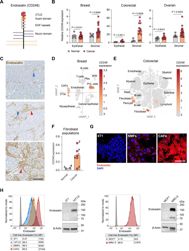
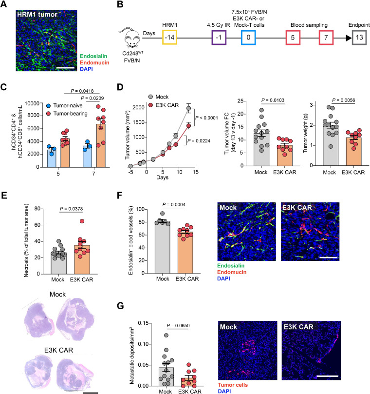
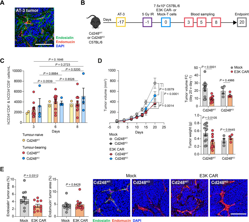
Background: Targeting of solid cancers with chimeric antigen receptor (CAR)-T cells is limited by the lack of suitable tumor-specific antigens and the immunosuppressive, desmoplastic tumor microenvironment that impedes CAR-T cell infiltration, activity and persistence. We hypothesized that targeting the endosialin (CD248) receptor, strongly expressed by tumor-associated pericytes and perivascular cancer-associated fibroblasts, would circumvent these challenges and offer an exciting antigen for CAR-T cell therapy due to the close proximity of target cells to the tumor vasculature, the limited endosialin expression in normal tissues and the lack of phenotype observed in endosialin knockout mice.

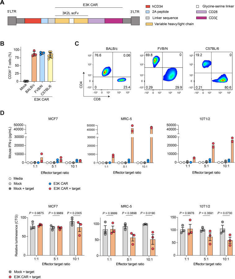
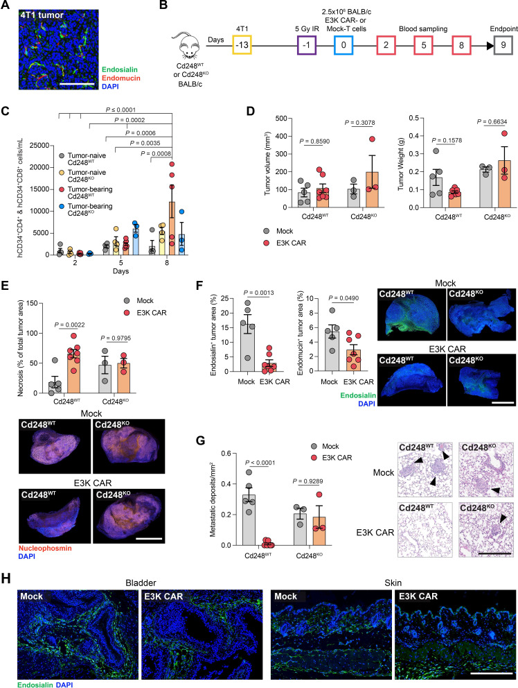
Methods: We generated endosialin-directed E3K CAR-T cells from three immunocompetent mouse strains, BALB/c, FVB/N and C57BL/6. E3K CAR-T cell composition (CD4+/CD8+ ratio), activity in vitro against endosialin+ and endosialin- cells, and expansion and activity in vivo in syngeneic tumor models as well as in tumor-naive healthy and wounded mice and tumor-bearing endosialin knockout mice was assessed.

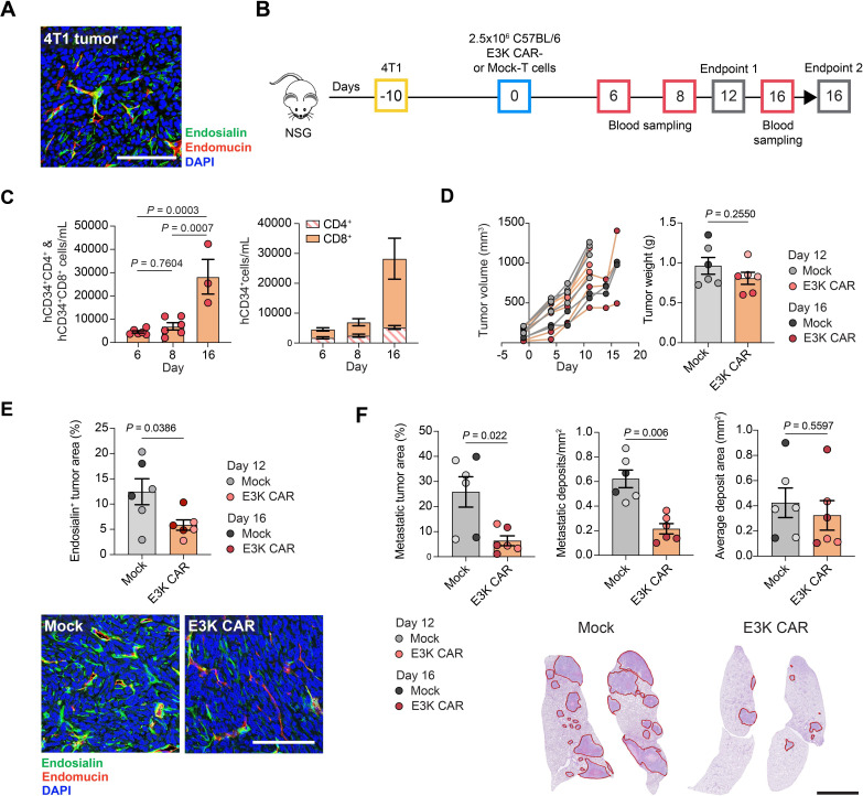
Results: E3K CAR-T cells were active in vitro against both mouse and human endosialin+, but not endosialin-, cells. Adoptively transferred E3K CAR-T cells exhibited no activity in endosialin knockout mice, tumor-naive endosialin wildtype mice or in wound healing models, demonstrating an absence of off-target and on-target/off-tumor activity. By contrast, adoptive transfer of E3K CAR-T cells into BALB/c, FVB/N or C57BL/6 mice bearing syngeneic breast or lung cancer lines depleted target cells in the tumor stroma resulting in increased tumor necrosis, reduced tumor growth and a substantial impairment in metastatic outgrowth.

Conclusions: Together these data highlight endosialin as a viable antigen for CAR-T cell therapy and that targeting stromal cells closely associated with the tumor vasculature avoids CAR-T cells having to navigate the harsh immunosuppressive tumor microenvironment. Further, the ability of E3K CAR-T cells to recognize and target both mouse and human endosialin+ cells makes a humanized and optimized E3K CAR a promising candidate for clinical development applicable to a broad range of solid tumor types.

Keywords: Breast Cancer; Chimeric antigen receptor - CAR; Lung Cancer; Tumor microenvironment - TME.


Conflict of interest statement

Competing interests: None declared.

Figures

Figure 1
Figure 1
Upregulation of endosialin expression in the tumor stroma. (A) Structure of the endosialin (CD248) protein. (B) Endosialin (CD248) expression in stroma microdissected from tumors and normal tissues in breast (n=5 normal, n=28 cancer), colon (n=4 normal, n=13 cancer) or ovary (n=6 normal epithelial, n=8 normal stromal, n=32 cancer epithelial, n=31 cancer stromal). Data represent mean values±SEM, two-way ANOVA. (C) Representative images of invasive human breast cancers stained with anti-human endosialin mAb B1/35. Arrowheads indicate endosialin+ pericytes (blue) and CAFs (red). Scale bar, 125 µm. (D,E) Endosialin (CD248) expression in scRNA-seq data sets from one of three primary human breast cancers analyzed (panel D), and in a merged set of 62 colorectal cancers (panel E). (F) Cluster-specific endosialin (CD248) expression in fibroblast populations identified from scRNA-seq analysis of normal (adjacent non-malignant tissue) and cancer (tumor tissue) samples collected from patients with lung cancer. Each number represents an individual fibroblast cluster. (G) Mouse cells stained with anti-endosialin mAb 3K2L (red) and counterstained with DAPI (blue). Scale bar, 100 µm. Representative images are shown of two independent assays. (H) Flow cytometry and western blot analysis using mAb 3K2L of endosialin levels in mouse (left panel) and human (right panel) cells. Data is representative of two independent assays. ANOVA, analysis of variance; CAFs, cancer-associated fibroblasts; CTLD, C-type lectin-like domain; DAPI, 4′,6-diamidino-2-phenylindole; EGF, epidermal growth factor; mAb, monoclonal antibodies; MFI, median fluorescence intensity; NMF, normal mammary gland fibroblasts; PVL, perivascular-like; scRNA-seq, single-cell RNA sequencing; t-SNE, t-distributed stochastic neighbor embedding; UMPA, uniform manifold approximation and projection.
Figure 2
Figure 2
E3K CAR-T cells demonstrate specific activity against human and mouse endosialin+ fibroblasts. (A) Diagram of the E3K CAR construct. (B) Transduction efficiency of BALB/c (n=4), FVB/N (n=3) and C57BL/6 (n=8) E3K CAR-T cells determined by hCD34 expression in combined CD4+ and CD8+ T cell populations (mean values±SD). Data points represent independent transductions with strain-matched Mock-T cells used as hCD34 controls. (C) Proportion of CD4+ and CD8+ E3K CAR-T cells in different mouse strains. Data is representative of all transductions shown in panel B. (D) E3K CAR-T cell activity in vitro. Mouse (2,000/well) or human (5,000/well) target cells were seeded in 96-well plates and the following day C57BL/6 Mock or E3K CAR-T cells were added at the indicated ratios. At 24 hours, mouse IFN-γ was detected in culture supernatants by ELISA (upper panels, n=2 independent assays). T cells were washed off and viability of target cells was measured by CellTiter-Glo with readouts from target cells cultured alone defined as 100% viability (lower panels; mean values±SD, two-way analysis of variance, n=3 independent assays). Data points represent independent assays with each assay performed with three technical replicates/condition. CAR, chimeric antigen receptor; hCD34, human CD34; IFN, interferon.
Figure 3
Figure 3
E3K CAR-T cells limit 4T1 tumor metastasis in NSG mice. (A) NSG mice were inoculated subcutaneously with 2.5×10⁵ 4T1 cells and sacrificed 13 days later. Shown is a representative image of a tumor section stained for endosialin (green), endomucin (red) and counterstained with DAPI (blue). Scale bar, 125 µm. (B) ACT experimental timeline. NSG mice were inoculated subcutaneously with 2.5×10⁵ 4T1 cells. Tumors were allowed to grow for 10 days to 30–200 mm3. Mice were inoculated intravenously with 2.5×10⁶ C57BL/6 Mock or E3K CAR-T cells (n=6 per group) and sacrificed on day 12 or day 16 post-ACT. (C) Number of circulating hCD34+CD4+ and hCD34+CD8+ E3K CAR-T cells monitored in venous blood (n=6, day 6 and 8; n=3, day 16). Left panel, hCD34+CD4+ and hCD34+CD8+ combined (mean values±SEM, one-way analysis of variance). Right panel, ratio of hCD34+CD4+ and hCD34+CD8+ cells (mean values±SEM). (D) Left panel, tumor growth curves for individual mice. Right panel, tumor weights at endpoint (mean values±SEM, unpaired t-test). (E) Tumor sections stained for endosialin (green), endomucin (red) and counterstained with DAPI (blue). Upper panel, percentage endosialin+ viable tumor area (mean values±SEM, unpaired t-test). Lower panel, representative tumor images from mice sacrificed on day 16. Scale bar, 125 µm. (F) Quantification of lung metastatic burden as per cent tumor area (left panel), number of metastases per mm2 (center panel) and average metastasis area (right panel) (mean values±SEM, unpaired t-test). Representative H&E-stained lung sections from mice sacrificed on day 16, red outlines indicate metastatic deposits. Scale bar, 2.5 mm. (D–F) Mice sacrificed on day 12 and day 16 are shown in light and dark circles, respectively. ACT, adoptive cell transfer; CAR, chimeric antigen receptor; DAPI, 4',6-diamidino-2-phenylindole; hCD34, human CD34.
Figure 4
Figure 4
E3K CAR-T cells limit 4T1 tumor progression in BALB/c mice. (A) BALB/c mice were inoculated subcutaneously with 2.5×10⁵ 4T1 tumor cells and sacrificed 13 days later. Shown is a representative tumor section stained for endosialin (green), endomucin (red) and counterstained with DAPI (blue). Scale bar, 125 µm. (B) ACT experimental timeline. Cd248WT and Cd248KO mice were inoculated subcutaneously with 2.5×10⁵ 4T1 cells or left tumor-naive. After 12 days, when the tumors were 20–200 mm3 in volume, mice were subjected to 5 Gy whole body X-irradiation 18 hours prior to intravenous injection with 2.5×10⁶ BALB/c Mock or E3K CAR-T cells. Mice were sacrificed 9 days post-ACT. Cd248WT tumor-bearing (Mock n=5, E3K CAR n=7), Cd248WT naive (n=4); Cd248KO tumor-bearing (n=3), Cd248KO naive (n=4). (C) Circulating combined hCD34+CD4+ and hCD34+CD8+ CAR-T cells monitored in venous blood. Samples from Mock-T cell treated mice were used as negative controls (mean values±SEM, three-way ANOVA). (D) Tumor volume (left panel) and tumor weight (right panel) at endpoint (mean values±SEM, two-way ANOVA). (E) Upper panels, tumor necrosis quantified from H&E-stained section (mean values±SEM, two-way ANOVA). Lower panels, representative images of tumor sections stained for the cell viability marker nucleophosmin illustrating loss of tumor cell viability in E3K CAR-T cell treated Cd248WT mice. Scale bar, 2.5 mm. (F) Quantification endosialin+ (left panel) and endomucin+ (center panel) areas in viable tumor tissue (mean values±SEM, unpaired t-test). Right panel, representative images of tumor sections showing absence of endosialin staining in Cd248KO mice and loss of endosialin staining in E3K CAR-T cell treated Cd248WT mice. Scale bar, 2.5 mm. (G) Quantification of lung metastatic burden from H&E-stained sections (mean values±SEM, two-way ANOVA). Right panel, representative images, arrowheads indicate metastatic lesions. Scale bar, 250 µm. (H) Representative images of bladder and skin sections from Cd248WT tumor-bearing mice treated with Mock or E3K CAR-T cells and stained for endosialin (green) and counterstained with DAPI (blue). Scale bar, 250 µm. ACT, adoptive cell transfer; ANOVA, analysis of variance; CAR, chimeric antigen receptor; DAPI, 4',6-diamidino-2-phenylindole; hCD34, human CD34.
Figure 5
Figure 5
E3K CAR-T cells limit HRM1 tumor growth and metastasis in FVB/N mice. (A) FVB/N mice were inoculated orthotopically with 2×10⁵ HRM1 tumor cells and sacrificed 14 days later. Shown is a representative tumor section stained for endosialin (green), endomucin (red) and counterstained with DAPI (blue). Scale bar, 100 µm. (B) ACT experimental timeline. Cd248WT FVB/N mice were inoculated orthotopically with 2×10⁵ HRM1 cells or left tumor-naive. After 13 days when the tumors were 80–280 mm3 in volume, mice were subjected to 5 Gy whole body X-irradiation 18 hours prior to intravenous injection with 7.5×10⁶ Mock or E3K FVB/N CAR-T cells. Mice were sacrificed 13 days post-ACT. Tumor-bearing (Mock n=13, E3K CAR n=9), tumor-naive (Mock n=3, E3K CAR n=3). (C) Circulating combined hCD34+CD4+ and hCD34+CD8+ CAR-T cells monitored in venous blood from a subset of mice (mean values±SEM, two-way ANOVA). (D) Left panel, tumor growth curves (mean values±SEM, two-way ANOVA); center panel, fold change in tumor volume from day −1 to endpoint (mean values±SEM, unpaired t-test); right panel, tumor weight at endpoint (mean values±SEM, unpaired t-test). (E) Quantification of tumor necrosis from H&E-stained sections (mean values±SEM, unpaired t-test). Lower panel, representative H&E-stained sections. Scale bar, 5 mm. (F) Percent endosialin+ blood vessels quantified in a subset of tumors stained for endosialin (green), endomucin (red) and counterstained with DAPI (blue) (mean values±SEM, Mann-Whitney U test). Right panel, representative images. Scale bar, 100 µm. (G) Quantification of lung metastatic burden in sections stained for the tumor cell marker Hmga2 (red) and counterstained with DAPI (blue) (mean values±SEM, unpaired t-test). Right panel, representative images. Scale bar, 250 µm. ACT, adoptive cell transfer; ANOVA, analysis of variance; CAR, chimeric antigen receptor; DAPI, 4',6-diamidino-2-phenylindole; hCD34, human CD34.
Figure 6
Figure 6
E3K CAR-T cells limit AT-3 tumor growth in C57BL/6 mice. (A) C57BL/6 mice were inoculated subcutaneously with 2.5×10⁵ AT-3 tumor cells and sacrificed 17 days later. Shown is a representative tumor section stained for endosialin (green), endomucin (red) and counterstained with DAPI (blue). Scale bar, 125 µm. (B) ACT experimental timeline. Cd248WT and Cd248KO mice were inoculated subcutaneously with 2.5×10⁵ AT-3 cells or left tumor-naive. After 16 days, when the tumors were 20–100 mm3, mice were subjected to 5 Gy whole body X-irradiation 18 hours prior to injection with 7.5×10⁶ Mock or C57BL/6 E3K CAR-T cells. Mice were sacrificed on day 20 post-ACT. Cd248WT tumor-bearing (Mock n=12, E3K CAR n=12), Cd248WT naive (Mock n=2, E3K CAR n=2); Cd248KO tumor-bearing (Mock n=4, E3K CAR n=4). (C) Circulating combined hCD34+CD4+ and hCD34+CD8+ CAR-T cells monitored in venous blood (mean values±SEM, two-way ANOVA). (D) Left panel, tumor growth curves (mean values±SEM, two-way ANOVA); upper right panel, fold change in tumor volume from day −1 to endpoint; lower right panel, tumor weight at endpoint. For right panels, data shows mean values±SEM, two-way ANOVA. (E) Tumor sections were stained for endosialin (green), endomucin (red) and counterstained with DAPI (blue). Quantification of endosialin+ (left panel) and endomucin+ (center panel) staining in viable tumor tissue (mean values±SEM). Left panel, unpaired t-test; an outlier (with a value of 15.01%) was identified via outlier analysis in GraphPad Prism (ROUT method, q=1%) and excluded from the analysis. Center panel, Mann-Whitney U test. Right panel, representative images of tumor sections showing lack of endosialin staining in Cd248KO mice and loss of endosialin staining in E3K CAR-T cell treated Cd248WT mice. Scale bar, 125 µm. ACT, adoptive cell transfer; ANOVA, analysis of variance; CAR, chimeric antigen receptor; DAPI, 4',6-diamidino-2-phenylindole; hCD34, human CD34.
Figure 7
Figure 7
E3K CAR-T cells limit Lewis lung carcinoma tumor growth and metastasis in C57BL/6 mice. (A) C57BL/6 mice were inoculated subcutaneously with 5×10⁵ LLC tumor cells and sacrificed 15 days later. Shown is a representative tumor section stained for endosialin (green), endomucin (red) and counterstained with DAPI (blue). Scale bar, 125 µm. (B) ACT experimental timeline. Cd248WT C57BL/6 mice were inoculated subcutaneously with 5×10⁵ LLC cells. After 14 days, when the tumors were 100–200 mm3, mice were subjected to 5 Gy whole body X-irradiation 18 hours prior to injection with 7.5×10⁶ C57BL/6 Mock (n=10) or E3K CAR (n=9) T cells. Mice were sacrificed on day 13 post ACT. (C) Circulating combined hCD34+CD4+ and hCD34+CD8+ CAR-T cells monitored in venous blood from a subset of mice (mean values±SEM, unpaired t-test). (D) Left panel, tumor growth curves (mean values±SEM, two-way analysis of variance; center, fold change in tumor volume from day −1 to endpoint; right panel, tumor weight at endpoint. For center and right panels, data shows mean values±SEM, Mann-Whitney U test (center), unpaired t-test (right). (E) Quantification of tumor necrosis from H&E-stained sections (mean values±SEM, unpaired t-test) and representative tumor sections. Scale bar 5 mm. (F) Quantification of lung metastatic lesions in H&E-stained sections (mean values±SEM, unpaired t-test) and representative images. Arrowheads indicate metastatic deposits. Scale bar, 500 µm. ACT, adoptive cell transfer; CAR, chimeric antigen receptor; DAPI, 4',6-diamidino-2-phenylindole; hCD34, human CD34; LLC, Lewis lung carcinoma.

Similar articles

Cited by

References

    1. Del Bufalo F, De Angelis B, Caruana I, et al. . GD2-CART01 for relapsed or refractory high-risk neuroblastoma. N Engl J Med 2023;388:1284–95. 10.1056/NEJMoa2210859 - DOI - PubMed
    1. Albelda SM. CAR T cell therapy for patients with solid tumours: key lessons to learn and unlearn. Nat Rev Clin Oncol 2024;21:47–66. 10.1038/s41571-023-00832-4 - DOI - PubMed
    1. Huynh D, Winter P, Märkl F, et al. . Beyond direct killing-novel cellular immunotherapeutic strategies to reshape the tumor microenvironment. Semin Immunopathol 2023;45:215–27. 10.1007/s00281-022-00962-4 - DOI - PMC - PubMed
    1. Xiao Z, Todd L, Huang L, et al. . Desmoplastic stroma restricts T cell extravasation and mediates immune exclusion and immunosuppression in solid tumors. Nat Commun 2023;14:5110. 10.1038/s41467-023-40850-5 - DOI - PMC - PubMed
    1. Hiltbrunner S, Britschgi C, Schuberth P, et al. . Local delivery of CAR T cells targeting fibroblast activation protein is safe in patients with pleural mesothelioma: first report of FAPME, a phase I clinical trial. Ann Oncol 2021;32:120–1. 10.1016/j.annonc.2020.10.474 - DOI - PubMed

Publication types