. 2024 Feb 27;16(1):18.
doi: 10.1038/s41368-023-00275-8.

The neutrophil-osteogenic cell axis promotes bone destruction in periodontitis

Affiliations

The neutrophil-osteogenic cell axis promotes bone destruction in periodontitis

Yutaro Ando et al. Int J Oral Sci. .

Abstract

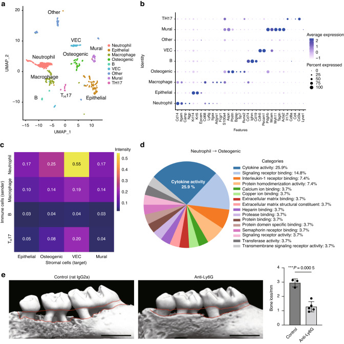
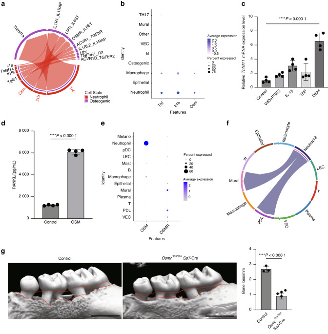
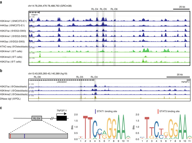
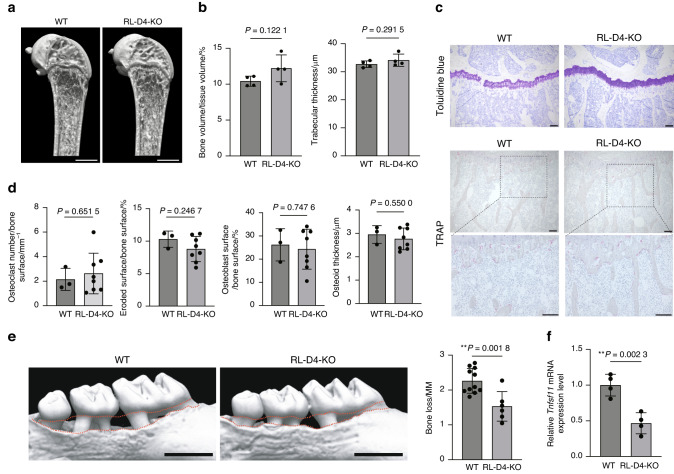
The immune-stromal cell interactions play a key role in health and diseases. In periodontitis, the most prevalent infectious disease in humans, immune cells accumulate in the oral mucosa and promote bone destruction by inducing receptor activator of nuclear factor-κB ligand (RANKL) expression in osteogenic cells such as osteoblasts and periodontal ligament cells. However, the detailed mechanism underlying immune-bone cell interactions in periodontitis is not fully understood. Here, we performed single-cell RNA-sequencing analysis on mouse periodontal lesions and showed that neutrophil-osteogenic cell crosstalk is involved in periodontitis-induced bone loss. The periodontal lesions displayed marked infiltration of neutrophils, and in silico analyses suggested that the neutrophils interacted with osteogenic cells through cytokine production. Among the cytokines expressed in the periodontal neutrophils, oncostatin M (OSM) potently induced RANKL expression in the primary osteoblasts, and deletion of the OSM receptor in osteogenic cells significantly ameliorated periodontitis-induced bone loss. Epigenomic data analyses identified the OSM-regulated RANKL enhancer region in osteogenic cells, and mice lacking this enhancer showed decreased periodontal bone loss while maintaining physiological bone metabolism. These findings shed light on the role of neutrophils in bone regulation during bacterial infection, highlighting the novel mechanism underlying osteoimmune crosstalk.


Conflict of interest statement

The Department of Osteoimmunology is an endowment department supported with an unrestricted grant from AYUMI Pharmaceutical Corporation, ELECOM, JCR Pharmaceuticals, Kondo Cotton Spinning, MIKIHOUSE, MITSUI FUDOSAN, Meiji, Noevir, TAKENAKA, TENNENBUTSU IKAGAKU KENKYU ZAIDAN and Yakult.

Figures

Fig. 1
Fig. 1
Neutrophils interact with osteogenic cells through cytokine production in periodontitis. a Uniform manifold approximation and projection (UMAP) plot of cells from the periodontal tissues of ligature-induced periodontitis mice (n = 5). b Dot plot of the expression of representative genes for each cluster. Each cell type was annotated using these marker genes. c Heat map showing the network intensity of interactions between immune cells (sender) and stromal cells (target). d Proportion of Gene Ontology terms on molecules mediating the effect of neutrophils on osteogenic cells. e Micro-CT analysis of periodontitis-induced bone loss in mice treated with isotype-control (rat IgG2a) and anti-Ly6G antibodies (n = 3 and n = 5, respectively). Scale bars, 1 mm. The upper red dotted line indicates the cement–enamel junction, and the lower red dotted line indicates the alveolar bone crest in the left panel. The periodontal bone loss was quantified in the right panel
Fig. 2
Fig. 2
OSM stimulates RANKL expression in osteogenic cells and promotes bone destruction in periodontitis. a Chord diagram showing cytokine signaling interaction pairs in the neutrophil-osteogenic network. b Dot plot showing the expression of TNF, IL-1β, and OSM in the scRNA-seq clusters of the mouse periodontitis lesion. c qPCR analysis of Tnfsf11 transcripts in calvaria-derived primary osteoblasts treated with TNF, IL-1β, OSM or Vitamin D plus Prostaglandin E2 (PGE2) (n = 4). The data were obtained from duplicate experiments. d RANKL concentration measured by ELISA in the lysate of primary osteoblasts treated with or without OSM. e Dot plot showing the expression of OSM and OSMR in scRNA-seq clusters of the human periodontitis lesion. f Chord diagram showing the OSM signaling interaction pairs in the cell-cell network of the human periodontitis lesion. g Micro-CT analysis of periodontitis-induced bone loss in the control (n = 3) and Osmrflox/flox Sp7-Cre mice (n = 5). Scale bars, 1 mm. The upper red dotted line indicates the cement–enamel junction and the lower red dotted line indicates the alveolar bone crest in the left panel. The periodontal bone loss was quantified in the right panel
Fig. 3
Fig. 3
Identification of the OSM-regulated RANKL enhancer region in osteogenic cells. a H3K4me1, H3K4me3, H4K5ac, and H3K27ac ChIP-seq and ATAC-seq profiles in mouse osteogenic cells (GSE51515, GSE54782, and GSE174045) and T cells (ENCFF021TWR). The gray shaded areas indicate the RANKL distal enhancer regions (RL-D4, RL-D5, and RL-D6). b H3K27ac, H3K4me1, and H3K4me2 ChIP-seq profiles in human osteogenic cells (GSE29611) and DNase-seq profile in human PDL fibroblasts (ENCFF021TWR). The gray shaded areas indicate the human homologous areas of the RL-D4, RL-D5, and RL-D6 regions. c Schematic depicting the STAT-binding motifs and the predicted binding sites within the human homologous region of RL-D4
Fig. 4
Fig. 4
The RANKL enhancer RL-D4 is involved in periodontitis-induced bone loss but not in physiological bone remodeling. Representative micro-CT images (a) and micro-CT parameters (b) of the femur in female WT and RL-D4-KO mice at the age of 12 weeks (n = 4 and n = 4). Scale bars, 1 mm. c Toluidine blue and TRAP staining of the proximal tibias of WT and RL-D4-KO mice at the age of 12 weeks. The data are representative of at least three independent experiments. Scale bars, 100 μm. d Bone histomorphometric analysis of the proximal tibias of WT and RL-D4-KO mice at the age of 12 weeks (n = 3 and n = 8). e Micro-CT analysis of periodontitis-induced bone loss in WT (n = 11) and RL-D4-KO mice (n = 5). The upper red dotted line indicates the cement–enamel junction and the lower red dotted line indicates the alveolar bone crest in the left panel. Scale bars, 1 mm. Periodontal bone loss was quantified in the right panel. f qPCR analysis of the Tnfsf11 transcripts in the calvaria-derived primary osteoblasts from WT mice and RL-D4-KO mice treated with OSM (n = 4). The data were obtained from duplicate experiments

References

    1. Lamont RJ, Koo H, Hajishengallis G. The oral microbiota: dynamic communities and host interactions. Nat. Rev. Microbiol. 2018;16:745–759. doi: 10.1038/s41579-018-0089-x. - DOI - PMC - PubMed
    1. Darveau RP. Periodontitis: a polymicrobial disruption of host homeostasis. Nat. Rev. Microbiol. 2010;8:481–490. doi: 10.1038/nrmicro2337. - DOI - PubMed
    1. Maekawa T, et al. Porphyromonas gingivalis manipulates complement and TLR signaling to uncouple bacterial clearance from inflammation and promote dysbiosis. Cell Host Microbe. 2014;15:768–778. doi: 10.1016/j.chom.2014.05.012. - DOI - PMC - PubMed
    1. Tsukasaki M, et al. Host defense against oral microbiota by bone-damaging T cells. Nat. Commun. 2018;9:701. doi: 10.1038/s41467-018-03147-6. - DOI - PMC - PubMed
    1. Tsukasaki M, Takayanagi H. Osteoimmunology: evolving concepts in bone–immune interactions in health and disease. Nat. Rev. Immunol. 2019;19:626–642. doi: 10.1038/s41577-019-0178-8. - DOI - PubMed

Publication types

Associated data