. 2024 Feb 27;15(1):1797.
doi: 10.1038/s41467-024-45946-0.

TopBP1 utilises a bipartite GINS binding mode to support genome replication

Affiliations

TopBP1 utilises a bipartite GINS binding mode to support genome replication

Matthew Day et al. Nat Commun. .

Abstract

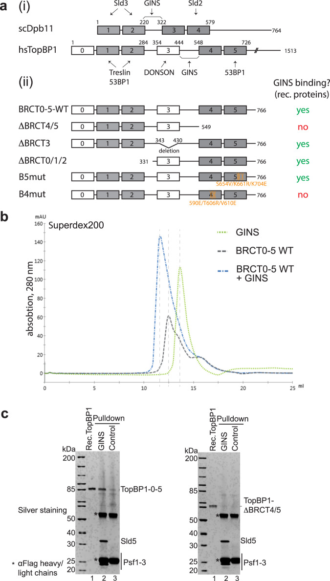
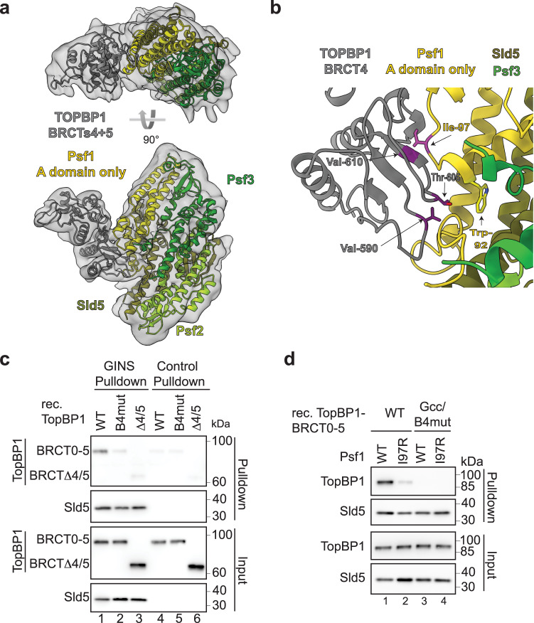
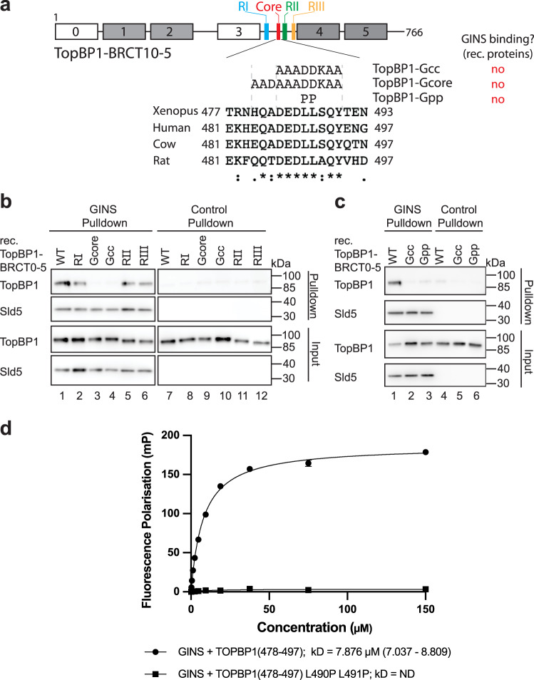
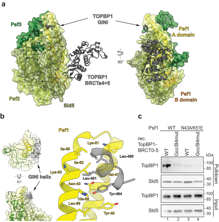
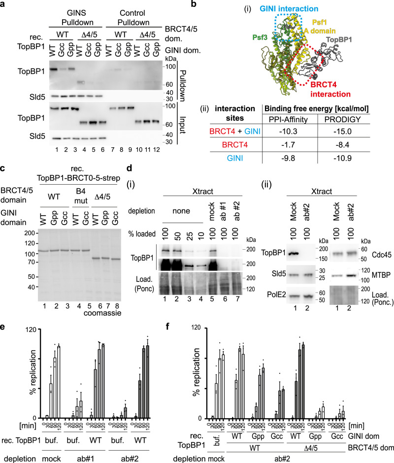
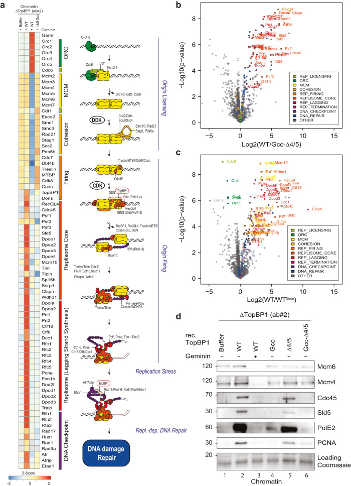
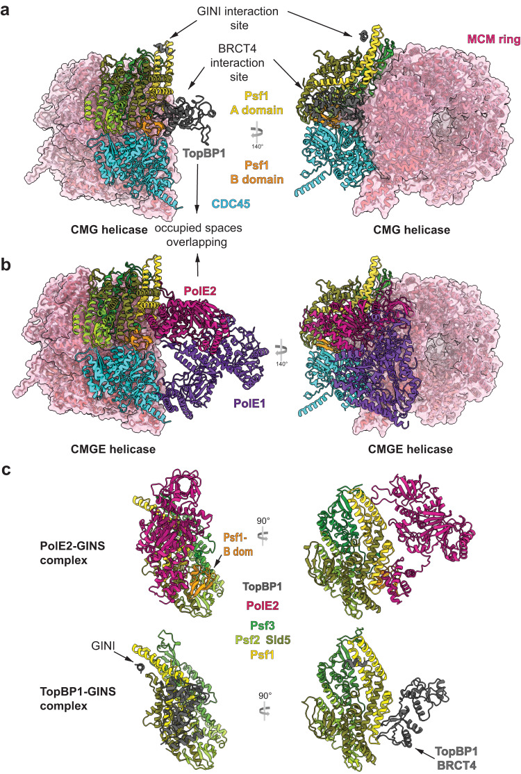
Activation of the replicative Mcm2-7 helicase by loading GINS and Cdc45 is crucial for replication origin firing, and as such for faithful genetic inheritance. Our biochemical and structural studies demonstrate that the helicase activator GINS interacts with TopBP1 through two separate binding surfaces, the first involving a stretch of highly conserved amino acids in the TopBP1-GINI region, the second a surface on TopBP1-BRCT4. The two surfaces bind to opposite ends of the A domain of the GINS subunit Psf1. Mutation analysis reveals that either surface is individually able to support TopBP1-GINS interaction, albeit with reduced affinity. Consistently, either surface is sufficient for replication origin firing in Xenopus egg extracts and becomes essential in the absence of the other. The TopBP1-GINS interaction appears sterically incompatible with simultaneous binding of DNA polymerase epsilon (Polε) to GINS when bound to Mcm2-7-Cdc45, although TopBP1-BRCT4 and the Polε subunit PolE2 show only partial competitivity in binding to Psf1. Our TopBP1-GINS model improves the understanding of the recently characterised metazoan pre-loading complex. It further predicts the coordination of three molecular origin firing processes, DNA polymerase epsilon arrival, TopBP1 ejection and GINS integration into Mcm2-7-Cdc45.


Conflict of interest statement

The authors declare no competing interests.

Figures

Fig. 1
Fig. 1. GINS and TopBP1 interact.
a (i) Domain model of human (hs) TopBP1, its conservation with BRCT1/2 and 3/4 domains (grey boxes) of budding yeast Dpb11 (scDpb11). Arrows point to binding domains for interacting proteins. (ii) Overview of TopBP1 wild type (WT) and BRCT deletion (Δ) and point mutants. GINS binding capabilities are labelled in red and green. Numbers, amino acid position; capital letters indicate amino acid substitutions. Amino acid substitutions of point mutants in BRCT4 (B4mut) and 5 (B5mut) are coloured orange. b Elution profiles of size exclusion chromatography (Superdex200) of individual recombinant TopBP1-BRCT0-5 protein (grey) and GINS tetramers (green), or of both upon incubation (blue). c Recombinant GINS immobilised on Flag-beads and control beads (Flag peptide-coupled beads) were used to pull down recombinant TopBP1-0-5-WT or TopBP1-ΔBRCT4/5 (compare a(ii)). SDS gels were silver stained. kDa specifies molecular weight marker bands. The experiment was done more than three times with similar results. Source data are provided as a Source data file.
Fig. 2
Fig. 2. Protein structure of the GINS-TopBP1-BRCT4/5 complex.
a Structural model of the GINS-TopBP1-BRCT4/5 complex. Crystal structures of the GINS subunits (PDB:2E9X) (shades of green, yellow), and the central BRCT4/5 domain of TopBP1 (PDB:3UEN) (grey) were docked into the cryo-EM volume shown as transparent volume. b Zoom-in of the BRCT4-Psf1 interface. The residues in stick representation appear crucial for the interface. Mutations to break the interaction are coloured pink (Psf1-I97R) and purple (TopBP1-B4mut; compare Fig. 1a(ii)). c Pulldown of the indicated recombinant TopBP1-BRCT0-5-strep versions (see Fig. 1a(ii)) by Flag-beads-immobilised recombinant GINS or Flag peptide-coupled control beads. Analysis was done by immunoblotting. The experiment was done more than three times with similar results. d Pulldown of the indicated recombinant TopBP1-BRCT0-5-WT-strep using immobilised GINS-WT or GINS carrying a Ile97 to arginine mutation in Psf1.TopBP1-Gcc/B4mut-strep (Figs. 1a(ii) and 3a) was used as a non-GINS binding control. The experiment was done twice with similar results. Source data are provided as a Source data file.
Fig. 3
Fig. 3. The conserved core of the TopBP1-GINI domain is required for a stable interaction with GINS.
a Domain model and T-COFFEE alignment (one-letter aa code) of TopBP1 proteins from humans (Q92547) Xenopus laevis (Q7ZZY3), Bos taurus (A0A3Q1LWE4) and Rattus norvegicus (A0A8I6GFZ6). Coloured boxes indicate GINI region mutants in sub-regions I (blue), II (green) and III (orange), and in the GINI core helix (red) (details in Supplementary Fig. 7). Amino acid substitutions, names of GINI core region mutations and corresponding GINS binding capabilities are shown. ., : and * indicate low conservation, high conservation and identical amino acids, respectively. b/c Pulldown of recombinant TopBP1-BRCT0-5-strep-WT or the indicated GINI region mutants (a) by bead-immobilised GINS or Flag peptide-coupled control beads. Analysis was done by immunoblotting. The experiments were done more than three times with similar results. Source data are provided as a Source data file. d Fluorescence polarisation measurements including dissociation constants (kD) calculations using recombinant GINS tetramer and fluorescently labelled TopBP1-GINI-core peptides (WT or Gpp (a)). For each peptide n = 3 independent experiments and data are presented as mean values +/−SEM.
Fig. 4
Fig. 4. The TopBP1-GINI domain interacts with Psf1-A domain.
a Structural model of TopBP1-BRCT3-5 complexed to the GINS tetramer predicted using AlphaFold2-Multimer. All chains are represented as cartoon. The transparent volume represents the surface of the GINS complex (not cryo-EM density). Of TopBP1, only the GINI helix (residues 485–496) and BRCT4/5 (residues 551-766), are shown. b Left: Cryo-EM volume showing extra helical density ascribed to the GINI helix (grey). Right: a structural model based on this cryo-EM and an AlphaFold2-Multimer predicted structure. c Pulldown of recombinant TopBP1-BRCT0-5-WT-strep using immobilised GINS-WT or GINS carrying the indicated Psf1 mutations. TopBP1-BRCT0-5-Gcc/B4mut-strep served as a non-GINS binding control. Analysis was done by immunoblotting. The experiment was done twice with similar results. Source data are provided as a Source data file.
Fig. 5
Fig. 5. Both GINS binding surfaces in TopBP1 cooperate in supporting genome replication.
a Immobilized recombinant Flag-GINS or Flag peptide-coupled beads were used to pulldown the indicated recombinant TopBP1-BRCT0-5-strep versions (Figs. 1a and 3a) using 100 mM NaAc in the binding buffer. The pulldown was analysed by immunoblotting. The experiment was done twice with similar results. b (i) shows the AlphaFold2-Multimer model of the GINS-TopBP1 complex that was used to estimate the binding free energy using PPI-Affinity and PRODIGY shown in (ii). Estimations were done for the binding interfaces involving TopBP1-GINI (blue) and TopBP1-BRCT4 (red), both together and alone. c Coomassie-stained SDS PAGE gel of the indicated recombinant TopBP1-BRCT0-5 proteins (Figs. 1a(ii) and 3a). d The indicated relative amounts (100%–10%) of immunodepleted and non-depleted Xenopus egg extracts (Xtract) were analysed by immunoblotting using anti-TopBP1 #2 and other indicated antibodies. TopBP1 antibodies #1 and #2, or unspecific IgG (mock) were used for immunodepletions. Ponc, ponceau staining. Error bars, SEM, n = 3. The experiments were done more than three times (i) or twice (ii) with similar results. e, f Replication analyses (radioactive nucleotide incorporation) using Xenopus egg extracts immunodepleted with antibodies anti-TopBP1#1, #2 or unspecific IgG (mock). Buffer (buf), recombinant wild-type (WT) or mutant TopBP1-BRCT0-5-strep (c) were added. Diamonds, Individual data points of n = 3 independent experiments, Error bars, SEM. Source data are provided as a Source data file.
Fig. 6
Fig. 6. GINS binding to TopBP1 is required for the origin firing step of replication.
ac Heat map showing the intensity of chromatin-bound proteins recovered from TopBP1-depleted interphase Xenopus egg extracts supplemented wild type or mutant TopBP1 determined by quantitative mass spectrometry. Chromatin was isolated 75 min after sperm addition. All extracts contained 50 mg/ml aphidicolin and, where indicated, 2.25 mM geminin. The heat map shows log2 transformed label-free quantification (LFQ) intensities z-score normalized across rows and mean averaged over four independent replicates (n = 4). For the intensities of all proteins see Supplementary Data 2. b, c Volcano plots highlighting a selection of differentially detected proteins in samples described in (a). Log2 transformed LFQ intensities from four replicates each were analysed by a two-sided Student’s t-test. Significantly changed proteins with an Benjamini–Hochberg adjusted p-value of equal or less than 0.05 are reported in Supplementary Data 2. For clarity, only proteins involved in DNA replication are labelled (all significantly enriched proteins are labelled in Supplementary Fig. 14). d Experiment as in (a) was analysed by chromatin isolation and immunoblotting with the indicated antibodies. Coomassie staining of the gel part containing histones served as a loading control.
Fig. 7
Fig. 7. Exclusive binding of TopBP1 and DNA polymerase epsilon to GINS in the CMG context.
a, b Structure of human CMG (PDB 7PFO), in panel (a) with our GINS-TopBP1 superposed, in panel (b) with DNA Polε (CMGE; PDB 7PFO) both shown from the same angle, demonstrating accessibility of the TopBP1 binding sites in CMG, and strong overlap between BRCT4 and PolE2 occupied spaces. The pink volume constitutes surface representation. c In-detail views of GINS-TopBP1 interaction sites from the cryo-EM model in this manuscript (8OK2) alongside the GINS-PolE2 interaction sites seen in the larger CMGE structure (7PFO).

References

    1. Siddiqui K, On KF, Diffley JF. Regulating DNA replication in eukarya. Cold Spring Harb. Perspect. Biol. 2013;5:a010108. - PMC - PubMed
    1. Remus D, et al. Concerted loading of Mcm2-7 double hexamers around DNA during DNA replication origin licensing. Cell. 2009;139:719–730. - PMC - PubMed
    1. Evrin C, et al. A double-hexameric MCM2-7 complex is loaded onto origin DNA during licensing of eukaryotic DNA replication. Proc. Natl Acad. Sci. USA. 2009;106:20240–20245. - PMC - PubMed
    1. Li N, et al. Structure of the eukaryotic MCM complex at 3.8 A. Nature. 2015;524:186–191. - PubMed
    1. Georgescu R, et al. Structure of eukaryotic CMG helicase at a replication fork and implications to replisome architecture and origin initiation. Proc. Natl Acad. Sci. USA. 2017;114:E697–E706. - PMC - PubMed

MeSH terms