. 2024 Feb 27;14(1):4715.
doi: 10.1038/s41598-024-52940-5.

MicroRNA-203a inhibits breast cancer progression through the PI3K/Akt and Wnt pathways

Affiliations

MicroRNA-203a inhibits breast cancer progression through the PI3K/Akt and Wnt pathways

Maryam Entezari et al. Sci Rep. .

Abstract

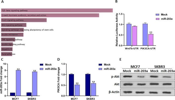
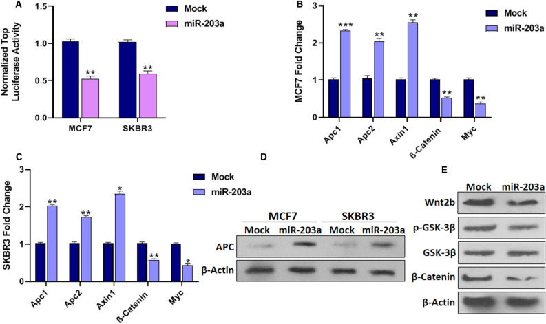
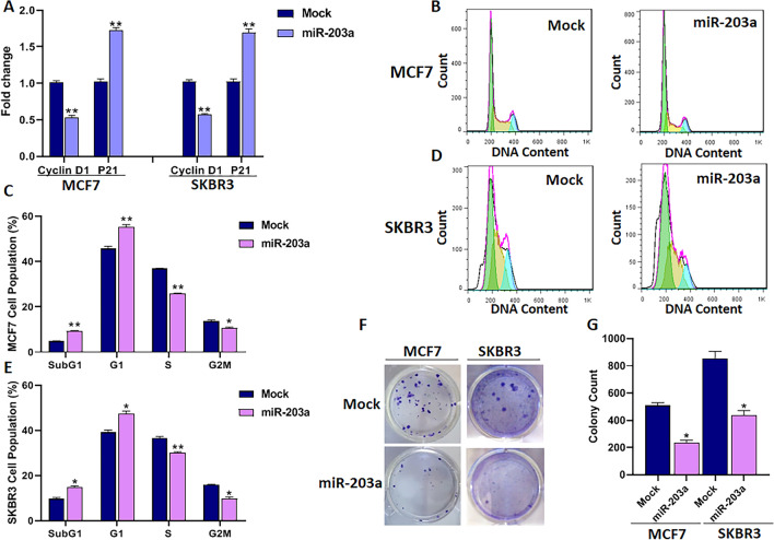
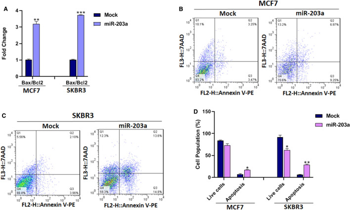
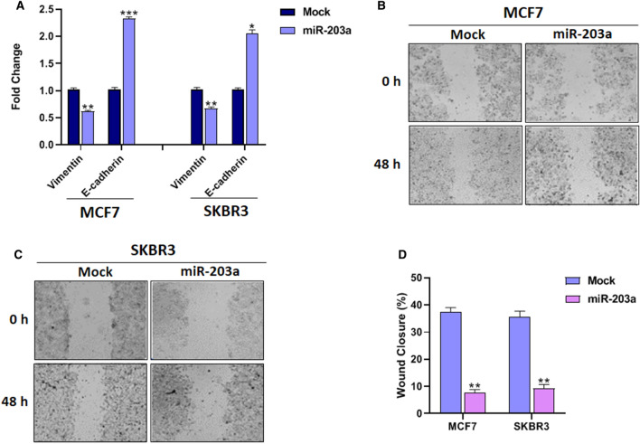
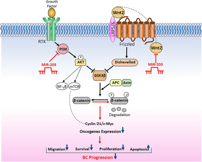
MicroRNA expression in breast cancer (BC) is explored both as a potential biomarker and for therapeutic purposes. Recent studies have revealed that miR-203a-3p is involved in BC, and importantly contributes to BC chemotherapy responses; however, the regulatory pathways of miR-203a in BC remain elusive. Hence, we aimed to investigate the miR-203a regulatory mechanisms and their potential functions in the progress of BC. To this end, the miR-203a potential involving pathways was predicted by databases analyzing its target genes. The relations between miR-203a, the phosphatidylinositol 3'-kinase (PI3K)-Akt, and Wnt signaling pathways were mechanistically investigated. Our results revealed that miR-203a inhibited the activation of the PI3K/Akt and Wnt pathways and reduced its downstream cell cycle signals, including Cyclin D1 and c-Myc. Moreover, the overexpression of miR-203a drastically arrested the cell cycle at subG1 and G1 phases, decreased the viability, proliferation, and migration, and increased apoptosis of BC cells. Therefore, miR-203a-3p may be considered a tumor suppressor factor and a potential biomarker or therapeutic target for BC.


Conflict of interest statement

The authors declare no competing interests.

Figures

Figure 1
Figure 1
MiR-203a regulates PI3K/Akt signaling through its ability to target PIK3CA. (A) Flowchart of enriched KEGG pathways (www.kegg.jp/kegg/kegg1.html) of the genes correlated with miR-203a expression categorized according to their P-value. (B) PIK3CA is a direct target of miR-203a shown by 3′UTR luciferase assay. The 3′UTR sequence of Wnt-7b was used as off-target in this experiment. (C) The transfection efficiency of miR-203a was measured by qRT-PCR 48h after transfection (D) miR-203a upregulation lowered PIK3CA mRNA level as measured by the qRT-PCR method. (D) Western blot of p-Akt and Akt showed that miR-203a inhibited Akt phosphorylation in BC cells. P-values as described in Fig. 1.
Figure 2
Figure 2
Association of miR-203a expression with Wnt signaling. (A) BC cells were co-transfected using TOP flash plasmid with either control or miR-203a expressing vectors. TOP flash luciferase activity indicated that overexpression of miR-203a was followed by significantly decreased Wnt activity in the BC cells. (B) miR-203a upregulation was followed by increased APC1, APC2, and Axin expression while decreased β-Catenin and c-Myc expression in MCF7 cells and (C) SKBR3 cells. (D) The western blot results confirmed the increased APC protein levels in BC cells by miR-203a overexpression. (E) Wnt2b, p-GSK-3β, and β-Catenin protein levels were decreased in MCF7 cells by overexpression of miR-203a as measured by Western blotting. *P < 0.05, **P < 0.01, ***P < 0.001.
Figure 3
Figure 3
The effect of miR-203a overexpression on cell cycle and proliferation of BC cells. (A) Cyclin D1 and P21 mRNA levels. (B) Histogram analysis of MCF7 and (D) SKBR3 cells cycles transfected with miR-203a overexpressing vector or mock vector 48h after transfection. (C), and (E) Bar plot analysis of BC cells percentage in each phase of the cell cycle, following the transfection. (F) Colony formation assay following miR-203a overexpression (G) Bar plot analysis of colony formation. *P < 0.05, **P < 0.01.
Figure 4
Figure 4
The effect of miR-203a overexpression on apoptosis of BC cells. (A) The ratio of mRNA expression of Bax to Bcl2 in BC cells with or without miR-203a overexpression. (B and C) Represents Annexin flow cytometry dot plots and (G) bar plot of BC cells percentage in different phases. *P < 0.05, **P < 0.01, ***P < 0.001.
Figure 5
Figure 5
The effect of miR-203a overexpression on migration BC cells. (A) qRT-PCR analysis of epithelial and mesenchymal markers in BC cells with or without miR-203a overexpression. (B) Wound healing analysis of MCF7 and (C) SKBR3 cells transfected with either miR-203a or mock vector at 0 and 48h post-scratching, Scale bar = 200 μm, (D) Quantitative analysis of scratch wound closure.*P < 0.05, **P < 0.01, ***P < 0.001.
Figure 6
Figure 6
A schematic representative of the molecular mechanisms by which miR-203a affects cancerous phenotypes of BC cells through the PI3K/Akt and Wnt signaling pathways.

Similar articles

Cited by

References

    1. Sung H, et al. Global Cancer Statistics 2020: GLOBOCAN estimates of incidence and mortality worldwide for 36 cancers in 185 countries. CA Cancer J. Clin. 2021;71:209–249. doi: 10.3322/caac.21660. - DOI - PubMed
    1. Jahangiri B, Khalaj-Kondori M, Asadollahi E, Purrafee Dizaj L, Sadeghizadeh M. MSC-Derived exosomes suppress colorectal cancer cell proliferation and metastasis via miR-100/mTOR/miR-143 pathway. Int. J. Pharm. 2022;627:122214. doi: 10.1016/j.ijpharm.2022.122214. - DOI - PubMed
    1. Garrido-Palacios A, et al. MicroRNA dysregulation in early breast cancer diagnosis: A systematic review and meta-analysis. Int. J. Mol. Sci. 2023;24:8270. doi: 10.3390/ijms24098270. - DOI - PMC - PubMed
    1. Mohammaddoust S, Sadeghizadeh M. MiR-183 functions as an oncogene via decreasing PTEN in breast cancer cells. Sci. Rep. 2023;13:8086. doi: 10.1038/s41598-023-35059-x. - DOI - PMC - PubMed
    1. Zhao S, Han J, Zheng L, Yang Z, Zhao L, Lv Y. MicroRNA-203 regulates growth and metastasis of breast cancer. Cell Physiol. Biochem. 2015;37:35–42. doi: 10.1159/000430331. - DOI - PubMed

MeSH terms

Grants and funding