. 2024 Feb;56(2):478-490.
doi: 10.1038/s12276-024-01170-w. Epub 2024 Feb 28.

Role of lung ornithine aminotransferase in idiopathic pulmonary fibrosis: regulation of mitochondrial ROS generation and TGF-β1 activity

Affiliations

Role of lung ornithine aminotransferase in idiopathic pulmonary fibrosis: regulation of mitochondrial ROS generation and TGF-β1 activity

Jong-Uk Lee et al. Exp Mol Med. 2024 Feb.

Abstract

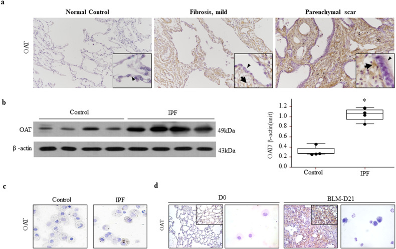
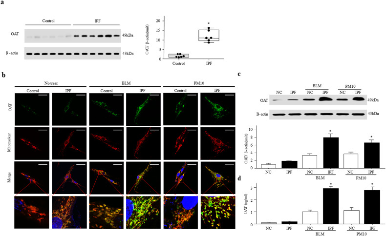
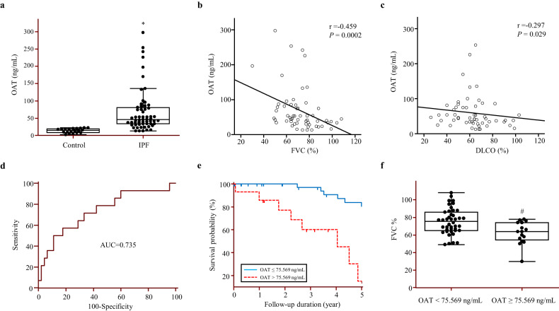
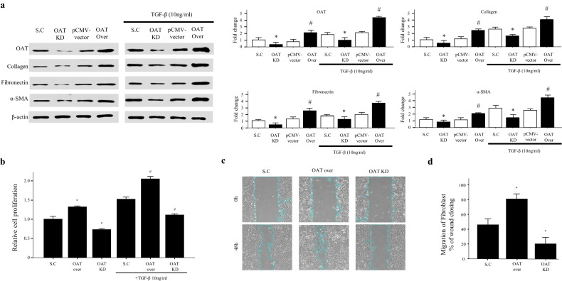
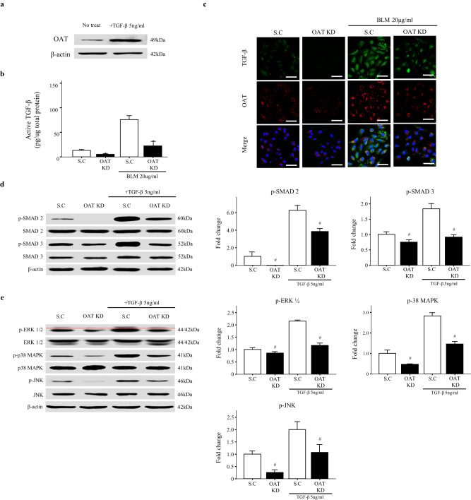
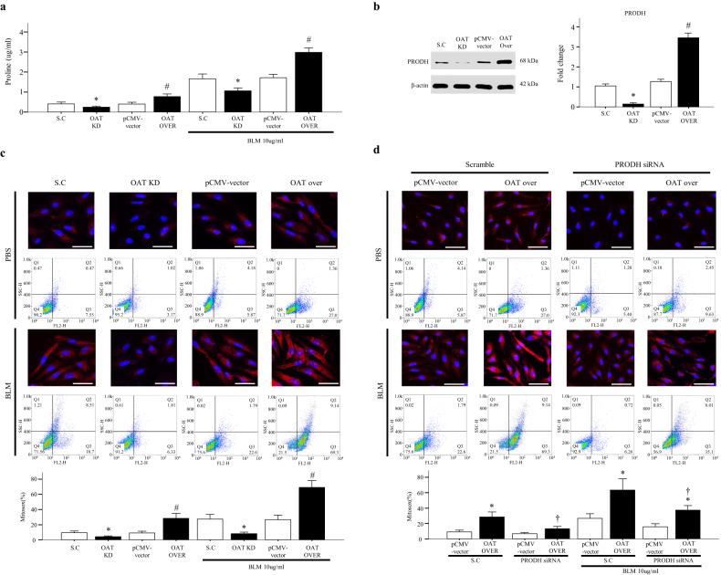
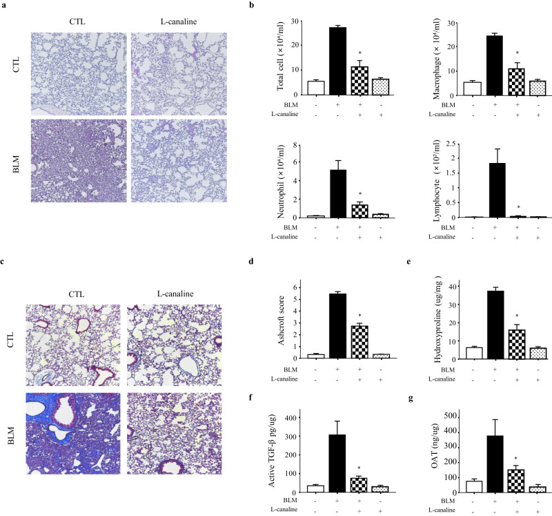
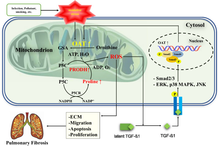
Idiopathic pulmonary fibrosis (IPF) is characterized by aberrant lung remodeling and the excessive accumulation of extracellular matrix (ECM) proteins. In a previous study, we found that the levels of ornithine aminotransferase (OAT), a principal enzyme in the proline metabolism pathway, were increased in the lungs of patients with IPF. However, the precise role played by OAT in the pathogenesis of IPF is not yet clear. The mechanism by which OAT affects fibrogenesis was assessed in vitro using OAT-overexpressing and OAT-knockdown lung fibroblasts. The therapeutic effects of OAT inhibition were assessed in the lungs of bleomycin-treated mice. OAT expression was increased in fibrotic areas, principally in interstitial fibroblasts, of lungs affected by IPF. OAT levels in the bronchoalveolar lavage fluid of IPF patients were inversely correlated with lung function. The survival rate was significantly lower in the group with an OAT level >75.659 ng/mL than in the group with an OAT level ≤75.659 ng/mL (HR, 29.53; p = 0.0008). OAT overexpression and knockdown increased and decreased ECM component production by lung fibroblasts, respectively. OAT knockdown also inhibited transforming growth factor-β1 (TGF)-β1 activity and TGF-β1 pathway signaling. OAT overexpression increased the generation of mitochondrial reactive oxygen species (ROS) by activating proline dehydrogenase. The OAT inhibitor L-canaline significantly attenuated bleomycin-induced lung injury and fibrosis. In conclusion, increased OAT levels in lungs affected by IPF contribute to the progression of fibrosis by promoting excessive mitochondrial ROS production, which in turn activates TGF-β1 signaling. OAT may be a useful target for treating patients with fibrotic lung diseases, including IPF.


Conflict of interest statement

The authors declare no competing interests.

Figures

Fig. 1
Fig. 1. Ornithine aminotransferase (OAT) expression is increased in the lungs of patients with idiopathic pulmonary fibrosis (IPF).
a Immunohistochemical staining for OAT in the lungs of control subjects compared to patients with IPF. Original magnification 100×; inset magnification 1000×. The arrowheads indicate alveolar epithelial cells (AECs) in the normal control tissues, hyperplastic AECs in mildly fibrotic tissues and metaplastic bronchiolar epithelial cells in parenchymal scars, and arrows indicate interstitial fibroblasts. b OAT expression in lung tissues was measured by immunoblotting. OAT bands were quantified using densitometry. *p < 0.05 versus the control subjects. An anti-OAT antibody was used to immunoblot equal quantities of proteins obtained from lung lysates. The blots were stripped and reprobed with an anti-β-actin antibody. Lung tissues were obtained from patients with IPF and control subjects who underwent thoracic surgery. c. OAT was not expressed in inflammatory cells in bronchoalveolar lavage fluid. Magnification, 1000×. d OAT expression was induced by BLM. BLM was administered into the tracheas of wild-type mice that were subsequently sacrificed on Day 21. Strong OAT expression was observed in fibrotic areas. Magnification, 100×; inset magnification, 1000×. OAT was not expressed in inflammatory cells. Magnification, 1000×.
Fig. 2
Fig. 2. OAT expression is increased in the cytoplasm and mitochondria of fibroblasts under oxidative stress.
a OAT expression levels in fibroblasts derived from the lung tissues of six IPF patients and six control subjects were measured by immunoblotting. OAT bands were quantified by densitometry. *p < 0.05 versus the control subjects. b Confocal analysis of MitoTracker Red and OAT colocalization in fibroblasts from lung tissues of patients with IPF and controls after treatment with 50 µg/mL PM 10 for 24 h. Magnification, 1000×. Scale bar, 50 μm. The insets show higher magnifications (10 × 50 μm rectangles) of the indicated regions of interest. c OAT levels in fibroblasts and d their culture supernatants after exposure of the cells to oxidative stress-inducing BLM and PM10. The data are presented as the means ± standard errors of the means (SEMs; n = 4). *p < 0.05 versus the BLM- or PM10-treated control group.
Fig. 3
Fig. 3. The OAT concentration in bronchoalveolar lavage fluid (BALF) is associated with lung function and the survival of patients with IPF.
a OAT protein levels in BALF were quantified using enzyme-linked immunosorbent assays (ELISAs). The data are expressed as medians with 25% and 75% quartiles. *p < 0.05 versus the control group. b Correlations between BALF OAT levels and forced vital capacity (FVC; % pred.) and c between BALF OAT levels and diffusing capacity of the lung for carbon monoxide (% pred.) in patients with IPF. d Receiver operating characteristic curve analysis to determine the optimal OAT concentration that could be used as cutoff values for predicting survival in 59 patients with IPF. e. Comparison of OAT level survival curves using Kaplan–Meier plots. The survival rate was markedly lower in the group with an OAT level ≥75.659 ng/mL (red line) than in the group with an OAT level <75.659 ng/mL (blue line; hazard ratio, 29.53; 95% confidence interval, 7.29–119.55; p < 0.0001). f Comparison of FVC in terms of BALF OAT levels. The data are expressed as medians with the 25% and 75% quartiles. #p < 0.05 versus the OAT < 75.659 ng/mL group.
Fig. 4
Fig. 4. OAT regulates major components of the extracellular matrix and fibroblast proliferation.
a Effect of knockdown and overexpression of OAT on collagen, fibronectin, and alpha-smooth muscle actin expression in lung fibroblasts. Protein expression was quantified using immunoblotting and densitometry (n = 4 per group). b Proliferation was assessed using a water-soluble tetrazolium assay. c Fibroblast migration was assessed using a wound-healing assay. d Quantification of migration. The data are expressed as the means ± SEMs (n = 4). *p < 0.05 versus the scramble or BLM-treated scramble group. #p < 0.05 versus the pCMV vector group or BLM-treated pCMV vector group.
Fig. 5
Fig. 5. OAT knockdown and overexpression alters transforming growth factor (TGF)-β1 activity in fibroblasts.
a Effect of TGF-β1 on OAT expression by fibroblasts. b Active TGF-β1 levels in fibroblast lysates were quantified using ELISAs. The data are expressed as the means ± SEMs (n = 4). *p = 0.05 versus the BLM+/OAT scramble group. c Expression of active TGF-β1 as determined by immunofluorescence staining. Magnification, 400×. Scale bar, 50 μm. d The levels of Smad-dependent signaling pathway proteins (i.e., Smad2 and Smad3) and e non-Smad-dependent signaling pathway proteins (i.e., extracellular signal-regulated kinase, c-Jun N-terminal kinases, and mitogen activated protein kinase) were quantified by immunoblotting. Red dotted lines are used to indicate the separation of p-ERK1 and ERK2. Independent experiments were analyzed using densitometry. The data are expressed as the means ± SEMs (n = 4). *p < 0.05 versus the scramble or BLM-treated scramble group, and #p < 0.05 versus the scramble group or the TGF-β1+/scramble group.
Fig. 6
Fig. 6. OAT knockdown and overexpression regulate proline dehydrogenase (PRODH)-mediated mitochondrial reactive oxygen species (ROS) production.
a Proline levels and b PRODH expression in OAT-knockdown and OAT-overexpressing lung fibroblasts. c, d Fluorescence intensities of mitochondria-derived ROS as measured using a MitoSOX Red probe. Magnification, 400×. Scale bar, 50 μm. MitoSOX Red specifically targets mitochondrial ROS, which appear red. The flow cytometry data are quantitative. The data are presented as the means ± standard errors of the means (SEMs; n = 4). *p < 0.05 versus the scrambled RNA (scramble) or BLM-treated scramble group. #p < 0.05 versus the pCMV vector group or BLM-treated pCMV vector group. p < 0.05 versus the PRODH knockdown/OAT-overexpressing cells or the BLM-treated PRODH knockdown/OAT-overexpressing cells.
Fig. 7
Fig. 7. The OAT inhibitor L-canaline attenuates BLM-induced lung inflammation and fibrosis in a mouse model.
a H&E staining of mouse lungs after 100 nM L-canaline was intranasally administered on Days 8–12. Lung samples were collected on Day 21. Magnification, 100×. b Cell counts in BALF collected on Day 21. Differences between BALF cell counts were analyzed based on 500 cells stained with Diff-Quik (n = 8 per group). c Masson’s trichrome staining. Magnification, 100×. d Lung fibrosis as quantified using the Ashcroft score (n = 8 per group). e Collagen measurements from hydroxyproline assays of control and BLM-treated mouse lungs, with and without L-canaline (n = 8 per group). f Active TGF-β1 and g OAT levels in control and BLM-treated mouse lungs, with and without L-canaline (n = 8 per group). Active TGF-β1 and OAT levels in lung lysates were quantified using ELISAs. The data are expressed as the means ± SEMs. *p < 0.05 versus the BLM-treated group.
Fig. 8
Fig. 8. Hypothetical model showing the role of OAT in IPF pathogenesis.
Increased expression of OAT activates the proline metabolism pathway and increases the level of PRODH, which generates reactive oxygen species (ROS). The increased ROS levels activate TGF-β1, stimulate fibrotic responses, such as fibroblast proliferation, and modulates ECM production.

Similar articles

Cited by

References

    1. American_Thoracic_Society. Idiopathic pulmonary fibrosis: diagnosis and treatment. International consensus statement. American Thoracic Society (ATS), and the European Respiratory Society (ERS) Am. J. Respir. Crit. Care Med. 2000;161:646–664. doi: 10.1164/ajrccm.161.2.ats3-00. - DOI - PubMed
    1. Booth AJ, et al. Acellular normal and fibrotic human lung matrices as a culture system for in vitro investigation. Am. J. Respir. Crit. Care Med. 2012;186:866–876. doi: 10.1164/rccm.201204-0754OC. - DOI - PMC - PubMed
    1. Hynes RO. The extracellular matrix: not just pretty fibrils. Science. 2009;326:1216–1219. doi: 10.1126/science.1176009. - DOI - PMC - PubMed
    1. Kulkarni T, O’Reilly P, Antony VB, Gaggar A, Thannickal VJ. Matrix remodeling in pulmonary fibrosis and emphysema. Am. J. Respir. Cell Mol. Biol. 2016;54:751–760. doi: 10.1165/rcmb.2015-0166PS. - DOI - PMC - PubMed
    1. Leeming DJ, et al. Serological investigation of the collagen degradation profile of patients with chronic obstructive pulmonary disease or idiopathic pulmonary fibrosis. Biomark. Insights. 2012;7:119–126. doi: 10.4137/BMI.S9415. - DOI - PMC - PubMed

LinkOut - more resources