Transanal opening of the intersphincteric space (TROPIS): a novel procedure on the horizon to effectively manage high complex anal fistulas
- PMID: 38414123
- PMCID: PMC10915533
- DOI: 10.3393/ac.2022.01263.0180
Transanal opening of the intersphincteric space (TROPIS): a novel procedure on the horizon to effectively manage high complex anal fistulas
Abstract
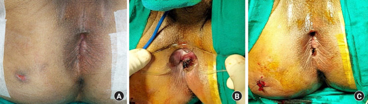
Anal fistulas, especially complex and high fistulas, are difficult to manage. The transanal opening of the intersphincteric space (TROPIS) procedure was first described in 2017, and a high success rate of over 90% was reported in high complex fistulas. Since then, more studies and even a meta-analysis have corroborated the high efficacy of this procedure in high fistulas. Conventionally, the main focus was to close the internal (primary) opening for the fistula to heal. However, most complex fistulas have a component of the fistula tract in the intersphincteric plane. This component is like an abscess (sepsis) in a closed space (2 muscle layers). It is a well-known fact that in the presence of sepsis, healing by secondary intention leads to better results than attempting to heal by primary intention. Therefore, TROPIS is the first procedure in which, instead of closing the internal opening, the opening is widened by laying open the fistula tract in the intersphincteric plane so that healing can occur by secondary intention. Although the drainage of high intersphincteric abscesses through the transanal route was described 5 decades ago, the routine utilization of TROPIS for the definitive management of high complex fistulas was first described in 2017. The external anal sphincter (EAS) is completely spared in TROPIS, as the fistula tract on either side of the EAS is managed separately-inner (medial) to the EAS by laying open the intersphincteric space and outer (lateral) to the EAS by curettage or excision.
Keywords: Anal fistula; Fistula in ano; Ligation of the intersphincteric tract; Rectal fistula; Transanal opening of the intersphincteric space (TROPIS).
Conflict of interest statement
No potential conflict of interest relevant to this article was reported.
Figures


Similar articles
-
Transanal opening of the intersphincteric space: a novel sphincter-sparing procedure to treat 325 high complex anal fistulas with long-term follow-up.Colorectal Dis. 2021 May;23(5):1213-1224. doi: 10.1111/codi.15555. Epub 2021 Feb 19. Colorectal Dis. 2021. PMID: 33529491
-
Transanal opening of intersphincteric space (TROPIS) - A new procedure to treat high complex anal fistula.Int J Surg. 2017 Apr;40:130-134. doi: 10.1016/j.ijsu.2017.02.095. Epub 2017 Mar 1. Int J Surg. 2017. PMID: 28259693
-
Transanal Opening of Intersphincteric Space for Fistula-in-Ano.Am Surg. 2022 Jun;88(6):1131-1136. doi: 10.1177/0003134821989048. Epub 2021 Jan 30. Am Surg. 2022. PMID: 33517706
-
Comparison between recent sphincter-sparing procedures for complex anal fistulas-ligation of intersphincteric tract vs transanal opening of intersphincteric space.World J Gastrointest Surg. 2022 May 27;14(5):374-382. doi: 10.4240/wjgs.v14.i5.374. World J Gastrointest Surg. 2022. PMID: 35734614 Free PMC article. Review.
-
Surgery of Simple and Complex Anal Fistulae in Adults: A Review of the Literature for Optimal Surgical Outcomes.Cureus. 2023 Mar 8;15(3):e35888. doi: 10.7759/cureus.35888. eCollection 2023 Mar. Cureus. 2023. PMID: 36911578 Free PMC article. Review.
Cited by
-
Comparative Evaluation Between Cutting of the Intersphincteric Space vs Cutting Seton in High Anal Fistula: A Randomized Controlled Trial.J Am Coll Surg. 2024 Dec 1;239(6):563-573. doi: 10.1097/XCS.0000000000001192. Epub 2024 Aug 21. J Am Coll Surg. 2024. PMID: 39166759 Free PMC article. Clinical Trial.
-
Recent advances in the diagnosis and treatment of complex anal fistula.Ann Coloproctol. 2024 Aug;40(4):321-335. doi: 10.3393/ac.2024.00325.0046. Epub 2024 Aug 30. Ann Coloproctol. 2024. PMID: 39228196 Free PMC article. Review.
-
Systematic review and meta-analysis of Transanal Opening of Intersphincteric Space (TROPIS) versus conventional treatments for anal fistula.Surg Open Sci. 2025 May 30;27:15-30. doi: 10.1016/j.sopen.2025.05.010. eCollection 2025 Sep. Surg Open Sci. 2025. PMID: 40607121 Free PMC article. Review.
-
Tissue engineering and regenerative medicine approaches in colorectal surgery.Ann Coloproctol. 2024 Aug;40(4):336-349. doi: 10.3393/ac.2024.00437.0062. Epub 2024 Aug 30. Ann Coloproctol. 2024. PMID: 39228197 Free PMC article. Review.
-
Achieving a high cure rate in complex anal fistulas: understanding the conceptual role of the Garg cardinal principles.Ann Coloproctol. 2024 Oct;40(5):521-523. doi: 10.3393/ac.2024.00430.0061. Epub 2024 Oct 25. Ann Coloproctol. 2024. PMID: 39448054 Free PMC article. No abstract available.
References
-
- Gaertner WB, Burgess PL, Davids JS, Lightner AL, Shogan BD, Sun MY, et al. The American Society of Colon and Rectal Surgeons clinical practice guidelines for the management of anorectal abscess, fistula-in-ano, and rectovaginal fistula. Dis Colon Rectum. 2022;65:964–85. - PubMed
-
- Garg P. Understanding understanding and treating supralevator fistula-in-ano: MRI analysis of 51 cases and a review of literature. Dis Colon Rectum. 2018;61:612–21. - PubMed
-
- Garg P, Kaur B. Transanal opening of intersphincteric space: a novel procedure to manage highly complex anal fistulas. Dis Colon Rectum. 2023;66:e292–3. - PubMed
-
- Li YB, Chen JH, Wang MD, Fu J, Zhou BC, Li DG, et al. Transanal opening of intersphincteric space for fistula-in-ano. Am Surg. 2022;88:1131–6. - PubMed
LinkOut - more resources
Full Text Sources