. 2024 May 1;32(5):1526-1539.
doi: 10.1016/j.ymthe.2024.02.031. Epub 2024 Feb 27.

Activation of the YAP/KLF5 transcriptional cascade in renal tubular cells aggravates kidney injury

Affiliations

Activation of the YAP/KLF5 transcriptional cascade in renal tubular cells aggravates kidney injury

Yang Liu et al. Mol Ther. .

Abstract

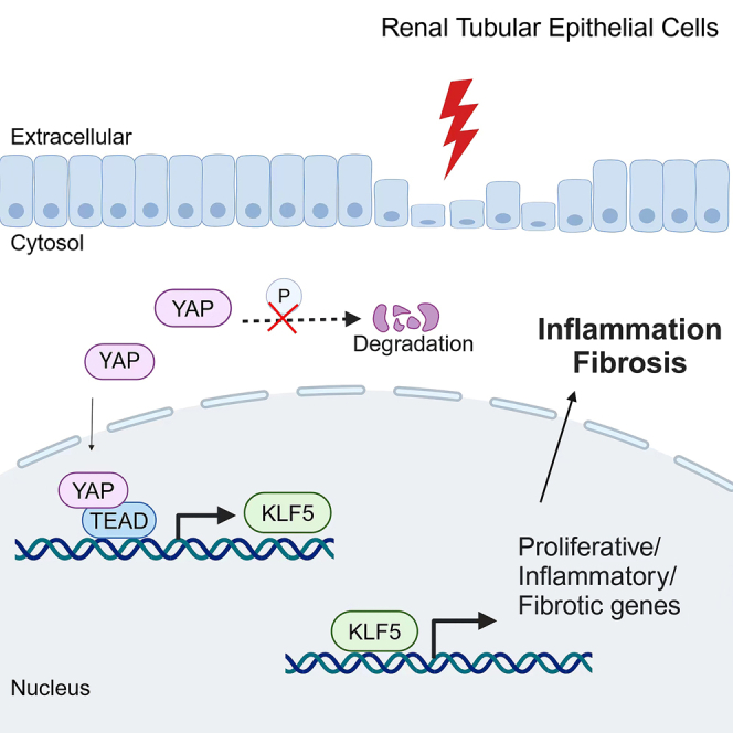
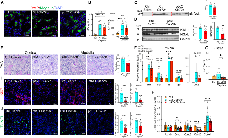
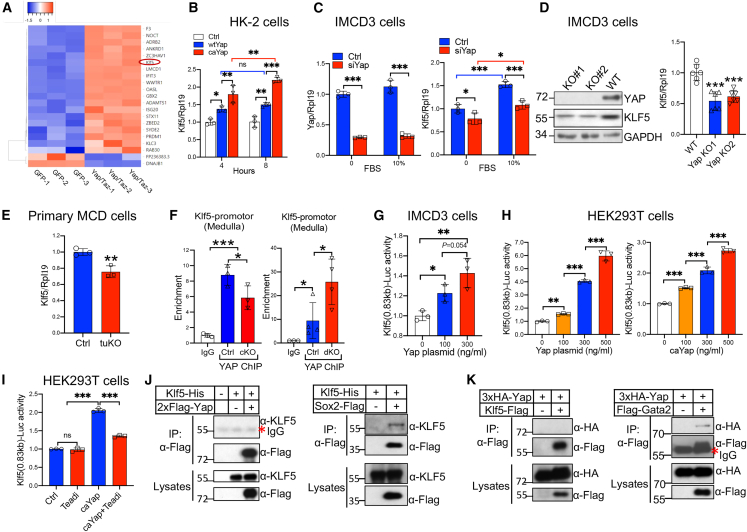
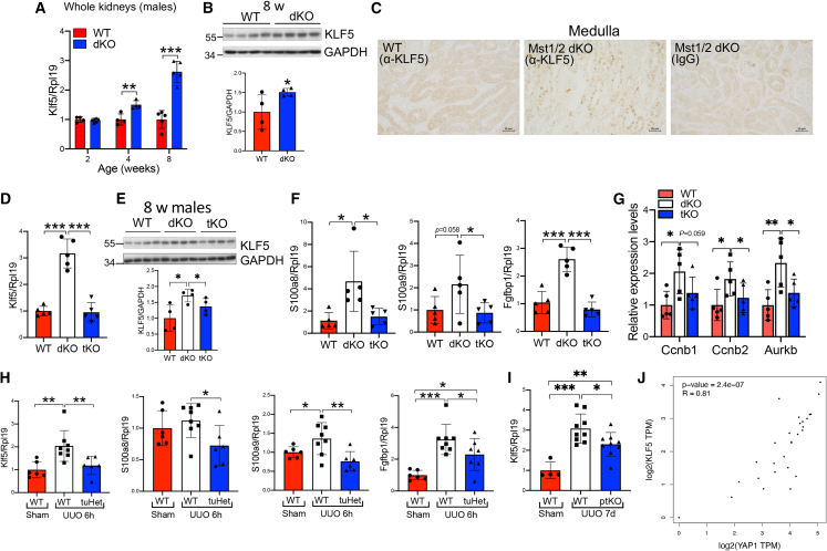
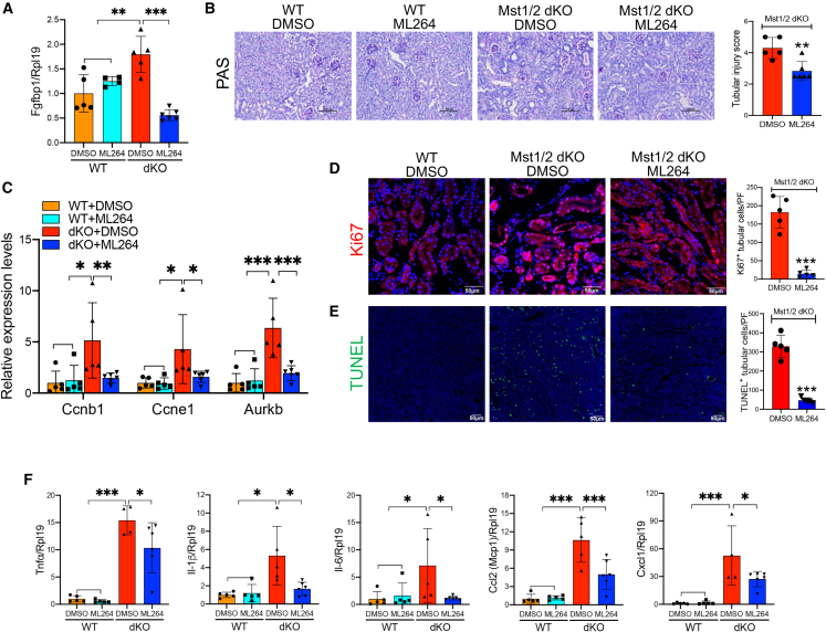
The Hippo/YAP pathway plays a critical role in tissue homeostasis. Our previous work demonstrated that renal tubular YAP activation induced by double knockout (dKO) of the upstream Hippo kinases Mst1 and Mst2 promotes tubular injury and renal inflammation under basal conditions. However, the importance of tubular YAP activation remains to be established in injured kidneys in which many other injurious pathways are simultaneously activated. Here, we show that tubular YAP was already activated 6 h after unilateral ureteral obstruction (UUO). Tubular YAP deficiency greatly attenuated tubular cell overproliferation, tubular injury, and renal inflammation induced by UUO or cisplatin. YAP promoted the transcription of the transcription factor KLF5. Consistent with this, the elevated expression of KLF5 and its target genes in Mst1/2 dKO or UUO kidneys was blocked by ablation of Yap in tubular cells. Inhibition of KLF5 prevented tubular cell overproliferation, tubular injury, and renal inflammation in Mst1/2 dKO kidneys. Therefore, our results demonstrate that tubular YAP is a key player in kidney injury. YAP and KLF5 form a transcriptional cascade, where tubular YAP activation induced by kidney injury promotes KLF5 transcription. Activation of this cascade induces tubular cell overproliferation, tubular injury, and renal inflammation.

Keywords: Hippo; KLF5; MST1; MST2; UUO; YAP; kidney; tubular injury.


Conflict of interest statement

Declaration of interests The authors declare no competing interests.

Figures

None
Graphical abstract
Figure 1
Figure 1
Tubular YAP activation in kidneys subjected to UUO for 6 h (UUO 6h) Male mice were subjected to UUO or sham operations. 6 h after surgery, kidneys were harvested. (A) Protein expression of phospho-YAP (p-YAP) and YAP. Western blotting was performed on the same mice as indicated. (B–D) Quantitative analysis of p-Erk1/2 levels relative to Erk1/2 levels (B), p-Smad1/5/8 levels relative to Smad1 levels (C), and p-Yap levels relative to YAP levels (D). Quantification of other kinases is presented in Figures S1A–S1H. (E) mRNA levels of the YAP target genes Ankrd1, Ctgf, and Cyr61. (F and G) Cellular localization of YAP in UUO kidneys. Frozen kidney sections were used for co-immunofluorescence staining of YAP (red) with AQP2 (green), THP (green), or megalin (green) (F). Tubular cells positive for nuclear YAP staining were counted in the cortex and medulla (G). n = 4/4 mice. (H) mRNA levels of the inflammatory factors TNF-α, IL-1β, IL-6, Mcp-1, Cxcl1, and Cxcl2. (I) mRNA levels of the proliferation genes Aurkb, Ccnb1, Ccnb2, Ccnd2, and Ccne1. (J) mRNA levels of the fibrotic genes Acta2, Col1, and Fn1. GAPDH was used as the loading control for western blotting. Rpl19 was used as the internal control for real-time PCR. ∗p < 0.05, ∗∗p < 0.01, ∗∗∗p < 0.001.
Figure 2
Figure 2
Effects of tubular Yap haploinsufficiency on the expression of inflammatory and fibrotic genes in kidneys subjected to UUO 6h Male WT (Yapf/w) and heterozygous Yap tuHet (Yapf/w;Ksp-cre) mice were subjected to UUO or sham operations. 6 h after surgery, kidneys were harvested, and real-time PCR assays were performed to measure mRNA levels of Yap (A); the Yap target genes Ankrd1, Ctgf, and Cyr61 (B); the inflammatory factors TNF-α, IL-1β, IL-6, Ccl2 (Mcp-1), and Cxcl1 (C); the proliferation gene Ccnd2 (D); the tubule injury markers Kim1 and Ngal (E); and the pro-fibrotic factors TGF-β1, TGF-β2 and PDGFβ (F). Rpl19 was used as the internal control. ∗p < 0.05, ∗∗p < 0.01, ∗∗∗p < 0.001.
Figure 3
Figure 3
Effects of inducible deletion of Yap in PTs on renal inflammation and fibrosis and tubular cell proliferation Male Yapwt/wt;GCE/WT and Yapf/f;GCE/WT mice were treated with tamoxifen by gavage once a day for 4 days. 3 days later, WT (Yapwt/wt;GCE/WT) and Yap ptKO (Yapf/f;GCE/WT) were subjected to UUO. 7 days after the surgery, kidney samples were collected. (A) Immunofluorescence for YAP. Frozen kidney sections were used for co-immunofluorescence staining of YAP (red) with megalin (green). (B–D) mRNA levels of the Yap target genes Ankrd1, Ctgf, and Cyr61 (B); the inflammatory factors Ccl2 (Mcp-1) and Ccl5 (C); and the fibrotic factors Acta2 (α-Sma), Col1, Col3, and Fn1 (D). (E) Protein levels of YAP, α-SMA, COL1, and FN-1. Kidney lysates were used for western blotting as indicated. Quantitative analysis of YAP, α-SMA, Col1, or Fn1 levels relative to GAPDH levels was performed. (F) Paraffin kidney sections were used for Masson trichrome staining (MTS) (left). The blue area relative to the whole area of a field was quantified to indicate the collagen deposition. n = 6/5 mice (right). (G) mRNA levels of the proliferation genes Ccnb2, Ccnd2, and Ccne1. (H) Immunofluorescence of Ki67. Ki67-positive tubular cells were counted. 4 WT and 4 Yap ptKO mice were used. Rpl19 was used as the internal control for real-time PCR, and GAPDH was used as the loading control for western blotting. ∗p < 0.05, ∗∗p < 0.01, ∗∗∗p < 0.001.
Figure 4
Figure 4
Effects of inducible deletion of Yap in PTs on tubular injury, tubular cell proliferation and death, renal inflammation and renal function 72 h after cisplatin injection Male Yapwt/wt;GCE/WT and Yapf/f;GCE/WT mice were treated with tamoxifen by gavage once a day for 4 days. 3 days later, WT (Yapwt/wt;GCE/WT) and Yap ptKO (Yapf/f;GCE/WT) mice were injected (i.p.) with cisplatin at a dose of 20 mg/kg body weight. 72 h after cisplatin injection, urine, serum, and kidney samples were collected. (A) Immunofluorescence for YAP. Frozen kidney sections were used for co-immunofluorescence staining of YAP (red) with megalin (green). (B) SCr and BUN levels. (C) Excreted NGAL in urine (uNGAL). Spot urine samples were collected from control mice and mice injected with cisplatin when they were sacrificed. 1 μL of urine was used for western blotting (left). uNGAL levels were quantified by densitometry (right). (D) Expression of KIM1 and NGAL in the kidneys of WT and Yap ptKO mice after cisplatin injection. Kidney lysates were subjected to western blotting for KIM1 and NGAL (left) and quantified (right). (E) Periodic acid-Schiff (PAS) staining of kidney sections from control and Yap ptKO mice after cisplatin injection. Representative images from the cortex and medulla are shown (left). Quantitative assessment of overall tubular injury is presented (right). (F–H) mRNA levels of the inflammatory factors TNF-α, IL-6, IL-1β, TGF-β1, and Cxcl2 (F); Col1 (G); and the proliferation genes Aurkb, Ccnb1, Ccnb2, Ccnd2, and Ccne1 (H). Real-time PCR analysis was performed as indicated. (I) Immunofluorescence for Ki67 in control and Yap ptKO kidneys after cisplatin injection. Frozen kidney sections were used for immunofluorescence staining of Ki67 (red) (left). Ki67-positive tubular cells in the cortex and medulla were counted (right). (J) TUNEL staining of kidney sections from control and Yap ptKO mice after cisplatin injection; nuclear localization of the TUNEL signal was demonstrated by cyan-colored nuclei after merging with DAPI (left). Positive tubular epithelial cells in the cortex and medulla were counted (right). Rpl19 was used as the internal control for real-time PCR, and GAPDH was used as the loading control for western blotting. ∗p < 0.05, ∗∗p < 0.01, ∗∗∗p < 0.001.
Figure 5
Figure 5
YAP promotes Klf5 transcription in renal tubular epithelial cells (A) Heatmap of the differentially expressed TFs (fold change > 2, p < 0.05, Q < 0.05) shows increases in Klf5 and the Yap/Taz target gene Ankrd1. HK-2 cells cultured in DMEM/F12 containing 1% FBS underwent electrotransfection with GFP or Yap and Taz plasmids. 5 h after electrotransfection, total RNA was extracted for RNA-seq. (B) YAP promoted Klf5 mRNA expression in HK-2 cells. HK-2 cells cultured in medium containing 1% FBS were electrically transfected with WT Yap or caYap. 4 or 8 h after electrotransfection, cells were collected to measure Klf5 mRNA levels by real-time PCR. (C) Inhibition of Yap reduced Klf5 mRNA expression in IMCD3 cells. Cells were transfected with scramble (Ctrl) or Yap siRNAs (siYap). 24 h after transfection, cells were cultured overnight with serum-free medium containing 0.1% BSA. Cells were then cultured in either serum-free medium or normal medium containing 10% FBS for 3 h before they were collected for analysis for Yap and Klf5 mRNA levels. (D) Yap deletion inhibited Klf5 expression. Two Yap KO clones (KO#1 and KO#2) were generated by CRISPR-Cas9. Absence of YAP protein and decreases in KLF5 protein in KO#1 and KO#2 were demonstrated by western blotting (left). Klf5 mRNA expression was analyzed by real-time PCR (right). (E) Expression of Klf5 mRNA in primary medullary collecting duct (MCD) cells isolated from control (Yapf/f) and Yap tuKO (Yapf/f; Ksp-Cre) kidneys. (F) YAP bound to the promoter of the Klf5 gene. Chromatin immunoprecipitation (ChIP) assays were performed by using anti-YAP or rabbit IgG as an isotype control in the medullae of 3 control and 3 Yap tuKO mice (left) or in 4 control or 4 Mst1/2 dKO mice (right). The enrichment of YAP binding to the promoter region of Klf5 was quantified by real-time PCR. (G) Effects of YAP on Klf5-Luc activity in IMCD3 cells. Cells were transfected with Klf5-Luc and pTK-RL in the presence of increasing amounts of Yap plasmids. 46 h later, luciferase assays were performed. (H) Effects of Yap and caYap on Klf5-Luc activity in HEK293T cells. Cells were transfected with Klf5-Luc and pTK-RL in the presence of increasing amounts of Yap (left) or caYap (right) plasmids. Luciferase assays were performed. (I) Effects of TEAD inhibition on Yap-induced Klf5-Luc activity in HEK293T cells. Cells were transfected with Klf5-Luc and pTK-RL, in the absence or presence of caYap plasmids, in combination with or without Teadi plasmids. Luciferase assays were performed. (J) KLF5 was not co-precipitated with YAP. HEK293T cells were transfected with Klf5-His in the absence or presence of 2×Flag-Yap (left) or Sox2-FLAG (right). Cell lysates were immunoprecipitated with anti-Flag to pull down YAP (left) or Sox2 (right). Western blotting was performed on whole lysates and precipitates as indicated. (K) YAP was not co-precipitated with KLF5. HEK293T cells were transfected with 3×hemagglutinin (HA)-Yap in the absence or presence of Klf5-Flag (left) or Flag-Gata2 (right). Cell lysates were immunoprecipitated with anti-Flag to pull down KLF5 (left) or GATA2 (right). Western blotting was performed on whole lysates and precipitates as indicated. Rpl19 was used as the internal control for real-time PCR. GAPDH was used as the loading control. ∗p < 0.05, ∗∗p < 0.01, ∗∗∗p < 0.001.
Figure 6
Figure 6
Impact of YAP on Klf5 expression in Mst1/2 dKO and UUO kidneys (A) Klf5 mRNA levels in the kidneys of WT and Mst1/2 dKO mice. Kidneys were collected from WT (Mst1f/f;Mst2f/f) and Mst1/2 dKO (Mst1f/f;Mst2f/f;Ksp-Cre) mice at 2, 4, and 8 weeks of age and analyzed for Klf5 mRNA levels by real-time PCR. (B) KLF5 protein levels in the kidneys of WT and Mst1/2 dKO mice. Kidneys were collected from WT and Mst1/2 dKO mice at 8 weeks of age and analyzed for KLF5 protein levels by western blotting (top). KLF5 levels relative to GAPDH levels were quantified (bottom). (C) Immunohistochemistry for KLF5 in the kidneys of WT and Mst1 dKO kidneys. Paraffin kidney sections from 8-week-old mice were used for immunohistochemistry with DAB staining. Normal IgG was used as the negative control. (D) Klf5 mRNA levels in the kidneys of WT, Mst1/2 dKO, and Mst1/2/Yap tKO mice. Kidneys were collected from WT, Mst1/2 dKO, and Mst1/2/Yap tKO (Mst1f/f;Mst2f/f;Yapf/f;Ksp-Cre) mice at 8 weeks of age and analyzed for Klf5 mRNA levels by real-time PCR. (E) KLF5 protein levels in the kidneys of WT, Mst1/2 dKO, and Mst1/2/Yap tKO mice. Kidneys were collected from WT, Mst1/2 dKO, and Mst1/2/Yap tKO mice at 8 weeks of age and analyzed for KLF5 protein levels by western blotting (top). KLF5 levels relative to GAPDH levels were quantified (bottom). (F) S100a8, S100a9, and Fgfbp1 mRNA expression in WT, Mst1/2 dKO, and Mst1/2/Yap tKO kidneys. Kidneys from 4-week-old mice were used for real-time PCR analysis. (G) Ccnb1, Ccnb2, and Aurkb mRNA expression in WT, Mst1/2 dKO, and Mst1/2/Yap tKO kidneys. Kidneys from 4-week-old mice were used for real-time PCR analysis. (H) Klf5, S1008a, S1009a, and Fgfbp1 mRNA expression in the kidneys of WT and Yap tuHet mice subjected to UUO for 6 h (UUO6h). Kidneys were collected from sham-operated WT (Yapf/w) mice or WT and Yap tuHet (Yapf/w;Ksp-Cre) mice with UUO6h and analyzed for Klf5, S1008a, S1009a, and Fgfbp1 mRNA levels by real-time PCR. (I) Klf5 mRNA expression in the kidneys of WT and ptKO mice subjected to UUO for 7 days (UUO7d). Male WT (Yapwt/wt;GCE/WT) and Yap ptKO (Yapf/f;GCE/WT) mice were treated with tamoxifen and then subjected to UUO. 7 days later, kidney samples were collected to measure Klf5 mRNA levels. (J) Correction in gene expression between Klf5 and Yap1 in the cortices of kidneys of healthy people (n = 28) was downloaded from the GTEx (Genotype-Tissue Expression) database. Rpl19 was used as the internal control for real-time PCR. GAPDH was used as the loading control for western blotting. ∗p < 0.05, ∗∗p < 0.01, ∗∗∗p < 0.001.
Figure 7
Figure 7
Effects of inhibition of KLF5 on tubular injury, tubular cell death, and proliferation and renal inflammation in Mst1/2 dKO mice The KLF5 inhibitor ML264 (25 mg/kg body weight) or the vehicle (DMSO) was injected (i.p.) every 12 h into control and Mst1/2 dKO mice at 7 weeks of age. Kidney samples were collected 6 h after the fifth injection. (A) Fgfbp1 mRNA expression. (B) PAS staining of kidney sections from Mst1/2 dKO mice treated with and without ML264. Representative images from the cortex are shown (left). Quantitative assessment of overall tubular injury is presented (right). (C) mRNA levels of Ccnb1, Ccne1, and Aurkb in the kidneys of WT and Mst1/2 dKO treated with and without ML264. (D) Immunofluorescence of Ki67 (red) on kidney sections from Mst1/2 dKO mice treated with and without ML264 (left). Tubular epithelial cells positive for Ki67 were counted (right). (E) TUNEL staining of kidney sections from Mst1/2 dKO mice treated with and without ML264 (left). Positive tubular epithelial cells were counted (right). (F) mRNA levels of Tnf-α, Ccl2 (Mcp-1), Il-6, Il-1β, and Cxcl1 in the kidneys of WT and Mst1/2 dKO mice treated with and without ML264. Rpl19 was used as the internal control for real-time PCR. ∗p < 0.05, ∗∗p < 0.01, ∗∗∗p < 0.001.

Similar articles

Cited by

References

    1. Humphreys B.D. Mechanisms of Renal Fibrosis. Annu. Rev. Physiol. 2018;80:309–326. - PubMed
    1. Levin A. Global challenges in kidney diseases. Nephrol. Dial. Transpl. 2018;33:371–372. - PubMed
    1. Yu S.M.W., Bonventre J.V. Acute kidney injury and maladaptive tubular repair leading to renal fibrosis. Curr. Opin. Nephrol. Hypertens. 2020;29:310–318. - PMC - PubMed
    1. Zhang L., Wang F., Wang L., Wang W., Liu B., Liu J., Chen M., He Q., Liao Y., Yu X., et al. Prevalence of chronic kidney disease in China: a cross-sectional survey. Lancet. 2012;379:815–822. - PubMed
    1. Liu B.C., Tang T.T., Lv L.L., Lan H.Y. Renal tubule injury: a driving force toward chronic kidney disease. Kidney Int. 2018;93:568–579. - PubMed

Publication types

MeSH terms

LinkOut - more resources