. 2024 Mar 1;28(2):129-143.
doi: 10.4196/kjpp.2024.28.2.129.

Effects of gas signaling molecule SO2 in cardiac functions of hyperthyroid rats

Affiliations

Effects of gas signaling molecule SO2 in cardiac functions of hyperthyroid rats

Qi Yang et al. Korean J Physiol Pharmacol. .

Abstract

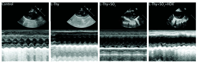
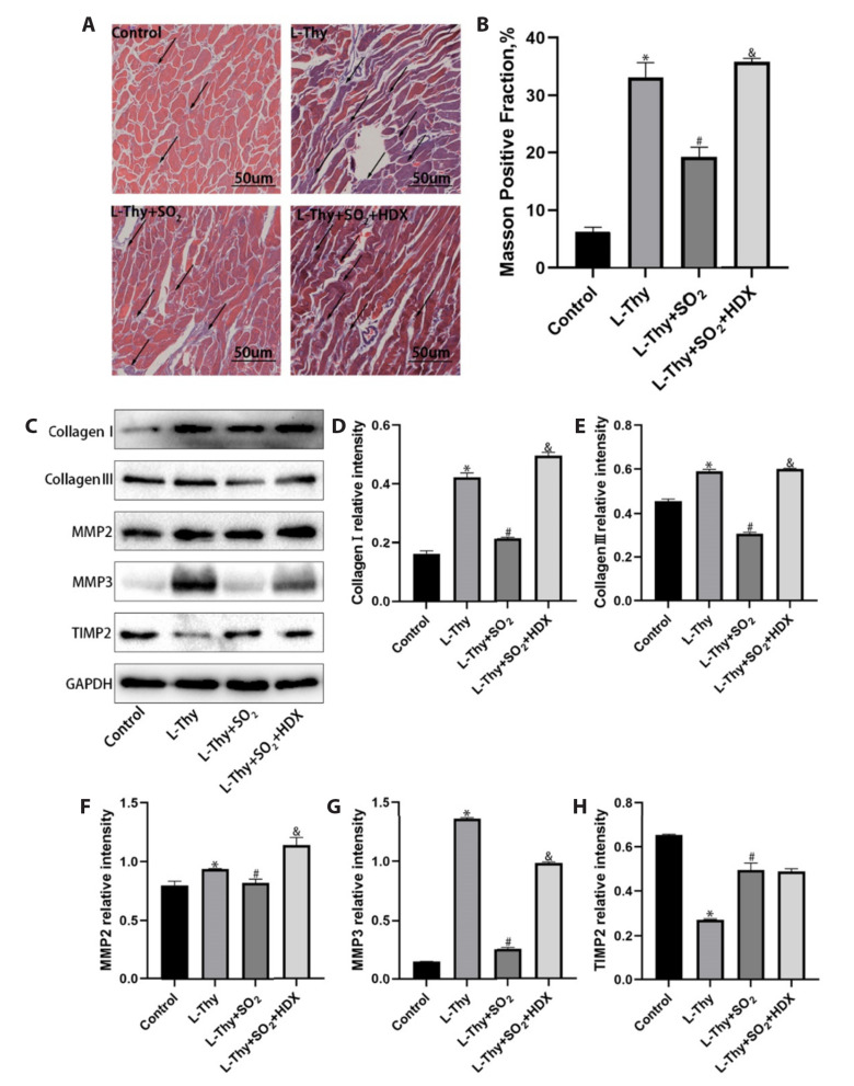
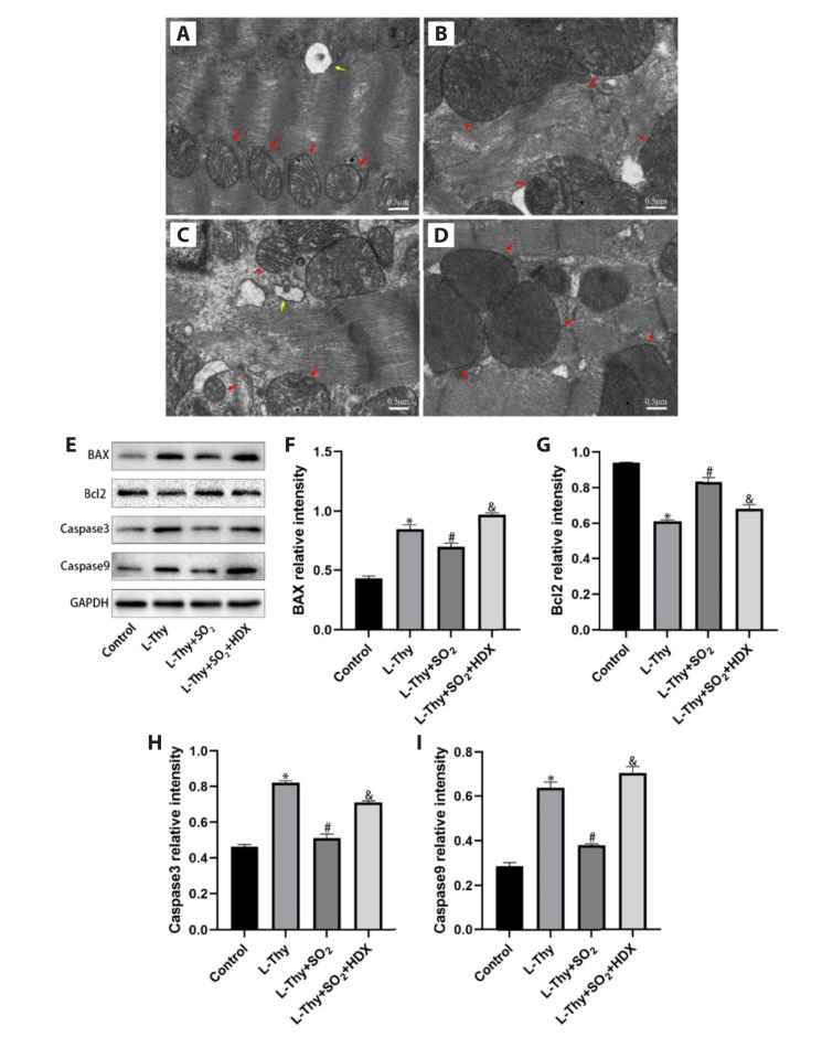
Sulfur dioxide (SO2), a novel endogenous gas signaling molecule, is involved in the regulation of cardiac function. Exerting a key role in progression of hyperthyroidism-induced cardiomyopathy (HTC), myocardial fibrosis is mainly caused by myocardial apoptosis, leading to poor treatment outcomes and prognoses. This study aimed to investigate the effect of SO2 on the hyperthyroidism-induced myocardial fibrosis and the underlying regulatory mechanisms. Elisa, Masson staining, Western-Blot, transmission electron microscope, and immunofluorescence were employed to evaluate the myocardial interstitial collagen deposition, endoplasmic reticulum stress (ERS), apoptosis, changes in endogenous SO2, and Hippo pathways from in vitro and in vivo experiments. The study results indicated that the hyperthyroidism-induced myocardial fibrosis was accompanied by decreased cardiac function, and down-regulated ERS, apoptosis, and endogenous SO2-producing enzyme aspartate aminotransferase (AAT)1/2 in cardiac myocytes. In contrast, exogenous SO2 donors improved cardiac function, reduced myocardial interstitial collagen deposition, up-regulated AAT1/2, antagonized ERS and apoptosis, and inhibited excessive activation of Hippo pathway in hyperthyroid rats. In conclusion, the results herein suggested that SO2 inhibited the overactivation of the Hippo pathway, antagonized ERS and apoptosis, and alleviated myocardial fibrosis in hyperthyroid rats. Therefore, this study was expected to identify intervention targets and new strategies for prevention and treatment of HTC.

Keywords: Apoptosis; Endoplasmic reticulum stress; Hippo signaling pathway; Hyperthyroidism-induced cardiomyopathy; Sulfur dioxide.


Conflict of interest statement

CONFLICTS OF INTEREST

The authors declare no conflicts of interest.

Figures

Fig. 1
Fig. 1. SO2 contents and AAT1/2 expressions in myocardial tissues of rats in each group.
(A) ELISA assay for SO2 contents. (B–D) Western-Blot results of changes in AAT1/2 expressions in myocardial tissues of hyperthyroid rats (n = 3); *, #, and & meant p < 0.05 vs. the values in the control, L-Thy, and L-Thy + SO2 groups, respectively. (E–G) Western-Blot results of changes in AAT1/2 expressions in myocardial tissues of each group of H9c2 cardiomyocytes (n = 3); *, #, and & meant p < 0.05 vs. the values in the control, L-Thy, and L-Thy + SO2 groups, respectively. SO2, sulfur dioxide; AAT, aspartate aminotransferase; L-Thy, levothyroxine; HDX, hydrogen deuterium exchange.
Fig. 2
Fig. 2. Echocardiograms of rats in each group.
L-Thy, levothyroxine; SO2, sulfur dioxide; HDX, hydrogen deuterium exchange.
Fig. 3
Fig. 3. Effect of SO2 on myocardial fibrosis in hyperthyroid rats.
(A) Masson staining results of myocardial tissues in the control, L-Thy, L-Thy + SO2, and L-Thy + SO2 + HDX groups under 10 × 40x field of view (blue-stained part referred to the collagen fibers, and black arrows marked the collagen fibers), n = 3. Scale bar = 50 um. (B) Masson staining results of collagen volume fraction, *,#, and & meant p < 0.05 vs. the values in the control, L-Thy, and L-Thy + SO2 groups, respectively. (C–H) Western-Blot results of changes in expressions of Collagen I, Collagen III, MMP2, MMP3, and TIMP2 in myocardial tissues of rats in each group, n = 3. *, #, and & meant p < 0.05 vs. the values in the control, L-Thy, and L-Thy + SO2 groups, respectively. L-Thy, levothyroxine; SO2, sulfur dioxide; HDX, hydrogen deuterium exchange.
Fig. 4
Fig. 4. SO2 reduces hyperthyroidism apoptosis in rat cardiomyocytes.
(A–D) TEM results of myocardial tissues of rats in control group, L-Thy group, L-Thy + SO2 group, and L-Thy + SO2 + HDX group, respectively (red arrow: mitochondria; ×7,000). Scale bar = 0.5 μM. (E–I) Western-Blot results for changes in expressions of BAX, Bcl2, Caspase3, and Caspase9 in myocardial tissues (n = 3). *, #, and & meant p < 0.05 vs. the values in the control, L-Thy, and L-Thy + SO2 groups, respectively. TEM, transmission electron microscopy; L-Thy, levothyroxine; SO2, sulfur dioxide; HDX, hydrogen deuterium exchange.
Fig. 5
Fig. 5. SO2 inhibits hyperthyroidism-induced apoptosis in cardiomyocytes.
(A) Immunofluorescence observation for the expressions of BAX and Caspase3 in each group of H9c2 cardiomyocytes. Scale bar = 50 μM. (B–D) Western-Blot results of changes in expressions of BAX and Bcl2 in each group of H9c2 cardiomyocytes (n = 3). *, #, and & meant p < 0.05 vs. the values in the control, L-Thy, and L-Thy + SO2 groups, respectively. L-Thy, levothyroxine; SO2, sulfur dioxide; HDX, hydrogen deuterium exchange.
Fig. 6
Fig. 6. SO2 reduces endoplasmic reticulum stress in cardiomyocytes of hyperthyroid rats.
(A–D) Western-Blot results for changes in expressions CHOP, GRP78/BIP, and ERP72 in cardiomyocytes of each group (n = 3). *, #, and & meant p < 0.05 vs. the values in the control, L-Thy, and L-Thy + SO2 groups, respectively. (E–G) Western-Blot results for changes in expressions of CHOP and ERP72 in each group of H9c2 cardiomyocytes (n = 3). *, #, and & meant p < 0.05 vs. the values in the control, L-Thy, and L-Thy + SO2 groups, respectively. L-Thy, levothyroxine; SO2, sulfur dioxide; HDX, hydrogen deuterium exchange.
Fig. 7
Fig. 7. Reversing effect of SO2 in up-regulation of Hippo pathway expression induced by hyperthyroid.
(A–D) Western-Blot results for changes in expressions of MST1, LATS1, and P-YAP in myocardial tissues of rats (with n = 3); *, #, and & meant p < 0.05 vs. the values in the control, L-Thy, and L-Thy + SO2 groups, respectively. (E–H) Western-Blot results for changes in expressions of MST1, LATS1, and P-YAP in H9c2 cardiomyocytes in each group; n = 3. *,#, and & meant p < 0.05 vs. the values in the control, L-Thy, and L-Thy + SO2 groups, respectively. (I, J) Cells were transfected with siRNA negative control (si-NC) or si-YAP1 for 24 h, and the expression of YAP1 was measured by Western blot analysis (I) and quantitatively analyzed (J). n = 3. * meant p < 0.05 vs. the values in the si-NC groups, respectively. (K, L) Western-Blot results for changes in expressions of ERP72 in H9c2 cardiomyocytes in each group; n = 3. * and # meant p < 0.05 vs. the values in the control, and L-Thy groups, respectively. L-Thy, levothyroxine; SO2, sulfur dioxide; HDX, hydrogen deuterium exchange.

References

    1. De Leo S, Lee SY, Braverman LE. Hyperthyroidism. Lancet. 2016;388:906–918. doi: 10.1016/S0140-6736(16)00278-6. - DOI - PMC - PubMed
    1. Garmendia Madariaga A, Santos Palacios S, Guillén-Grima F, Galofré JC. The incidence and prevalence of thyroid dysfunction in Europe: a meta-analysis. J Clin Endocrinol Metab. 2014;99:923–931. doi: 10.1210/jc.2013-2409. - DOI - PubMed
    1. Osuna PM, Udovcic M, Sharma MD. Hyperthyroidism and the heart. Methodist Debakey Cardiovasc J. 2017;13:60–63. doi: 10.14797/mdcj-13-2-60. - DOI - PMC - PubMed
    1. Danzi S, Klein I. Thyroid disease and the cardiovascular system. Endocrinol Metab Clin North Am. 2014;43:517–528. doi: 10.1016/j.ecl.2014.02.005. - DOI - PubMed
    1. Razvi S, Jabbar A, Pingitore A, Danzi S, Biondi B, Klein I, Peeters R, Zaman A, Iervasi G. Thyroid hormones and cardiovascular function and diseases. J Am Coll Cardiol. 2018;71:1781–1796. doi: 10.1016/j.jacc.2018.02.045. - DOI - PubMed