. 2024 Feb 23:19:1827-1842.
doi: 10.2147/IJN.S444470. eCollection 2024.

Localized Administration of Bcar3 siRNA via Nano-Self-Assembly to Treat Idiopathic Pulmonary Fibrosis by Disrupting Macrophage-Fibroblast Crosstalk

Affiliations

Localized Administration of Bcar3 siRNA via Nano-Self-Assembly to Treat Idiopathic Pulmonary Fibrosis by Disrupting Macrophage-Fibroblast Crosstalk

Chenxi Zeng et al. Int J Nanomedicine. .

Abstract

Background: Idiopathic pulmonary fibrosis (IPF) is a severe interstitial lung disease characterized by chronic lung injury leading to macrophage infiltration and fibroblast activation. However, there is no effective therapeutic strategy targeting the crucial crosstalk between macrophages and fibroblasts to halt IPF progression.

Methods: Studies were conducted in IPF patients and fibrotic mice models to elucidate the role of Bcar3 in the pathogenesis of pulmonary fibrosis. The effect of Bcar3 on macrophage polarization, fibroblast activation, and related signaling pathways were next investigated to unravel the underlying mechanisms.

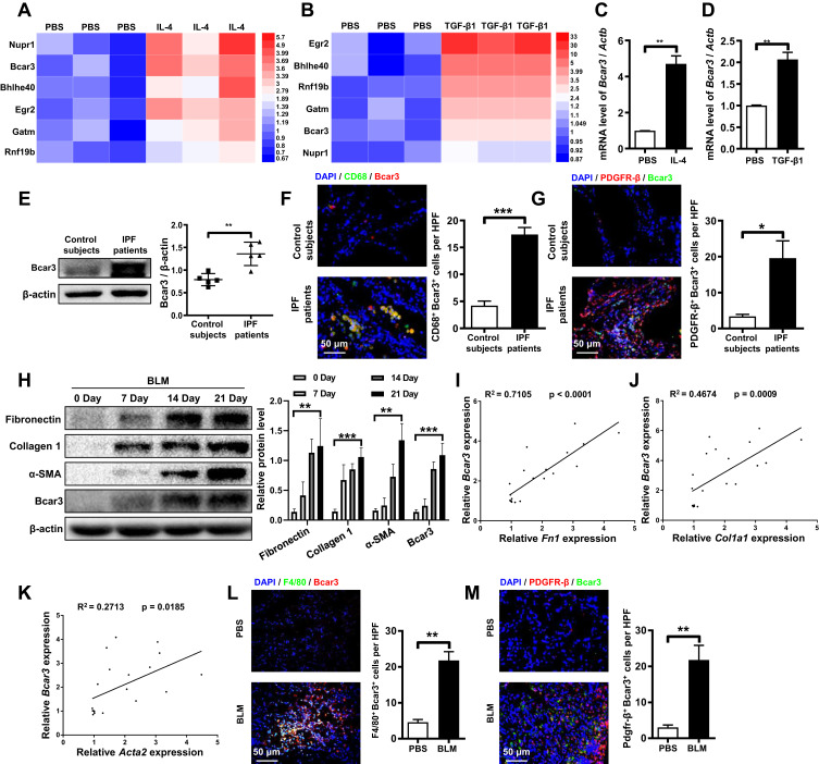
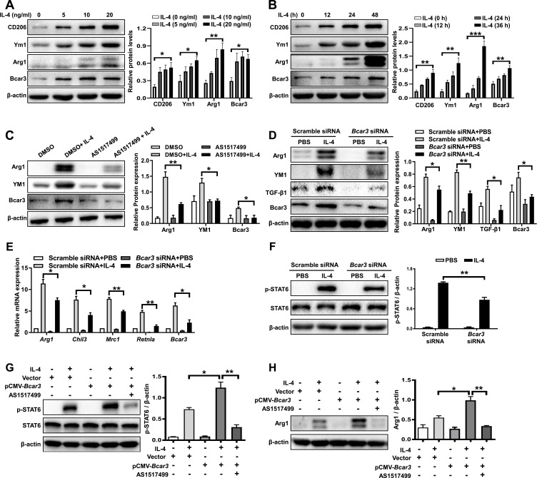
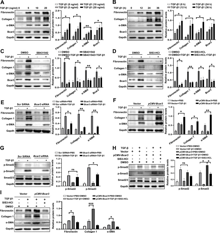
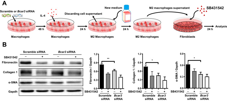
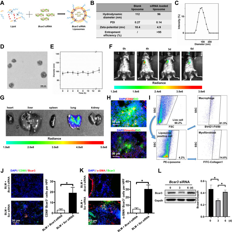
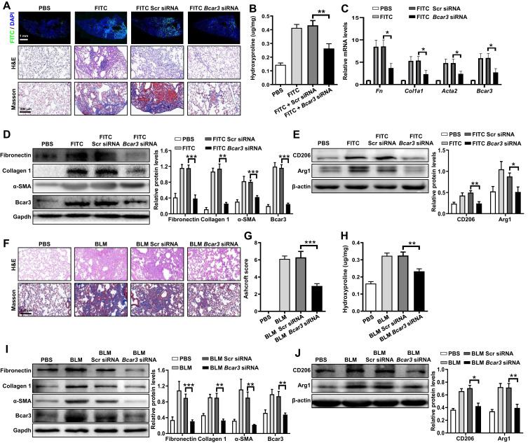
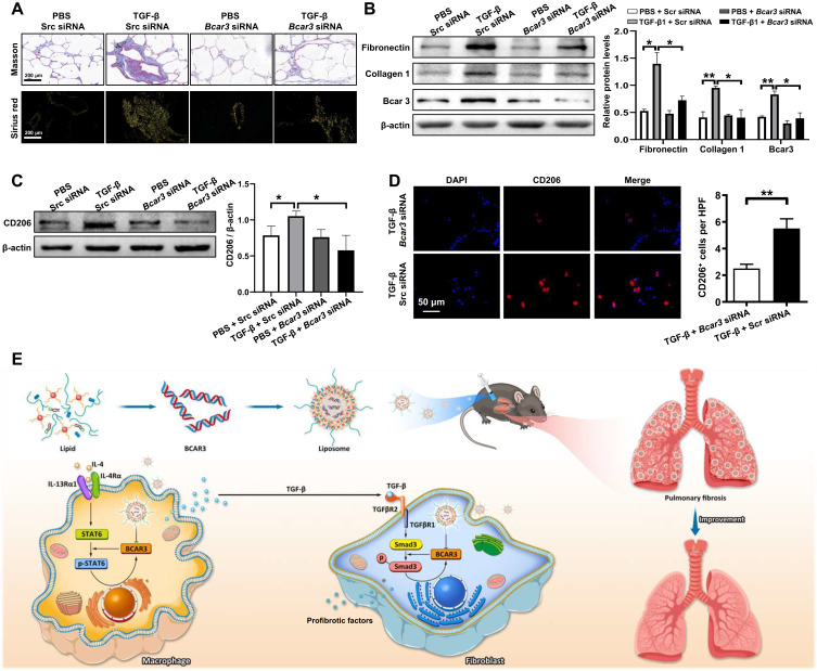
Results: Our study elucidates a marked increase in Bcar3 expression in lung tissues from IPF patients and fibrotic mice, recording 1.7 and 7.8-fold increases compared to control subjects, respectively. Additionally, Bcar3 was found to significantly enhance macrophage activation and fibroblast differentiation, observable in both in vivo and in vitro settings. Mechanistically, the upregulation of Bcar3 in macrophages was reliant on Stat6, while in fibroblasts, it depended on TGFβR1/Smad3. Furthermore, Bcar3 augmented IL-4/Stat6 pathway in macrophages and TGF-β/Smad3 pathway in fibroblasts, supporting a synergistic activation loop that expedited lung fibrogenesis. Notably, intratracheal injection of liposomes containing Bcar3 siRNA precisely delivered gene therapeutics to lung macrophages and fibroblasts, effectively reducing Bcar3 expression to 59% of baseline levels. Importantly, this intervention protected mice from lung fibrosis induced by either FITC or bleomycin, as well as human precision-cut lung slices against TGF-β1 stimulation.

Conclusion: Our study underscores the pivotal role of Bcar3 in orchestrating the macrophage-fibroblast crosstalk during pulmonary fibrosis progression. Targeting Bcar3 emerges as a novel therapeutic avenue to halt IPF progression and enhance patient prognosis.

Keywords: Bcar3; fibroblasts; idiopathic pulmonary fibrosis; liposomes; macrophages.


Conflict of interest statement

The authors declare that they have no competing interests in this work.

Figures

Figure 1
Figure 1
Differential expression of Bcar3 in IPF patient samples and pulmonary fibrosis mouse model. (A and B) show heatmaps displaying co-overexpressed genes in IL-4- and TGF-β1-treated macrophage and fibroblast, respectively, with colors representing the fold enrichment. (C and D) present the RT-PCR data of Bcar3 in macrophage and fibroblast after IL-4 and TGF-β1 induction, respectively. (E) depicts the Western blot results of Bcar3 in the lung homogenates of IPF patients (n=5) and control subjects (n=5). (F and G) display representative coimmunostaining data of Bcar3 and CD68 or PDGFR-β in the lung tissues of IPF patients and healthy controls. The nuclei exhibited a blue with DAPI. Magnification ×400. (H) shows the Western blot results of fibronectin, Collagen 1, a-SMA, and Bcar3 in the lung tissues of mice (n=5) at different time points after BLM stimulation. (IK) demonstrate RT-PCR assays of the correlations among Bcar3, Acta2, Col1a1, and Fn1 upon TGF-β1 stimulation. (L and M) exhibit representative coimmunostaining data of Bcar3 and PDGFR-β or F4/80 in the lung tissues of mice exposed to BLM or PBS. The nuclei exhibited a blue staining with DAPI. Magnification ×400. Mean±SEM. *p<0.05; **p<0.01; ***p<0.001.
Figure 2
Figure 2
The role of Bcar3 in promoting M2 macrophage polarization through enhanced p-Stat6 signaling. (A and B) show the Western blot results of Bcar3, Arg1, Ym1, and CD206 in BMDMs treated with various concentrations of IL-4 (A) and at various time points (B). (C) displays the Western blot results of Bcar3, Arg1, and Ym1 in BMDMs treated with AS1517499 after IL-4 induction. (D) demonstrates the attenuation of M2 macrophage polarization after Bcar3 knockdown, as shown by the Western blot results of Bcar3, Ym1, and Arg1. The corresponding mean data for each group are presented in the bar graph. (E) shows the RT-PCR data of Mrc1, Chil3, Arg1, and Retnla in BMDMs transfected with Bcar3 siRNA following IL-4 induction. (F) illustrates the Western blot results of Stat6 and p-Stat6 in BMDMs transfected with Bcar3 siRNA after 1 hour of IL-4 induction. (G and H) demonstrate the impact of Bcar3 overexpression on p-Stat6 and Arg1 in BMDMs treated with AS1517499 and induced with IL-4 for 1 hour and 24 hours, respectively. Mean±SEM. *p<0.05; **p<0.01; ***p<0.001.
Figure 3
Figure 3
The involvement of Bcar3 in promoting fibroblast-to-myofibroblast differentiation through the TGF-β1/Smad3 pathway. (A and B) show the Western blot results of Bcar3, fibronectin, Collagen 1, and a-SMA in fibroblasts treated with different doses of TGF-β1 (A) and at various time points (B). (C and D) depict Western blot results of Bcar3, fibronectin, Collagen 1, and a-SMA in fibroblasts treated with SB431542 (C) or SIS3-HCl (D) after TGF-β1 induction for 24 hours. (E) shows the Western blot results of Bcar3, fibronectin, Collagen 1, TGF-β1, and a-SMA in fibroblasts transfected with Bcar3 siRNA after 24 hours of TGF-β1 stimulation. (F) presents the Western blot results of fibronectin, Collagen 1, and a-SMA in Bcar3-overexpressing fibroblasts. (G and H) display Western blot results of Bcar3, Smad2/3 and p-Smad2/3 in fibroblasts transfected with Bcar3 siRNA after 1 hour of TGF-β1 stimulation, and in Bcar3-overexpressing fibroblasts exposed to SIS3-HCl and induced with TGF-β1 for 1 hour, respectively. (I) presents the Western blot results of fibronectin, Collagen 1, and a-SMA in Bcar3-overexpressing fibroblasts treated with SIS3-HCl after TGF-β1 induction for 24 hours. Mean±SEM. *p<0.05; **p<0.01; ***p<0.001.
Figure 4
Figure 4
The inhibitory effect of Bcar3 on reciprocal macrophage-fibroblast crosstalk. (A) presents a diagram illustrating the experimental setup of macrophage induction of fibroblast differentiation. (B) shows the Western blot results of fibronectin, Collagen 1, and a-SMA in fibroblasts treated with BMDM supernatant. Mean±SEM. *p<0.05; **p<0.01.
Figure 5
Figure 5
The distribution pattern of Bcar3 siRNA liposomes following intratracheal administration. (A) shows the preparation of liposomes loaded with Bcar3 siRNA. (B) presents the zeta potential, PDI, hydrodynamic diameter, and entrapment efficiency of liposomes (Bcar3 siRNA-loaded or empty). (C) displays a representative TEM image of liposomes loaded with Bcar3 siRNA. (D) shows the hydrodynamic diameter distribution of liposomes loaded with Bcar3 siRNA. (E) demonstrates the colloidal stability of liposomes loaded with Bcar3 siRNA in PBS. (F) shows the time-lapse IVIS imaging depicting the mouse’s response to intratracheally injected liposomes labeled with DiR at different time points. (G) presents ex vivo IVIS images of the main organs of mice. (H) displays immunofluorescence images illustrating the distribution of Vimentin+ cells (red), CD68+ cells (red), and DiO-labeled liposomes (green) in the mouse lungs. (I) presents the flow cytometry of liposome distribution in the lungs. (J and K) show immunofluorescent staining of CD68 (green) and Bcar3 (red), and Bcar3 (green) and a-SMA (red) in mice exposed to BLM followed by intravenous injection of liposomes containing Bcar3 siRNA. (L) presents the Western blot results of Bcar3 in mice exposed to BLM followed by intravenous injection of liposomes containing Bcar3 siRNA. Mean±SEM. *p<0.05.
Figure 6
Figure 6
The protective effects of intratracheal injection of liposomes containing Bcar3 siRNA in pulmonary fibrosis mice. (A) shows the FITC (top), H&E (middle), and Masson (bottom) staining of the lung tissues following FITC simulation. Magnification 200×. (B) presents a bar graph illustrating hydroxyproline levels in following FITC stimulation. (C) displays the RT-PCR data of Bcar3, Acta2, Col1a1, and Fn1 following FITC stimulation. (D) shows the Western blot results of fibronectin, Collagen 1, a-SMA, and Bcar3 in mice exposed to FITC. (E) illustrates Western blot results of Arg1 and CD206 in mice exposed to FITC. (F) shows H&E (up) and Masson (down) staining of the lungs in after BLM stimulation. (G) presents a bar graph revealing the semi-quantitative Ashcroft scores of fibrosis severity. Magnification 200×. (H) shows a bar graph of hydroxyproline levels in the lungs following BLM stimulation. (I) presents the Western blot results of fibronectin, Collagen 1, a-SMA, and Bcar3 in mice exposed to BLM. (J) illustrates the Western blot results of Arg1 and CD206 following BLM stimulation. Mean±SEM. *p<0.05; **p<0.01; ***p<0.001.
Figure 7
Figure 7
The attenuation of fibrosis-related phenotypes in TGF-β-exposed hPCLS after treatment with liposomes containing Bcar3 siRNA. (A) shows Sirius red staining (bottom) and Masson staining (top) of fibrosis-related phenotypes in TGF-β-exposed hPCLS after treatment with liposomes containing Bcar3 siRNA. (B) presents the Western blot results of Bcar3, fibronectin, and Collagen 1 in hPCLS after treatment with TGF-β and liposomes containing Bcar3 siRNA. The left panel shows representative Western blot data, and the right panel presents the bar graph of Western blot data. (C) displays the Western blot results of CD206 in hPCLS after treatment with TGF-β and liposomes containing Bcar3 siRNA. The left panel shows representative Western blot data, and the right panel presents the bar graph of Western blot data. (D) shows the immunofluorescence of CD206 in hPCLS after treatment with TGF-β and liposomes containing Bcar3 siRNA. (E) depicts a schematic of the mechanism by which Bcar3 regulates pulmonary fibrosis. Mean±SEM. *p<0.05; **p<0.01.

Similar articles

Cited by

References

    1. Prele CM, Miles T, Pearce DR, et al. Plasma cell but not CD20-mediated B-cell depletion protects from bleomycin-induced lung fibrosis. Eur Respir J. 2022;60(5):2101469. doi:10.1183/13993003.01469-2021 - DOI - PMC - PubMed
    1. Wang Y, Zhao J, Zhang H, Wang CY. Arginine is a key player in fibroblasts during the course of IPF development. Mol Ther. 2021;29(4):1361–1363. doi:10.1016/j.ymthe.2021.02.023 - DOI - PMC - PubMed
    1. Hu Y, Wang Q, Yu J, et al. Tartrate-resistant acid phosphatase 5 promotes pulmonary fibrosis by modulating beta-catenin signaling. Nat Commun. 2022;13(1):114. doi:10.1038/s41467-021-27684-9 - DOI - PMC - PubMed
    1. Wang Y, Zhang L, Wu GR, et al. MBD2 serves as a viable target against pulmonary fibrosis by inhibiting macrophage M2 program. Sci Adv. 2021;7(1):eabb6075. - PMC - PubMed
    1. Wang Y, Zhang L, Huang T, et al. The methyl-CpG-binding domain 2 facilitates pulmonary fibrosis by orchestrating fibroblast to myofibroblast differentiation. Eur Respir J. 2022;60(3):2003697. doi:10.1183/13993003.03697-2020 - DOI - PMC - PubMed

Substances