

Topical Ophthalmic Liposomes Dual-Modified with Penetratin and Hyaluronic Acid for the Noninvasive Treatment of Neovascular Age-Related Macular Degeneration
- PMID: 38414529
- PMCID: PMC10898604
- DOI: 10.2147/IJN.S446425
Topical Ophthalmic Liposomes Dual-Modified with Penetratin and Hyaluronic Acid for the Noninvasive Treatment of Neovascular Age-Related Macular Degeneration
Abstract
Introduction: Since intrinsic ocular barrier limits the intraocular penetration of therapeutic protein through eye drops, repeated intravitreal injections of anti-vascular endothelial growth factor (anti-VEGF) agents are the standard therapy for neovascular age-related macular degeneration (nAMD), which are highly invasive and may cause particular ocular complications, leading to poor patient compliance.
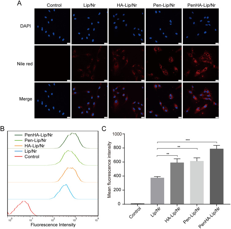
Methods: Using Penetratin (Pen) as the ocular penetration enhancer and hyaluronic acid (HA) as the retina-targeting ligand, a dual-modified ophthalmic liposome (Penetratin hyaluronic acid-liposome/Conbercept, PenHA-Lip/Conb) eye drop was designed to non-invasively penetrate the ocular barrier and deliver anti-VEGF therapeutic agents to the targeted intraocular tissue.
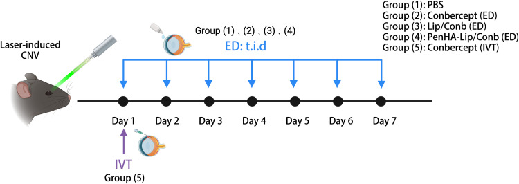
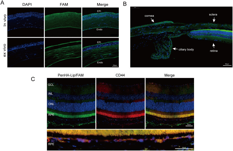
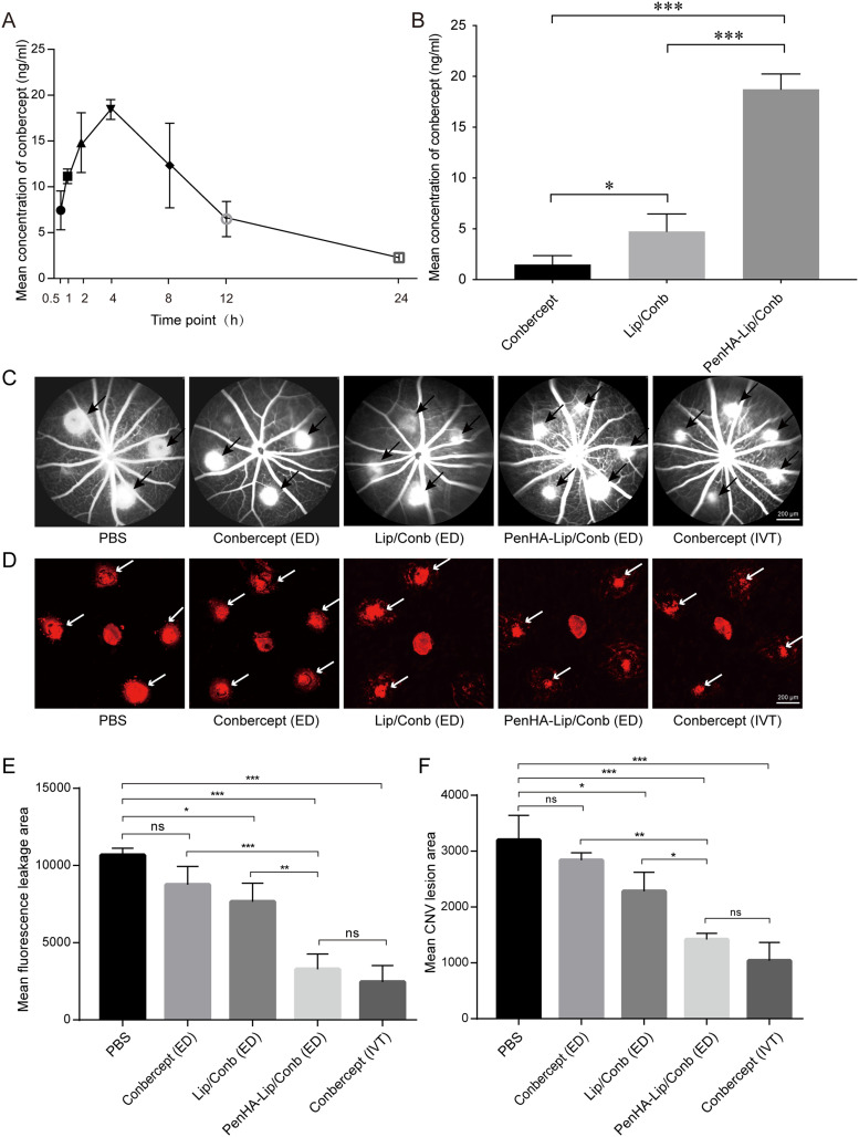
Results: PenHA-Lip effectively penetrates the ocular barrier and targets the retinal pigment epithelium via corneal and non-corneal pathways. After a single topical administration of conbercept-loaded PenHA-Lip (PenHA-Lip/Conb), the intraocular concentration of conbercept peaked at 18.74 ± 1.09 ng/mL at 4 h, which is 11.55-fold higher than unmodified conbercept. In a laser-induced choroidal neovascularization (CNV) mouse model, PenHA-Lip/Conb eye drops three times daily for seven days inhibited CNV formation and progression without any significant tissue toxicity and achieved an equivalent effect to a single intravitreal conbercept injection.
Conclusion: PenHA-Lip efficiently and safely delivered conbercept to the posterior eye segment and may be a promising noninvasive therapeutic option for nAMD.
Keywords: Penetratin; conbercept; dual-modified liposomes; hyaluronic acid; ocular drug delivery.
© 2024 Sun et al.
Conflict of interest statement
The authors declare that they have no competing interests in this work.
Figures


Similar articles
-
Peptide-Bound Aflibercept Eye Drops for Treatment of Neovascular Age-Related Macular Degeneration in Nonhuman Primates.Adv Sci (Weinh). 2025 Mar;12(11):e2410744. doi: 10.1002/advs.202410744. Epub 2025 Jan 30. Adv Sci (Weinh). 2025. PMID: 39888276 Free PMC article.
-
Topical Application of Cell-Penetrating Peptide Modified Anti-VEGF Drug Alleviated Choroidal Neovascularization in Mice.Int J Nanomedicine. 2024 Jan 3;19:35-51. doi: 10.2147/IJN.S428684. eCollection 2024. Int J Nanomedicine. 2024. PMID: 38187905 Free PMC article.
-
Topical Delivery of Anti-VEGF Drugs to the Ocular Posterior Segment Using Cell-Penetrating Peptides.Invest Ophthalmol Vis Sci. 2017 May 1;58(5):2578-2590. doi: 10.1167/iovs.16-20072. Invest Ophthalmol Vis Sci. 2017. PMID: 28494491
-
Profile of conbercept in the treatment of neovascular age-related macular degeneration.Drug Des Devel Ther. 2015 Apr 22;9:2311-20. doi: 10.2147/DDDT.S67536. eCollection 2015. Drug Des Devel Ther. 2015. PMID: 25960634 Free PMC article. Review.
-
Updates on the Management of Ocular Vasculopathies with VEGF Inhibitor Conbercept.Curr Eye Res. 2020 Dec;45(12):1467-1476. doi: 10.1080/02713683.2020.1781193. Epub 2020 Jul 7. Curr Eye Res. 2020. PMID: 32631094 Review.
Cited by
-
Topical Ocular Drug Delivery: The Impact of Permeation Enhancers.Pharmaceutics. 2025 Mar 31;17(4):447. doi: 10.3390/pharmaceutics17040447. Pharmaceutics. 2025. PMID: 40284442 Free PMC article. Review.
-
The Role of Reactive Oxygen Species in Age-Related Macular Degeneration: A Comprehensive Review of Antioxidant Therapies.Biomedicines. 2024 Jul 16;12(7):1579. doi: 10.3390/biomedicines12071579. Biomedicines. 2024. PMID: 39062152 Free PMC article. Review.
-
Utilization of Nanoparticles for Treating Age-Related Macular Degeneration.Pharmaceuticals (Basel). 2025 Jan 25;18(2):162. doi: 10.3390/ph18020162. Pharmaceuticals (Basel). 2025. PMID: 40005976 Free PMC article. Review.
-
Age-Related Macular Degeneration (AMD): Pathophysiology, Drug Targeting Approaches, and Recent Developments in Nanotherapeutics.Medicina (Kaunas). 2024 Oct 8;60(10):1647. doi: 10.3390/medicina60101647. Medicina (Kaunas). 2024. PMID: 39459435 Free PMC article. Review.
-
Frontier applications of retinal nanomedicine: progress, challenges and perspectives.J Nanobiotechnology. 2025 Feb 25;23(1):143. doi: 10.1186/s12951-025-03095-6. J Nanobiotechnology. 2025. PMID: 40001147 Free PMC article. Review.
References
-
- Steinmetz JD, Bourne RRA, Briant PS. Causes of blindness and vision impairment in 2020 and trends over 30 years, and prevalence of avoidable blindness in relation to VISION 2020: the right to sight: an analysis for the global burden of disease study. Lancet Glob Health. 2021;9(2):e144–e60. doi:10.1016/S2214-109X(20)30489-7 - DOI - PMC - PubMed
MeSH terms
Substances
LinkOut - more resources
Full Text Sources
Medical
