. 2024 Feb 13:15:1309691.
doi: 10.3389/fneur.2024.1309691. eCollection 2024.

Endoscopic endonasal transsphenoidal surgery for unusual sellar lesions: eight cases and review of the literature

Affiliations

Endoscopic endonasal transsphenoidal surgery for unusual sellar lesions: eight cases and review of the literature

Jiandong Wu et al. Front Neurol. .

Abstract

Background: Preoperative imaging for some unusual lesions in the sellar region can pose challenges in establishing a definitive diagnosis, impacting treatment strategies.

Methods: This study is a retrospective analysis of eight cases involving unusual sellar region lesions, all treated with endoscopic endonasal transsphenoidal surgery (EETS). We present the clinical, endocrine, and radiological characteristics, along with the outcomes of these cases.

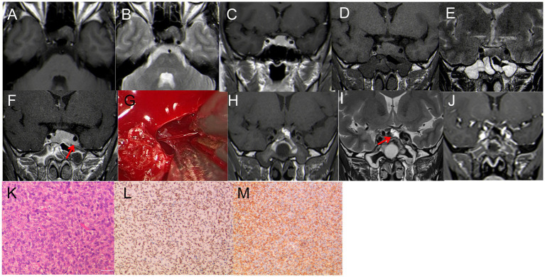
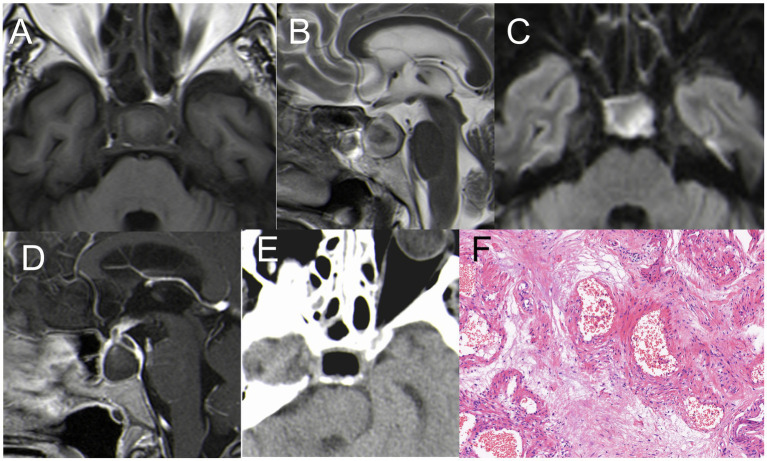
Results: Among the eight cases, the lesions were identified as follows: Solitary fibrous tumor (SFT) in one case, Lymphocytic hypophysitis (LYH) in one case, Cavernous sinus hemangiomas (CSH) in one case, Ossifying fibroma (OF) in two cases; Sphenoid sinus mucocele (SSM) in one case, Pituitary abscess (PA) in two cases. All patients underwent successful EETS, and their diagnoses were confirmed through pathological examination. Postoperatively, all patients had uneventful recoveries without occurrences of diabetes insipidus or visual impairment.

Conclusion: Our study retrospectively analyzed eight unusual lesions of the sellar region. Some lesions exhibit specific imaging characteristics and clinical details that can aid in preoperative diagnosis and inform treatment strategies for these unusual sellar diseases.

Keywords: Mucocele; cavernous sinus hemangiomas; endoscopic endonasal surgery; lymphocytic hypophysitis; ossifying fibroma; pituitary abscess; sellar lesions; solitary fibrous tumor.


Conflict of interest statement

The authors declare that the research was conducted in the absence of any commercial or financial relationships that could be construed as a potential conflict of interest.

Figures

Figure 1
Figure 1
(A–C) First MRI. (A) T1WI: isodense signal, (B) T2-MRI: isodense signal. (C) Enhanced T1WI: Significant enhancement, size 20 mm*18 mm*15 mm. (D–F) Review 3 months after the first surgery. (D) T1WI: isodense signal; (E) T2WI: isodense signal; (F) enhanced T1WI: Significant enhancement, size 30 mm*20 mm*18 mm. (G) During the operation, it was found that the tumor invaded the left cavernous sinus, and the blood vessels supplying the tumor bleeding was obvious, and the hemostasis was effectively stopped by bipolar electrocoagulation. (H–J) Three days after the second surgery. (H–I) T1WI; I: T2WI,The high signal indicated by the red arrow is the fat tissue; (J) Enhanced T1WI, satisfactory tumor resection with no residual. (K–L) Pathological examination, (K) H&E staining (×200); (L–M) immunohistochemistry (×200); CD 99 (+), STAT6 (+).
Figure 2
Figure 2
(A,B) T1WI and T2WI showed iso-hyper signal; (C,D) enhancement on T1WI and no enhancement in the cystic area, showing the “snowman” sign, 30 mm*22 mm in size. (E) The surrounding tumor tissue presented with pronounced fibrosis and a firm texture, markedly different from a typical pituitary tumor. (F–I) MRI showed that the tumor had disappeared after two months; (F) T1WI showed inhomogeneous hyperintense signal, red arrow showed cystic hypersignal; (G) T2WI: inhomogeneous hypointense signal. (H,I) Enhanced T1WI: Significant enhancement, red arrow showed pituitary. (K–M) Pathological examination, (J) H&E staining (×40) The disorder and destruction of pituitary tissue caused by lymphoplasmic cell inflammation and fibrosis; (K–M) immunohistochemistry (×40): (K) CD 20 (+), (L) CD 38 (+), (M) CD 43 (+).
Figure 3
Figure 3
(A) T1WI iso-signal, the red arrow indicated that the tumor is located in the right cavernous sinus (B) T2WI iso-hyper mixed signal. (C) T1WI enhancement: The tumor did not significant enhancement, the margin was enhanced, 30mm*30mm in size; (D) Normal pituitary and tumor tissue were fully exposed during the operation, the red arrow indicated the pituitary, the black arrow indicated the tumor, the white arrow indicated the internal carotid artery. (E–F) Postoperative MRI, the red arrow indicated satisfactory resection of the tumor. (G): H&E staining (×40); (H) immunohistochemistry (×40), CD 34(+).
Figure 4
Figure 4
Case 4 CT + MRI. (A) CT scan showed uneven density in the left sphenoid bone (B) hyper-signal on T1WI; (C) hyper-signal on T2WI; (D) Postoperative CT indicated satisfactory resection of the tumor. (E) Postoperative MRI (T1WI) indicated satisfactory resection of the tumor. (F) H&E staining demonstrating features typical of OF (×200).
Figure 5
Figure 5
Case 5 CT+ MRI (A) CT scan showed uneven density in the left sphenoid bone (B) hypo-signal on T1WI; (C) inhomogeneous hyperintense signal on T2WI; (D) T1WI enhancement: The tumor significant enhancement, 45 mm*30 mm in size. (E) The calcified lesions were removed with a drill. (F) Postoperative CT indicated small residual tumor in the left sphenoid bone. (G) H&E staining demonstrating features typical of OF (×200).
Figure 6
Figure 6
(A) CT: a lesion in the sphenoid sinus, with obvious dilation and bone destruction on the left side (B) hyper-signal on T1WI; (C) hypo-signal on T2WI; (D,E) Coronal enhancement of T1WI showed obvious enhancement, size 35*30 mm. (F) Postoperative CT indicated satisfactory resection of the tumor. (F) H&E staining demonstrating features typical of mucocele (×200).
Figure 7
Figure 7
(A) Hypointense on T1WI; (B) inhomogeneous hyperintense on T2WI; (C) hyperintense on DWI; (D) The pituitary gland and pituitary stalk were significantly strengthened. (E) Postoperative CT indicated satisfactory resection of the PA. (F) H&E staining (×200): A large number of inflammatory cells infiltrated with abnormal hyperplasia of blood vessels.
Figure 8
Figure 8
(A) Hypointense on T1WI; (B) inhomogeneous hyperintense on T2WI; (C) hyperintense on DWI. (D) The pituitary gland and pituitary stalk were significantly strengthened. (E) Postoperative CT indicated satisfactory resection of the PA. (F) H&E staining (×200): A large number of inflammatory cells infiltrated with abnormal hyperplasia of blood vessels.

Similar articles

References

    1. Dai C, Yu R, Wang H, Castano JP. Editorial: the progress of rare lesions of the sellar region. Front Endocrinol (Lausanne). (2022) 13:978284. doi: 10.3389/fendo.2022.978284, PMID: - DOI - PMC - PubMed
    1. Somma T, Solari D, Beer-Furlan A, Guida L, Otto B, Prevedello D, et al. . Endoscopic Endonasal Management of Rare Sellar Lesions: clinical and surgical experience of 78 cases and review of the literature. World Neurosurg. (2017) 100:369–80. doi: 10.1016/j.wneu.2016.11.057, PMID: - DOI - PubMed
    1. Abushamat LA, Kerr JM, Lopes BS, Kleinschmidt-DeMasters BK. Very unusual Sellar/Suprasellar region masses: a review. J Neuropathol Exp Neurol. (2019) 78:673–84. doi: 10.1093/jnen/nlz044, PMID: - DOI - PubMed
    1. Bove I, Franca RA, Ugga L, Solari D, Elefante A, de Caro MLDB, et al. . The "chameleon" sellar lesions: a case report of unexpected sellar lesions. Front Neurol. (2023) 14:1149858. doi: 10.3389/fneur.2023.1149858, PMID: - DOI - PMC - PubMed
    1. Zhu J, Cong Z, Ma C. Endoscopic endonasal transsphenoidal surgery for the cavernous sinus hemangioma: surgical application and review of the literature. World Neurosurg X. (2023) 18:100179. doi: 10.1016/j.wnsx.2023.100179, PMID: - DOI - PMC - PubMed