

Microglia-endothelial cross-talk regulates diabetes-induced retinal vascular dysfunction through remodeling inflammatory microenvironment
- PMID: 38414848
- PMCID: PMC10897849
- DOI: 10.1016/j.isci.2024.109145
Microglia-endothelial cross-talk regulates diabetes-induced retinal vascular dysfunction through remodeling inflammatory microenvironment
Abstract
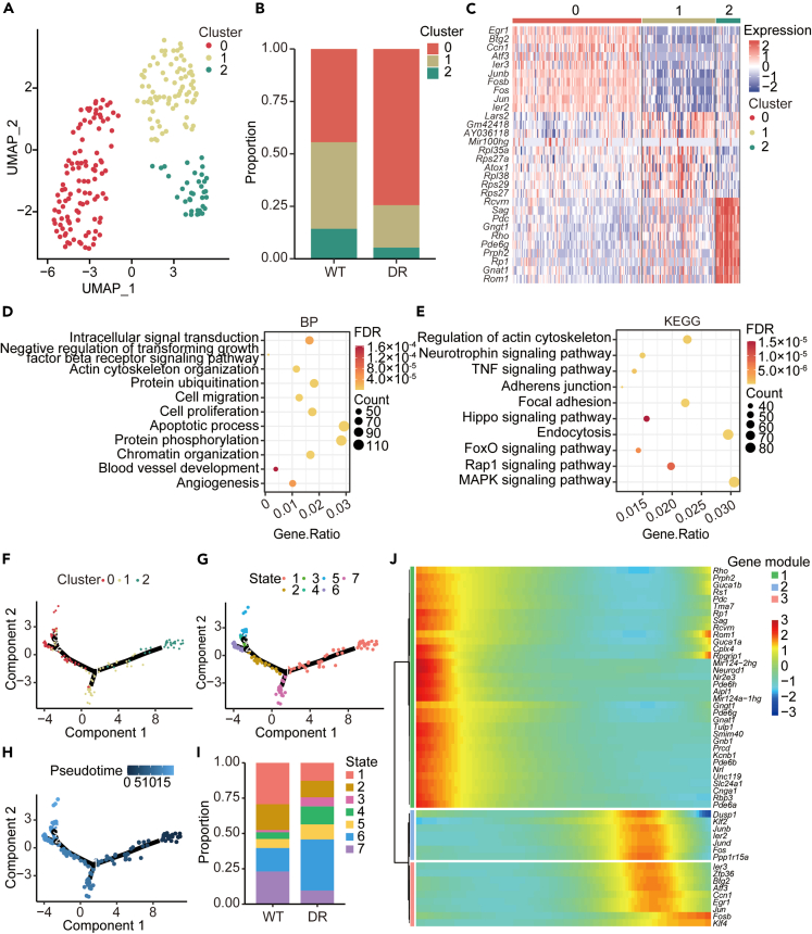
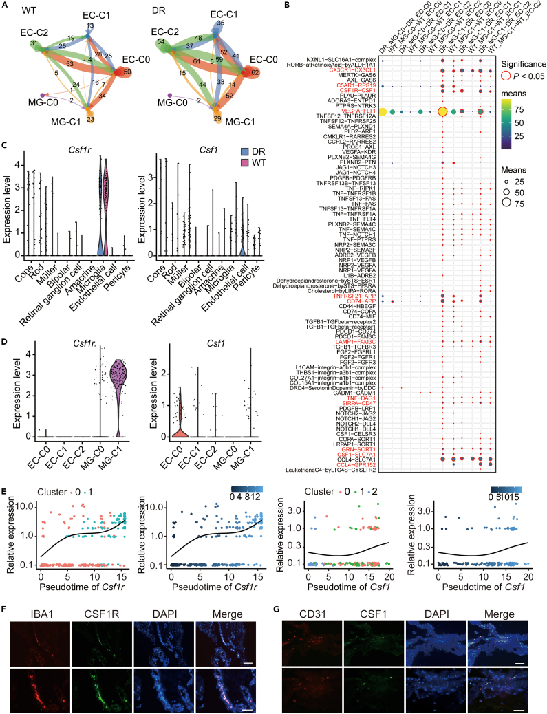
Inflammation-mediated crosstalk between neuroglial cells and endothelial cells (ECs) is a fundamental feature of many vascular diseases. Nevertheless, the landscape of inflammatory processes during diabetes-induced microvascular dysfunction remains elusive. Here, we applied single-cell RNA sequencing to elucidate the transcriptional landscape of diabetic retinopathy (DR). The transcriptome characteristics of microglia and ECs revealed two microglial subpopulations and three EC populations. Exploration of intercellular crosstalk between microglia and ECs showed that diabetes-induced interactions mainly participated in the inflammatory response and vessel development, with colony-stimulating factor 1 (CSF1) and CSF1 receptor (CSF1R) playing important roles in early cell differentiation. Clinically, we found that CSF1/CSF1R crosstalk dysregulation was associated with proliferative DR. Mechanistically, ECs secrete CSF1 and activate CSF1R endocytosis and the CSF1R phosphorylation-mediated MAPK signaling pathway, which elicits the differentiation of microglia and triggers the secretion of inflammatory factors, and subsequently foster angiogenesis by remodeling the inflammatory microenvironment through a positive feedback mechanism.
Keywords: Biological sciences; Diabetology; Endocrinology; Natural sciences.
© 2024 The Authors.
Conflict of interest statement
The authors declare no competing interests.
Figures


References
-
- Klemm F., Maas R.R., Bowman R.L., Kornete M., Soukup K., Nassiri S., Brouland J.-P., Iacobuzio-Donahue C.A., Brennan C., Tabar V., et al. Interrogation of the Microenvironmental Landscape in Brain Tumors Reveals Disease-Specific Alterations of Immune Cells. Cell. 2020;181:1643–1660.e17. doi: 10.1016/j.cell.2020.05.007. - DOI - PMC - PubMed
LinkOut - more resources
Full Text Sources
Research Materials
Miscellaneous
