

Deciphering non-canonical ubiquitin signaling: biology and methodology
- PMID: 38414868
- PMCID: PMC10897730
- DOI: 10.3389/fmolb.2023.1332872
Deciphering non-canonical ubiquitin signaling: biology and methodology
Abstract
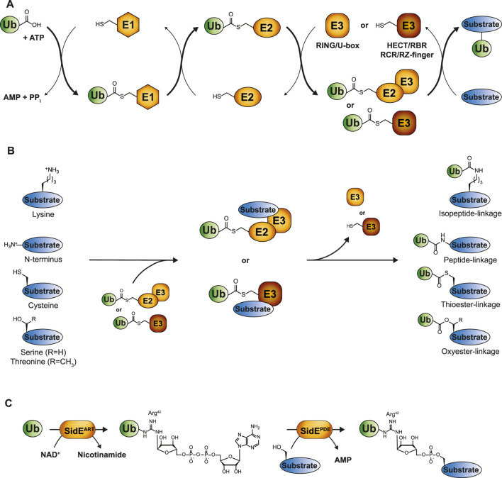
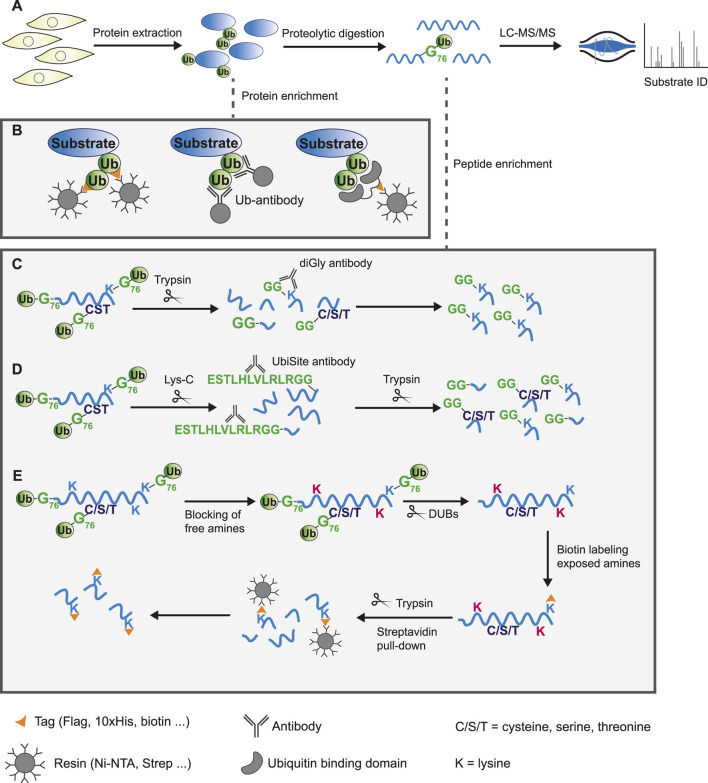
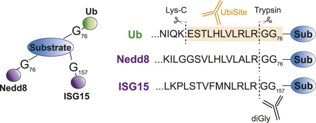
Ubiquitination is a dynamic post-translational modification that regulates virtually all cellular processes by modulating function, localization, interactions and turnover of thousands of substrates. Canonical ubiquitination involves the enzymatic cascade of E1, E2 and E3 enzymes that conjugate ubiquitin to lysine residues giving rise to monomeric ubiquitination and polymeric ubiquitination. Emerging research has established expansion of the ubiquitin code by non-canonical ubiquitination of N-termini and cysteine, serine and threonine residues. Generic methods for identifying ubiquitin substrates using mass spectrometry based proteomics often overlook non-canonical ubiquitinated substrates, suggesting that numerous undiscovered substrates of this modification exist. Moreover, there is a knowledge gap between in vitro studies and comprehensive understanding of the functional consequence of non-canonical ubiquitination in vivo. Here, we discuss the current knowledge about non-lysine ubiquitination, strategies to map the ubiquitinome and their applicability for studying non-canonical ubiquitination substrates and sites. Furthermore, we elucidate the available chemical biology toolbox and elaborate on missing links required to further unravel this less explored subsection of the ubiquitin system.
Keywords: E3 ligase; affinity purification; mass spectrometry; non-canonical ubiquitination; oxyester; proteomics; thioester; ubiquitin.
Copyright © 2024 van Overbeek, Aguirre, van der Heden van Noort, Blagoev and Vertegaal.
Conflict of interest statement
UbiSite is patented by the University of Southern Denmark (patent number US9476888B2). The author(s) declared that they were an editorial board member of Frontiers, at the time of submission. This had no impact on the peer review process and the final decision.
Figures
Similar articles
-
Structural insights into the conformation and oligomerization of E2~ubiquitin conjugates.Biochemistry. 2012 May 22;51(20):4175-87. doi: 10.1021/bi300058m. Epub 2012 May 14. Biochemistry. 2012. PMID: 22551455 Free PMC article.
-
Deciphering the ubiquitin proteome: Limits and advantages of high throughput global affinity purification-mass spectrometry approaches.Int J Biochem Cell Biol. 2013 Oct;45(10):2136-46. doi: 10.1016/j.biocel.2013.05.031. Epub 2013 Jun 10. Int J Biochem Cell Biol. 2013. PMID: 23764619 Review.
-
Mechanisms of mono- and poly-ubiquitination: Ubiquitination specificity depends on compatibility between the E2 catalytic core and amino acid residues proximal to the lysine.Cell Div. 2010 Aug 13;5:19. doi: 10.1186/1747-1028-5-19. Cell Div. 2010. PMID: 20704751 Free PMC article.
-
On the Possibility That Bond Strain Is the Mechanism of RING E3 Activation in the E2-Catalyzed Ubiquitination Reaction.J Chem Inf Model. 2022 Dec 26;62(24):6475-6481. doi: 10.1021/acs.jcim.2c00423. Epub 2022 Jun 7. J Chem Inf Model. 2022. PMID: 35671046
-
[Advances in the application of affinity separation for analyzing protein ubiquitination].Se Pu. 2021 Jan;39(1):26-33. doi: 10.3724/SP.J.1123.2020.07005. Se Pu. 2021. PMID: 34227356 Free PMC article. Review. Chinese.
Cited by
-
The Molecular Toolbox for Linkage Type-Specific Analysis of Ubiquitin Signaling.Chembiochem. 2025 May 27;26(10):e202500114. doi: 10.1002/cbic.202500114. Epub 2025 Apr 21. Chembiochem. 2025. PMID: 40192223 Free PMC article. Review.
References
-
- Akimov V., Olsen L. C. B., Hansen S. V. F., Barrio-Hernandez I., Puglia M., Jensen S. S., et al. (2018b). StUbEx PLUS-A modified stable tagged ubiquitin exchange system for peptide level purification and in-depth mapping of ubiquitination sites. J. Proteome Res. 17 (1), 296–304. 10.1021/acs.jproteome.7b00566 - DOI - PubMed
Publication types
LinkOut - more resources
Full Text Sources
Miscellaneous