

Sub-lethal concentrations of chlorhexidine inhibit Candida albicans growth by disrupting ROS and metal ion homeostasis
- PMID: 38415078
- PMCID: PMC10898817
- DOI: 10.1080/20002297.2023.2278937
Sub-lethal concentrations of chlorhexidine inhibit Candida albicans growth by disrupting ROS and metal ion homeostasis
Abstract
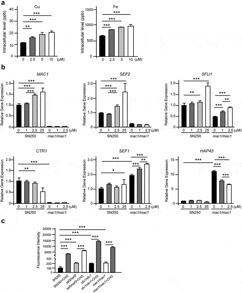
Candida albicans is a normal resident of the human oral cavity. It is also the most common fungal pathogen, causing various oral diseases, particularly in immunocompromised individuals. Chlorhexidine digluconate (CHG) is a broad-spectrum antimicrobial agent widely used in dental practice and has been recommended to treat oral candidiasis. However, its action mechanism against the fungal pathogen C. albicans remains poorly understood. The aim of the present study was to investigate the effect of CHG at sub-lethal concentrations against C. albicans. CHG inhibited the growth of C. albicans in a dose- and time-dependent manner. Cells treated with CHG exhibited altered membrane permeability, reduced metabolic activity, and enhanced metal ion and reactive oxygen species (ROS) accumulation. Copper-sensing transcription factor Mac1, iron-sensing transcription factors Sfu1 and Sef2, and copper transporter Ctr1 regulated intracellular metal ion and ROS homeostasis in response to CHG. Deletion of MAC1, SFU1, or SEF2 increased intracellular ROS production and cell susceptibility to CHG. This study revealed a novel mechanism by which CHG induced apoptosis of C. albicans cells through the disruption of metal ion and ROS homeostasis, which may help to identify new targets for fungal infections.
Keywords: Candida albicans; antifungal activity; chlorhexidine; metal ion homeostasis; reactive oxygen species.
© 2023 The Author(s). Published by Informa UK Limited, trading as Taylor & Francis Group.
Conflict of interest statement
No potential conflict of interest was reported by the author(s).
Figures


Similar articles
-
The effects of clioquinol in morphogenesis, cell membrane and ion homeostasis in Candida albicans.BMC Microbiol. 2020 Jun 16;20(1):165. doi: 10.1186/s12866-020-01850-3. BMC Microbiol. 2020. PMID: 32546212 Free PMC article.
-
The Antifungal Peptide MCh-AMP1 Derived From Matricaria chamomilla Inhibits Candida albicans Growth via Inducing ROS Generation and Altering Fungal Cell Membrane Permeability.Front Microbiol. 2020 Jan 21;10:3150. doi: 10.3389/fmicb.2019.03150. eCollection 2019. Front Microbiol. 2020. PMID: 32038583 Free PMC article.
-
Anti-biofilm activity of chlorhexidine digluconate against Candida albicans vaginal isolates.PLoS One. 2020 Sep 17;15(9):e0238428. doi: 10.1371/journal.pone.0238428. eCollection 2020. PLoS One. 2020. PMID: 32941438 Free PMC article.
-
Harnessing Metal Homeostasis Offers Novel and Promising Targets Against Candida albicans.Curr Drug Discov Technol. 2020;17(4):415-429. doi: 10.2174/1570163816666190227231437. Curr Drug Discov Technol. 2020. PMID: 30827249 Review.
-
Increasing usage of chlorhexidine in health care settings: blessing or curse? A narrative review of the risk of chlorhexidine resistance and the implications for infection prevention and control.Eur J Clin Microbiol Infect Dis. 2022 Mar;41(3):349-362. doi: 10.1007/s10096-022-04403-w. Epub 2022 Jan 19. Eur J Clin Microbiol Infect Dis. 2022. PMID: 35048278 Review.
Cited by
-
Synergistic antibacterial effect of ginsenoside Rh2 and calcium hydroxide on Enterococcus faecalis.Odontology. 2025 Jan;113(1):111-125. doi: 10.1007/s10266-024-00951-z. Epub 2024 May 19. Odontology. 2025. PMID: 38762821
-
Microbiological and toxicity analyses of the synthetic polymer polyhexamethylene guanidine hydrochloride against endodontic microorganisms.Braz J Microbiol. 2025 Mar;56(1):475-486. doi: 10.1007/s42770-024-01603-8. Epub 2025 Jan 15. Braz J Microbiol. 2025. PMID: 39812973
-
Are Mouthwashes Really Effective against Candida spp.?J Fungi (Basel). 2024 Jul 29;10(8):528. doi: 10.3390/jof10080528. J Fungi (Basel). 2024. PMID: 39194854 Free PMC article. Review.
References
-
- Calderone RA. Candida and candidiasis. Washington DC: ASM Press; 2002.
-
- Brailsford SR, Shah B, Simons D, et al. The predominant aciduric microflora of root-caries lesions. J Dent Res. 2001;80:1828–1833. - PubMed
LinkOut - more resources
Full Text Sources
Research Materials