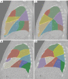
Deep learning-based bronchial tree-guided semi-automatic segmentation of pulmonary segments in computed tomography images
- PMID: 38415134
- PMCID: PMC10895116
- DOI: 10.21037/qims-23-1251
Deep learning-based bronchial tree-guided semi-automatic segmentation of pulmonary segments in computed tomography images
Abstract
Background: Pulmonary segments are valuable because they can provide more precise localization and intricate details of lung cancer than lung lobes. With advances in precision therapy, there is an increasing demand for the identification and visualization of pulmonary segments in computed tomography (CT) images to aid in the precise treatment of lung cancer. This study aimed to integrate multiple deep-learning models to accurately segment pulmonary segments in CT images using a bronchial tree (BT)-based approach.
Methods: The proposed segmentation method for pulmonary segments using the BT-based approach comprised the following five essential steps: (I) segmentation of the lung using a U-Net (R231) (public access) model; (II) segmentation of the lobes using a V-Net (self-developed) model; (III) segmentation of the airway using a combination of a differential geometric approach method and a BronchiNet (public access) model; (IV) labeling of the BT branches based on anatomical position; and (V) segmentation of the pulmonary segments based on the distance of each voxel to the labeled BT branches. This five-step process was applied to 14 high-resolution breath-hold CT images and compared against manual segmentations for evaluation.
Results: For the lung segmentation, the lung mask had a mean dice similarity coefficient (DSC) of 0.98±0.03. For the lobe segmentation, the V-Net model had a mean DSC of 0.94±0.06. For the airway segmentation, the average total length of the segmented airway trees per image scan was 1,902.8±502.1 mm, and the average number of the maximum airway tree generations was 8.5±1.3. For the segmentation of the pulmonary segments, the proposed method had a DSC of 0.73±0.11 and a mean surface distance of 6.1±2.9 mm.
Conclusions: This study demonstrated the feasibility of combining multiple deep-learning models for the auxiliary segmentation of pulmonary segments on CT images using a BT-based approach. The results highlighted the potential of the BT-based method for the semi-automatic segmentation of the pulmonary segment.
Keywords: Lung cancer; airway segmentation; lobe segmentation; lung segmentation; pulmonary segments segmentation.
2024 Quantitative Imaging in Medicine and Surgery. All rights reserved.
Conflict of interest statement
Conflicts of Interest: All authors have completed the ICMJE uniform disclosure form (available at https://qims.amegroups.com/article/view/10.21037/qims-23-1251/coif). The authors have no conflicts of interest to declare.
Figures


References
-
- Wild C, Weiderpass E, Stewart BW. World cancer report: cancer research for cancer prevention. IARC Press; 2020.
-
- Saji H, Okada M, Tsuboi M, Nakajima R, Suzuki K, Aokage K, et al. Segmentectomy versus lobectomy in small-sized peripheral non-small-cell lung cancer (JCOG0802/WJOG4607L): a multicentre, open-label, phase 3, randomised, controlled, non-inferiority trial. Lancet 2022;399:1607-17. 10.1016/S0140-6736(21)02333-3 - DOI - PubMed
-
- Baisden JM, Romney DA, Reish AG, Cai J, Sheng K, Jones DR, Benedict SH, Read PW, Larner JM. Dose as a function of lung volume and planned treatment volume in helical tomotherapy intensity-modulated radiation therapy-based stereotactic body radiation therapy for small lung tumors. Int J Radiat Oncol Biol Phys 2007;68:1229-37. 10.1016/j.ijrobp.2007.03.024 - DOI - PubMed
-
- Yuan ST, Frey KA, Gross MD, Hayman JA, Arenberg D, Cai XW, Ramnath N, Hassan K, Moran J, Eisbruch A, Ten Haken RK, Kong FM. Changes in global function and regional ventilation and perfusion on SPECT during the course of radiotherapy in patients with non-small-cell lung cancer. Int J Radiat Oncol Biol Phys 2012;82:e631-8. 10.1016/j.ijrobp.2011.07.044 - DOI - PMC - PubMed
LinkOut - more resources
Full Text Sources
