Review
. 2024 Dec 12;12(1):eesp00012023.
doi: 10.1128/ecosalplus.esp-0001-2023. Epub 2024 Jan 4.

Infection biology of Salmonella enterica

Affiliations
Review

Infection biology of Salmonella enterica

Jing Han et al. EcoSal Plus. .

Abstract

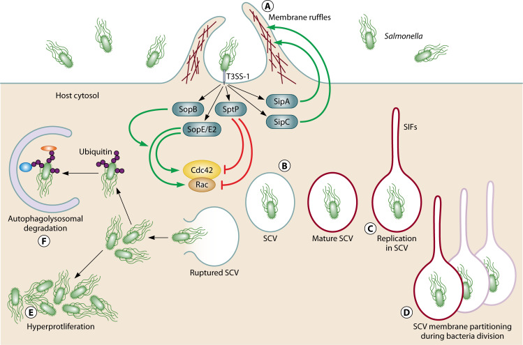
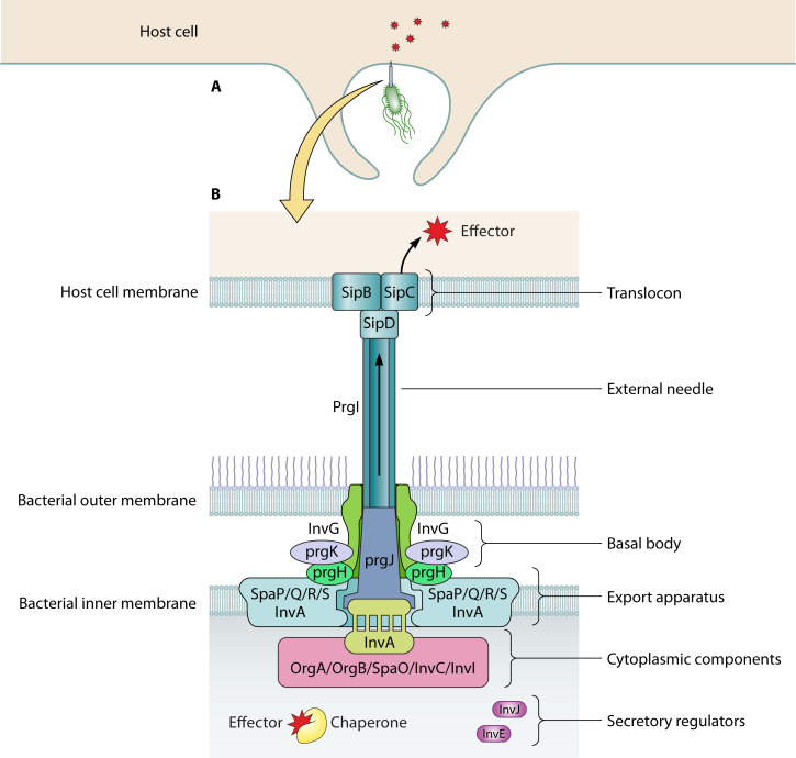
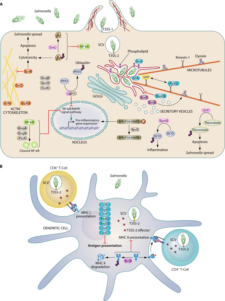
Salmonella enterica is the leading cause of bacterial foodborne illness in the USA, with an estimated 95% of salmonellosis cases due to the consumption of contaminated food products. Salmonella can cause several different disease syndromes, with the most common being gastroenteritis, followed by bacteremia and typhoid fever. Among the over 2,600 currently identified serotypes/serovars, some are mostly host-restricted and host-adapted, while the majority of serotypes can infect a broader range of host species and are associated with causing both livestock and human disease. Salmonella serotypes and strains within serovars can vary considerably in the severity of disease that may result from infection, with some serovars that are more highly associated with invasive disease in humans, while others predominantly cause mild gastroenteritis. These observed clinical differences may be caused by the genetic make-up and diversity of the serovars. Salmonella virulence systems are very complex containing several virulence-associated genes with different functions that contribute to its pathogenicity. The different clinical syndromes are associated with unique groups of virulence genes, and strains often differ in the array of virulence traits they display. On the chromosome, virulence genes are often clustered in regions known as Salmonella pathogenicity islands (SPIs), which are scattered throughout different Salmonella genomes and encode factors essential for adhesion, invasion, survival, and replication within the host. Plasmids can also carry various genes that contribute to Salmonella pathogenicity. For example, strains from several serovars associated with significant human disease, including Choleraesuis, Dublin, Enteritidis, Newport, and Typhimurium, can carry virulence plasmids with genes contributing to attachment, immune system evasion, and other roles. The goal of this comprehensive review is to provide key information on the Salmonella virulence, including the contributions of genes encoded in SPIs and plasmids during Salmonella pathogenesis.

Keywords: Salmonella; pathogenicity; virulence factors.


Conflict of interest statement

The authors declare no conflict of interest.

Figures

Fig 1
Fig 1
Attachment and internalization of Salmonella in host cell. Rho subfamily of GTPases Cdc42 and Rac are required in Salmonella-induced membrane ruffling. Following attachment to the host cell surface, Salmonella can express T3SS-1 that facilitates endothelial uptake and invasion through the translocation of effector proteins from the bacterial cell to the host cell cytosol. Salmonella translocates three proteins (SopB, SopE, and SopE2) that are mimetics of small Rho GTPase regulatory enzymes. SopE and SopE2, which have guanine nucleotide exchange factor (GEF) activity, activate Cdc42 and Rac directly. SopB, an inositol polyphosphatase, impacts indirectly on Cdc42 activation and is, furthermore, important for later steps of intracellular persistence. Effectors SipA and SipC are cytoskeleton-altering effectors and they interact directly with actin. SipA binds directly to F-actin to modulate the actin-bundling (crosslinking) activity of T-plastin. SipC is involved in the nucleation of actin polymerization. The membrane ruffling response is down-regulated by the GTPase-activating protein (GAP) SptP, which inactivates Cdc42 and Rac. The coordinate action of these effectors promotes membrane ruffling and bacterial invasion (A). Membrane ruffling allows Salmonella to be surrounded by the host cell membrane and internalized in the membrane-bound Salmonella-containing vacuole (SCV) (B). Effector proteins expressed by Salmonella in the SCV, including both T3SS-1 effectors (SipA, SopB, SopD, and SopD2) and T3SS-2 effectors (SseF, SseG, PipB2, and SifA), promote SCV mature (B) and also interact with the cytoskeleton and associated motor proteins leading to the formation of Salmonella-induced filaments (SIF) which extend from SCV (C). In the mature SCV, Salmonella replication is initiated 4–6  h post invasion (C). The SCV membrane frequently divides along with vacuolated Salmonella, resulting in a single bacterium per SCV (D). Instead of undergoing this maturation process, some of the SCVs will rupture, resulting in the release of Salmonella into host cytosol. In this case, Salmonella are either exposed to the nutrient rich host cytosol in which they can hyperproliferate (E) or can be targeted by the host cell pathogen defense system, which leads to their ubiquitylation and degradation through the autophagic pathway (F). Modified from models described by Herhaus and Dikic (99).
Fig 2
Fig 2
Schematic of the T3SS-1 structure and function. (A) Bacteria utilize T3SS-1 to inject bacterial effector proteins into eukaryotic host cells. (B) Structure T3SS-1 and its effectors/chaperones. The base structure of the T3SS-1 comprised the inner ring structure which is composed of inner membrane ring (PrgH and PrgK protein subunits) and outer membrane ring (InvG protein), the export apparatus which is composed of InvA and Spa PQRS, and the cytoplasmic component of the export machinery which is composed of SpaO, OrgAB, InvI, and InvC. The completed base structure allows for the assembly of the needle (sipD), needle filament (PrgI), and inner rod (PrgJ). Inside the bacteria cell, the chaperone molecules bind to the effector proteins and accompany the molecule to ATPase complex allowing the effector protein to be released from the chaperone and enter the inner rod of needle structure for translocation into the host cell. The needle complex, either directly or facilitated by accessory proteins at the needle tip, interacts with the translocation complex to allow for delivery of bacterial proteins into the host cytosol. Modified from models described by Lou et al. (119) and Wagner et al. (122).
Fig 3
Fig 3
Function of T3SS-2 effectors. T3SS-2 is induced after invasion and functions mainly in manipulating SCV trafficking and maturation, promoting intracellular survival and replication, and causing the systemic phase of infection. (A) The function of T3SS-2 in manipulating SCV membrane dynamics is through the action of the effectors SifA, SopD2, SseJ, and PipB2 (highlighted in turquoise color). The localization of Samonella-containing SCV is mediated by effectors SseF and SseG (highlighted in lavender). Targeting the host cytoskeletons is mediated by effectors SteC, SpvB, SspH2, and SrfH (highlighted in orange). Effectors (SspH1, SspH2, SlrP, and SseL, highlighted in pink) interfere with the host cell ubiquitylation system. SspH1, SspH2, and SlrP cause ubiquitylation, whereas SseL deubiquitylate various host proteins to favor Salmonella proliferation inside the host. Effectors SspH1, GogB, SpvC, SseK, GtgA, GogA, PipA, and SpvD (highlighted in brown) inhibit the innate immune signaling, and subsequently diminish the production of pro-inflammatory mediators and result in an inefficient clearance of phagocytized bacteria. Figure A is modified from the model described by Figueira and Holden (198). (B) T3SS-2 effectors modulating the adaptive immune signaling. After being delivered into DCs, effectors SifA, SspH2, SlrP, PipB2, and SopD2 inhibit DC chemotaxis toward CCL19, therefore compromising the antigen presentation in DC. Effector SteD stimulates ubiquitination of mature MHCII, leading to its degradation and directly preventing MHCII antigen presentation. Figure 2 is modified from the model described by Cerny and Holden.(2019) (212).

References

    1. Mead PS, Slutsker L, Dietz V, McCaig LF, Bresee JS, Shapiro C, Griffin PM, Tauxe RV. 1999. Food-related illness and death in the United States. Emerg Infect Dis 5:607–625. doi:10.3201/eid0505.990502 - DOI - PMC - PubMed
    1. CDC . 2019. Antibiotic resistance threats in the United States, 2019
    1. (IFSAC), T.I.F.S.A.C . 2019. Foodborne illness source attribution estimates for 2017 for Salmonella, Escherichia coli O157,Listeria monocytogenes, and Campylobacter using multi-year outbreak surveillance data,United States
    1. Scharff RL. 2012. Economic burden from health losses due to foodborne illness in the United States. J Food Prot 75:123–131. doi:10.4315/0362-028X.JFP-11-058 - DOI - PubMed
    1. Brenner FW, Villar RG, Angulo FJ, Tauxe R, Swaminathan B. 2000. Salmonella nomenclature. J Clin Microbiol 38:2465–2467. doi:10.1128/JCM.38.7.2465-2467.2000 - DOI - PMC - PubMed

Substances

LinkOut - more resources